
ASSOCIATE PROFESSOR DEPT OF MICROBIOLOGY CIMSH LKO DR MONIKA RAJANI CLASS SPOROZOA SUB CLASS COCCIDIA GENUS Cryptosporidium Isosporacyclospora Toxoplasma SUB ORDER EIMERIINA SUB ORDER ID: 1043276
Download Presentation The PPT/PDF document "TOXOPLASMA GONDII DR MONIKA RAJANI" is the property of its rightful owner. Permission is granted to download and print the materials on this web site for personal, non-commercial use only, and to display it on your personal computer provided you do not modify the materials and that you retain all copyright notices contained in the materials. By downloading content from our website, you accept the terms of this agreement.


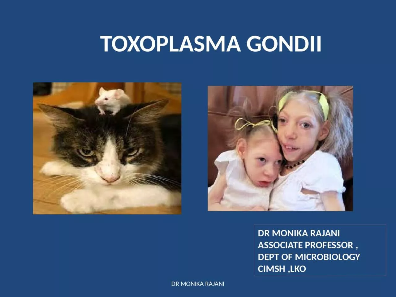
1. TOXOPLASMA GONDIIDR MONIKA RAJANI ASSOCIATE PROFESSOR ,DEPT OF MICROBIOLOGYCIMSH ,LKODR MONIKA RAJANI
2. CLASS SPOROZOASUB CLASSCOCCIDIAGENUSCryptosporidiumIsospora,cyclosporaToxoplasmaSUB ORDER EIMERIINASUB ORDERHAEMOSPORINAPHYLUM APICOMPLEXAGENUSPLASMODIUMBABESIADR MONIKA RAJANI
3. DR MONIKA RAJANIGeneral features CoccidiaThey possess a structure called apical complex by means of which they attach to and penetrate host cells.They live intracellularlyHave a sexual sporogenic phase and an asexual shizogenic phase,(alternation of generation).Alternation of hosts.Frequent association with HIV infection.
4. DR MONIKA RAJANIToxoplasma GondiiObligate intracellular coccidian parasite.It was first described in a small North American rodent called gondii(Ctenodactylus gondii).First recognised in a cyst in retina of childName “Toxo” is derived from Greek word “Toxon”meaning arc or brow referring to curved shape of trophozoite.most common parasite globally with widest range of hosts
5. MORPHOLOGYT gondii occures in three forms:TrophozoiteTissue cyst shizogonyOocyst - sporogonyDefinitive host: cats and other felines :all three forms are seenIntermediate host: humans,mammals (sheep,cattle,mice) and birds :only asexual forms are seen.All three forms are infectious to man.DR MONIKA RAJANI
6. DR MONIKA RAJANIMORPHOLOGY: TrophozoitesCrescent shaped structures with one end pointed and other end rounded.Apical complex at pointed end.intracellularReplicates by endogonyTacyzoites:proliferating trophozoites in acute infectionPseudocyst: proliferating trophozoites within cell may appear rounded by host cell membrane.Susceptible to gastric digestion.Giemsa stain: cytoplasm blue and nucleus red.
7. Tachyzoites DR MONIKA RAJANI
8. MORPHOLOGY: Tissue cystResting forms of parasite found in chronic stage of infection.Sites: brain(most common),muscles and other organs.Contain -Bradyzoite:slow multiplying parasites within cyst. Cysts may get reactivated in immunodeficient hostsCysts reach various organs through blood and lymphatics.susceptible to peptic or tryptic digestion.Infection occurs when raw or undercooked meat is eaten.DR MONIKA RAJANI
9. MORPHOLOGY: OocystSeen only in definitive hosts(intestine of cats).Oval,10-12 um, ,thick resistant wall.Formed by gametogony or sporogony.Millions of oocysts are shed by cats in their feces during primary infection.Freshly passed oocysts are not infectious.Cysts undergo sporulation in soil to form two sporocysts,each containing four sporozoites.Oocysts are resistant to environmental conditions.Sporulated oocyst is infective.When sporulated oocyst is ingested it releases sporozoites in intestine which initiates infection.DR MONIKA RAJANI
10. Life cycleTwo hosts required.Definitive host: cats and other felines :both sexual and asexual cycle takes place. :three morphological forms are seen.Intermediate host: man and other mammmals-mice,rats sheep,cattle pigs. :Only asexual cycle occurs. :Only trophozoites and tissue cysts are seen.Natural cycle occurs in mice and cats.Mice acquire infection by eating materials contaminated with cats feces containing oocysts.Two types of cycle:e nteric cycle and exo enteric cycleDR MONIKA RAJANI
11. DR MONIKA RAJANI
12. Life cycleDR MONIKA RAJANI
13. Enteric cycleOccurs in definitive host.Cats acquire infection by ingestion of tissue cysts in meat of rats and other animals.Tissue cysts ingested bradyzoites merozoitesmale and female gametocytes oocyst. In fecesSporulating oocyst in soil-infectiveDR MONIKA RAJANIHUMAN INFECTION IS THE DEAD END OF PARASITE.HUMAN TOXOPLASMOSIS IS A ZOONOSIS
14. Exo enteric cycleOccurs in intermediate host.1-Humans acquire infection by eating uncooked or undercooked meat particularly lamb and pork containing tissue cysts.2-Ingestion of oocysts in contaminated food and water containing oocysts.3-Intrauterine infection(congenital toxoplasmosis).4-Blood transfusion or organ transplant.DR MONIKA RAJANI
15. DR MONIKA RAJANIExo enteric cycleIngestion of oocystsSporozoites released in intestine.Shizogony occurs tacyzoites formedTacyzoites multiply by endogony.Tacyzoites move via blood and lymphatics to various organs.Tissue cyst tissue cysts can get reactivated Occurs in intermediate host.
16. Pathogenicity and clinical featuresMost human infections are asymptomatic.Active progression of disease is seen in immunodeficient and HIV pts.Clinical toxoplasmosis:congenital toxoplasmosis :Acquired toxoplasmosis.DR MONIKA RAJANI
17. Congenital toxoplasmosisTransplacental infection.Occurs when mother gets a primary infection in pregnancy.Risk of fetal infection rises with progress of gestation25% in first trimester65% in third trimesterBut severity of fetal damage is highest when infection is transmitted in early pregnancy.Acute infection in mothers lead to infection in babies.DR MONIKA RAJANI
18. Congenital toxoplasmosisMost infected newborns are asymptomatic at birth and remain so.Some develop manifestations subsequently.Chorioretinitis,cerebral calcifications, convulsions,microcephaly and hydrocephalus. Strabismus, deafness, blindness,MRacute condition: fever,jaundice,rash,cataract,glaucoma,chorioretinitis,HSM,lymphadenopathy,myocarditis.DR MONIKA RAJANI
19. Congenital toxoplasmosisDR MONIKA RAJANI
20. Acquired toxoplasmosisLymphadenopathy: most commonly cervical LNFever,headache,myalgia,spleenomegalyMild flu like symptoms -Self limitingTyphus like exanthema Pneumonitis,myocarditis,meningoencephalitisDR MONIKA RAJANI
21. OCULAR TOXOPLASMOSISUveitis,choroiditis,chorioretinitisMay be severe requiring enucleation.DR MONIKA RAJANI
22. DR MONIKA RAJANIToxoplasmosis in Immunocompromised patientsMost serious and often fatalCon occur either as reactivation or acquisition of new infection.Brain involvement: most commonEncephalitis,altered mental state,seizures,cerebellar signs,meningesmusMultiple organs involved.Toxoplasmosis pneumonia
23. Lab diagnosis Microscopy:Tachyzoites detected in blood,BMA,sputum,Tissue cyst:biopsy from LN,spleen,brain,placentaStains:Giemsa ,PAS ,GMSDR MONIKA RAJANI
24. Lab diagnosis: Animal inoculationBy inoculating blood,BF or tissue specimensIntraperitonial inoculation in mice or tissue culture.Mice examined for Toxoplasma in peritonial exudate after 7-10 days.DR MONIKA RAJANI
25. Serology Main stay of diagnosisAntibody detection IgG :after 2-3 weeks :By ELISA, IFAT ,LA :indicates past infection :Sabin Feldman Dye TestIgM :Indicates early primary infection :Congenital infection :By double sandwich IgM ELISAIg G avidity test: for recent infectionDR MONIKA RAJANI
26. Serology Antigen detectionBy ELISAIndicates recent toxoplasma infectionDetected in blood and BFDetection of antigen in amniotic fluid is used to diagnose congenital toxoplasmosisDR MONIKA RAJANI
27. Skin test of Frenkel Diluted toxoplasmin is injected intradermallyDelayed hypersenstivity reaction is seen.Not very reliable for diagnosisDR MONIKA RAJANI
28. Sabin Feldman Dye TestFirst serological test described for Toxoplasma antibody by Sabin and Feldman.The test is based on specific inhibition by antibody of staining of trophozoites by alkaline methylene blue.Equal vol of pt serum+live trophozoites+human serum incubated at 37 C for one hour.Later one drop of methylene blue added to each tube and seen under microscope.Interpretation:less than 50% trophozoites take up stain :POSITIVE90-100% trophozoites take up stain:NEGATIVEDenotes absence of Toxoplasma antibodies.Limitation: false positive reactions with Sarcocystis and Trichomonas vaginalisDR MONIKA RAJANI
29. Molecular testsPCRDNA hybridisation techniqueDR MONIKA RAJANI
30. Imaging In case of CNS involvement:CT scan :MRIUSG of fetus in utero at 20-24 weeks of pregnancy is useful for diagnosis of congenital toxoplasmosis.DR MONIKA RAJANI
31. Treatment Congenital toxoplasmosis: oral pyrimethamine+Sulfadiazine+Folinic acid :Systemic corticosteroidsOcular toxoplasmosis: pyrimethamine+sulfadiazine/clindamycinImmunocompromised pts:AIDS pts that are seropositive for toxoplasma and have CD4 counts less than 100 should receive primary prophylaxis against encephalitis DOC:Trimethoprim-sulphamethoxazole DR MONIKA RAJANI
32. Prophylaxis Individuals at risk should avoid contact with cat and its feces.Proper cooking of mealProper washing of hands ,fruits and vegetables before cooking and eatingScreening of blood before transfusionOrgan safetyNo vaccine available for humansDR MONIKA RAJANI
33. THANK YOUDR MONIKA RAJANI