
1943 Disorders Jigar V Shah 1 Sharadchandra Shah 2 ORIGINAL RESEARCH Introduction Endoscopy of gastrointestinal diseases is an effective tool for the diagnostic evaluation and management of patient ID: 953177
Download Pdf The PPT/PDF document "Upper Gastrointestinal Endoscopy in Earl..." is the property of its rightful owner. Permission is granted to download and print the materials on this web site for personal, non-commercial use only, and to display it on your personal computer provided you do not modify the materials and that you retain all copyright notices contained in the materials. By downloading content from our website, you accept the terms of this agreement.


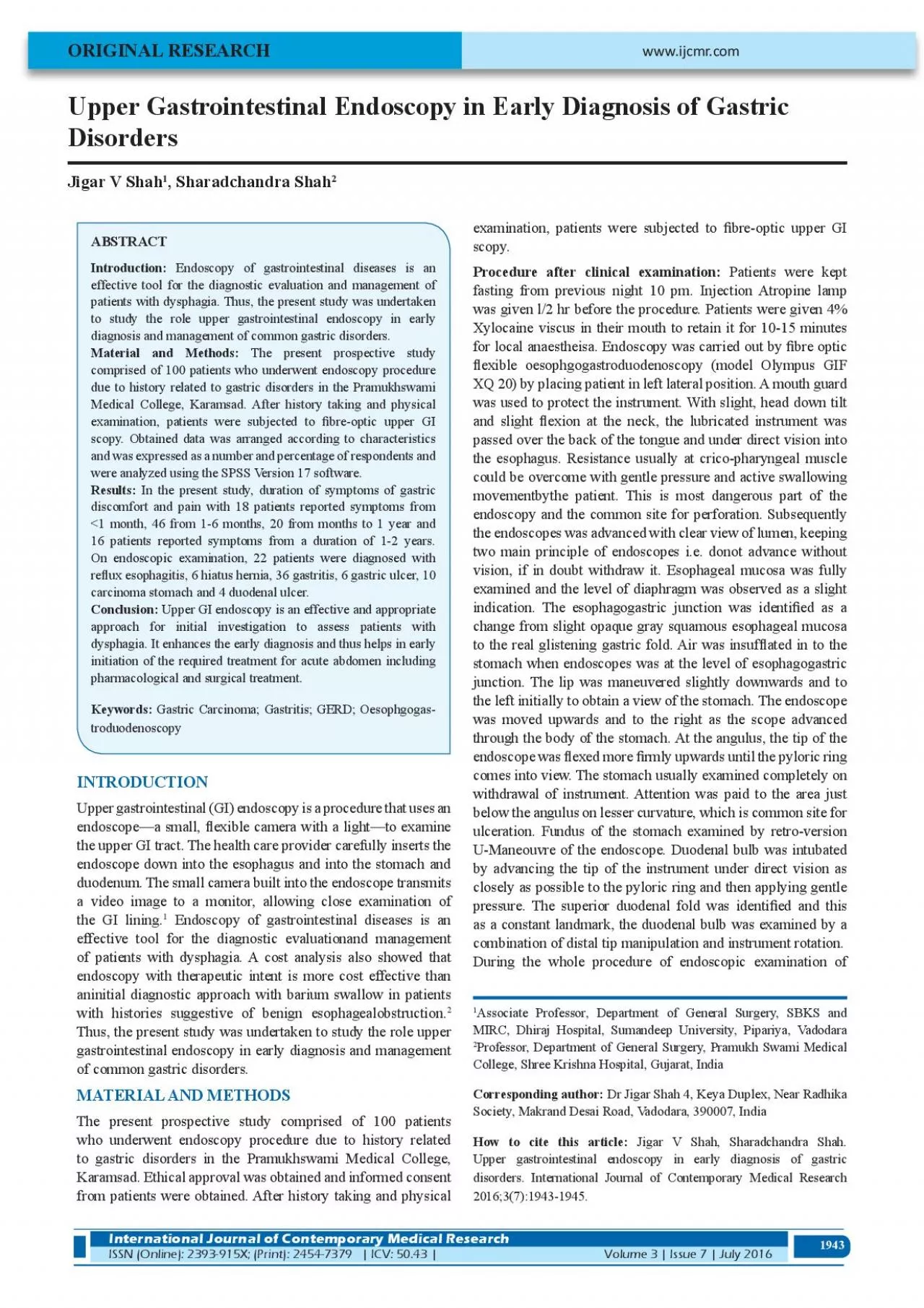
1943 Upper Gastrointestinal Endoscopy in Early Diagnosis of Gastric Disorders Jigar V Shah 1 , Sharadchandra Shah 2 ORIGINAL RESEARCH Introduction: Endoscopy of gastrointestinal diseases is an effective tool for the diagnostic evaluation and management of patients with dysphagia. Thus, the present study was undertaken to study the role upper gastrointestinal endoscopy in early Material and Methods: The present prospective study due to history related to gastric disorders in the Pramukhswami Medical College, Karamsad. After history taking and physical examination, patients were subjected to �bre-optic upper GI scopy. Obtained data was arranged according to characteristics and was expressed as a number and percentage of respondents and were analyzed using the SPSS Version 17 software. INTRODUCTION Upper gastrointestinal (GI) endoscopy is a procedure that uses an endoscope—a small, �exible camera with a light—to examine the upper GI tract. The health care provider carefully inserts the endoscope down into the esophagus and into the stomach and duodenum. The small camera built into the endoscope transmits a video image to a monitor, allowing close examination of the GI lining. 1 Endoscopy of gastrointestinal diseases is an effective tool for the diagnostic evaluationand management of patients with dysphagia. A cost analysis also showed that endoscopy with therapeutic intent is more cost effective than aninitial diagnostic approach with barium swallow in patients with histories suggestive of benign esophagealobstruction. Thus, the present study was undertaken to study the role upper gastrointestinal endoscopy in early diagnosis and management of common gastric disorders. MATERIAL AND METHODS The present prospective study comprised of 100 patients who underwent endoscopy procedure due to history related to gastric disorders in the Pramukhswami Medical College, Karamsad. Ethical approval was obtained and informed consent from patients were obtained. After history taking and physical examination, patients were subjected to �bre-optic upper GI scopy. Procedure after clinical examination: Patients were kept fasting from previous night 10 pm. Injection Atropine lamp was given l/2 hr before the procedure. Patients were given 4% for local anaestheisa. Endoscopy was carried out by �bre optic �exible oesophgogastroduodenoscopy (model Olympus GIF XQ 20) by placing patient in left lateral position. A mouth guard was used to protect the instrument. With slight, head down tilt and slight �exion at the neck, the lubricated instrument was passed over the back of the tongue and under direct vision into the esophagus. Resistance usually at crico-pharyngeal muscle could be overcome with gentle pressure and active swallowing movementbythe patient. This is most dangerous part of the endoscopy and the common site for perforation. Subsequently the endoscopes was advanced with clear view of lumen, keeping vision, if in doubt withdraw it. Esophageal mucosa was fully examined and the level of diaphragm was observed as a slight indication. The esophagogastric junction was identi�ed as a change from slight opaque gray squamous esophageal mucosa to the real glistening gastric fold. Air was insuf�ated in to the stomach when endoscopes was at the level of esophagogastric junction. The lip was maneuvered slightly downwards and to the left initially to obtain a view of the stomach. The endoscope was moved upwards and to the right as the scope advanced through the body of the stomach. At the angulus, the tip of the endoscope was �exed more �rmly upwards until the pyloric ring comes into view. The stomach usually examined completely on withdrawal of instrument. Attention was paid to the area just below the angulus on lesser curvature, which is common site for ulceration. Fundus of the stomach examined by retro-version U-Maneouvre of the endoscope. Duodenal bulb was intubated by advancing the tip of the instrument under direct vision as closely as possible to the pyloric ring and then applying gentle pressure. The superior duodenal fold was identi�ed and this as a constant landmark, the duodenal bulb was examined by a combination of distal tip manipulation and instrument rotat
ion. During the whole procedure of endoscopic examination of 1 Associate Professor, Department of General Surgery, SBKS and MIRC, Dhiraj Hospital, Sumandeep University, Pipariya, Vadodara www.ijcmr.com International Journal of Contemporary Medical Research ISSN (Online): 2393-915X; (Print): 2454-7379 | ICV: 50.43 |Volume 3 | Issue 7 | July 2016 Professor, Department of General Surgery, Pramukh Swami Medical Corresponding author: Dr Jigar Shah 4, Keya Duplex, Near Radhika Society, Makrand Desai Road, Vadodara, 390007, India How to cite this article: Jigar V Shah, Sharadchandra Shah. Upper gastrointestinal endoscopy in early diagnosis of gastric disorders. International Journal of Contemporary Medical Research 1944 esophagus stomach and duodenum, abnormal area either swelling, ulcer, growth, �brosis, bile re�ux, varices, gasto- esophageal re�ux, duodeno-gastric re�ux were properly evaluated and if doubt exists, biopsy was taken of pathological areas. All areas visualized and studied were recorded in form of drawing or written or by photography for comparison in future. Before withdrawal of instrument from stomach, air and gastric contents were aspirated. After completion of endoscopic examination, instrument was cleaned. STATISTICAL ANALYSIS Obtained data was arranged according to characteristics and was expressed as a number and percentage of respondents and were analyzed using the SPSS Version 17 software. RESULTS In the present study clinical diagnosis was carried out after history and Clinical examination and patients were subjected to endoscopic examination. Table-1 shows duration of symptoms of gastric discomfort and pain with 18 patients reported symptoms from <1 month, 46 from 1-6 months, 20 from months to 1 year and 16 patients reported symptoms from a duration of On physical examination, 68 patients showed tenderness, 12 scar, 10 hepatosplenomegly, 6 abdominal lump and 2 slenomegaly ( On endoscopic examination, 22 patients were diagnosed withre�ux esophagitis, 6 hiatus hernia, 36 gastritis, 6 gastric ulcer, 10 carcinoma stomach and 4 duodenal ulcer. In 16 patients, no abnormality in gastrointestinal tract was detected on endoscopic examination ( DISCUSSION The clinical indications for endoscopy of the uppergastrointestinal tract include symptoms typical of GERD but that are refractorytotreatment, alarmsigns (dysphagia,bleeding,weight loss, anemia), or symptoms in patients older than 50 years. There is no absolute contraindication for upper gastrointestinalendoscopy. Major complications such as perforation oraspiration are rare, occurring in less than 1 per 1000 cases. The present study found that maximum patients with gastrointestinal pain and discomfort were diagnosed with gastritis followed by re�ux esophagitis, carcinoma stomach, hiatus hernia, gastric ulcer and duodenal ulcer. Common reasons attributing to gastritis are Helicobacter pylori (H. pylori) infection, damage to the stomach lining, which leads to reactive gastritis sand an autoimmune response. The factors attributing to reactive gastritis are drinking alcohol, using cocaine, exposure to radiation or having radiation treatments, re�ux of bile from the small intestine into the stomach and a reaction to stress. This type of reactive gastritis is considered as stress gastritis. 1 Re�ux esophagitis or gastroesophageal re�ux disease (GERD) is a major digestive health problem and is de�ned as a condition that involves gastric content re�ux with ensuing symptoms or complications. It is one of the most frequent causes of gastroenterological consultations in out-patients and compromises the quality of life of the patients signi�cantly. 4 Gastric cancer is the second leading cause of death from malignant disease worldwide, with especially high mortality rates in East, South, and Central Asia; Central and Eastern Europe; and South America. Stomach cancer refers to any malignant neoplasm that arises from the region extending between the gastroesophageal junction and the pylorus. Approximately 95 percent of stomach tumours are epithelial in origin and designated as adenocarcinomas. Adenosquamous, squamous, and undifferentiated carcinomas are however rare. Even
though the multiple factors play role in etiology of gastric cancer, more than 80% of cases have been associated with H. pylori infection. Moreover, diet, lifestyle, genetic, socioeconomic and other factors also attributes to gastric carcinogenesis. 6 Qureshi NA et al 7 conducted a study to evaluate the diagnostic potential of endoscopy in 913 patients with dysphagia and found abnormal oesophagus in 678 cases (74%) and biopsies were taken in 428 patients (47%) with super�cial oesophagitis, Barrett's oesophagus, oesophageal cancer and oesophageal ulcer were The occurrence of hiatus hernia increases with age and body mass index. Herniation of the contents of the abdominal cavity most commonly the stomach is referred as Hiatus hernia, through the esophageal hiatus of the diaphragm into the mediastinum. Gastroesophageal re�ux disease is the main clinical manifestation of hiatus hernia. Endoscopy, high resolution manometry or radiology with barium swallow can help in diagonsis of hiatus hernia. Brewer BJ 8 conducted a study and revealed factors that help in identi�cation of the high risk patient with an acute surgical abdomen includes pain for less than 48 hours, pain followed by vomiting, guarding and rebound tenderness on physical examination, advanced age and a prior surgical procedure. The presence of these features demands careful evaluation by the No. Duration No. Percentage 1 18 18 46 46 4 16 16 Table-1: No. Findings No. Percentage 1 Tenderness 68 68 Scar Abdominal Lump 6 6 4 10 10 Slenomegaly Table-2: Patients with Positive Findings on Abdominal Examina tion No. Disease No. of Lesions 1 6 4 6 Carcinoma Stomach 10 6 4 7 16 Total 100 Table-3: pain 1945 physician for early diagnosis of the disease. Good pro�ciency in early diagnosis requires proper history taking, physical examination of the abdomen and knowledge of basic anatomy and physiology of gastrointestinal tract and. Therapeutic endoscopy, interventional radiology treatment and therapy using adult laparoscopy are the common modalities for treating patients with acute abdomen. CONCLUSION Upper GI endoscopy is an effective and appropriate approach for initialinvestigation to assess patients with dysphagia. It enhances the early diagnosis and thus helps in early initiation of the required treatment for acute abdomen including pharmacological and surgical treatment. REFERENCES 1. Gastritis. National Institute of Diabetes and Digestive and Kidney Diseases. Available at www.digestive.niddk.nih. gov . Pasha SF, Acosta RD, Chandrasekhara V et al. The role of endoscopy in the evaluation and management of dysphagia. Gastrointestinal Endoscopy. 2014;79:191-201. Roman S, KahrilasPJ.The diagnosis and management of 4. Coelho de ArrudaHMA.Diagnosis and management of gastroesophageal re�ux disease. Arq Bras Cir Dig. Takahashi T, Saikawa Y, Kitagawa Y.Gastric Cancer: Current Status of Diagnosis and Treatment Cancers 6. Nagini S. Carcinoma of the stomach: A review of epidemiology, pathogenesis, molecular genetics and chemoprevention. World J GastrointestOncol. 2012;4:156- 7. Qureshi NA, Hallissey MT, Fielding JW. Outcome of index upper gastrointestinal endoscopy in patients presenting with dysphagia in a tertiary care hospital-A 10 years. BMC Gastroenterology. 2007,7:43. 8. Brewer BJ, Golden GT, Hitch DC, Rudolf LE, Wangensteen SL. Abdominal pain. An analysis of 1,000 consecutive cases in a University Hospital emergency room. Am J Surg. Abdullah M, Firmansyah MA. Diagnostic Approach and Management of Acute Abdominal Pain. The Indonesian 10. Harsh Trivedi, Ronak Vyas, Kashyap Vyas, Shweta Sharma Evalution of etiological and high risk factors in the patients of acute pancreatitis. International Journal of Contemporary Source of Support: Con�ict of Interest: Submitted: Published online Shah, et al.Upper Gastrointestinal Endoscopy in Early Diagnosis of Gastric Disorders International Journal of Contemporary Medical Research Volume 3 | Issue 7 | July 2016 | ICV: 50.43 |ISSN (Online): 2393-915X; (Print): 2454-7379 Shah, et al.Upper Gastrointestinal Endoscopy in Early Diagnosis of Gastric Disorders International Journal of Contemporary Medical Research ISSN (Online): 2393-915X; (Print): 2454-7379 | ICV: 50.43 |Volume 3 | Issue 7 | July 2016