
The Double Helix DNA Structural model Model proposed by Watson amp Crick 1953 Two sugarphosphate strands next to each other but running in opposite directions Specific Hydrogen bonds occur among bases from one chain to the other ID: 916588
Download Presentation The PPT/PDF document "Nucleic Acids DNA & RNA" is the property of its rightful owner. Permission is granted to download and print the materials on this web site for personal, non-commercial use only, and to display it on your personal computer provided you do not modify the materials and that you retain all copyright notices contained in the materials. By downloading content from our website, you accept the terms of this agreement.


Slide1
Nucleic Acids
DNA & RNA
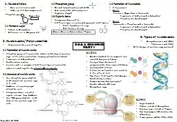
Slide2The Double Helix (DNA)
Structural model:
Model proposed by Watson & Crick, 1953
Two sugar-phosphate strands, next to each other, but running in opposite directions.
Specific Hydrogen bonds
occur among bases from one chain to the other:
A---T
,
C---G
Due to this specificity, a certain base on one strand indicates a certain base in the other
.
The 2 strands intertwine, forming a double-helix that winds around a central axis
Slide3Untwisted it looks like this:
The
sides
of the ladder are:
P =
phosphate S = sugar moleculeThe steps of the ladder are C, G, T, A = nitrogenous bases (Nitrogenous means containing the element nitrogen.) A = Adenine T = Thymine A always pairs with T in DNA C = Cytosine G = Guanine C always pairs with G in DNA
Nucleotide
(
A
pples are Tasty)
(
C
ookies are
G
ood)
Slide4Secondary Structure: DNA Double Helix
In DNA there are two strands of nucleotides that wind together in a
double helix
- the strands run in opposite directions
- the bases are are arranged in step-like pairs
- the
base pairs are held together by hydrogen bondingThe pairing of the bases from the two strands is very specificThe complimentary base pairs are A-T and G-C - two hydrogen bonds form between A and T - three hydrogen bonds form between G and CEach pair consists of a purine and a pyrimidine, so they are the same width, keeping the two strands at equal distances from each other
Slide5Model of
DNA
:
The model was developed by
Watson
and
Crick
in 1953. They received a nobel prize in 1962 for their work. The model looks like a twisted ladder – double helix.
Slide6Nucleic Acid Structure
“Base Pairing”
T
A
A
G
C
C3’TCGGTA3’
5’
5’
DNA base-pairing is
antiparalleli.e. 5’ - 3’ (l-r) on top : 5’ - 3’ (r-l) on
Slide7Discovering the structure of DNA
Erwin Chargaff – (1905-2002)
Columbia University, NY
Investigated the composition of DNA
His findings by 1950 strongly
suggested the base-pairings of A-T & G-C
Met with Watson and Crick in 1952 and shared his findings “Chargaff’s rule” A = T & C = G
Slide8Discovering the structure of DNA
DNA
=
Deoxyribose
nucleic acid Present in all living cells Contains all the information
Nucleotides: a subunit that consists of: a sugar (deoxyribose) a phosphate and one nitrogen base – 4 different basesAdenine (A) and Thymine (T)Guanine (G) and Cytosine (C)
Slide9PO
4
PO
4
PO
4
PO
4PO4PO4PO4PO4
PO
4
PO
4
PO
4
PO
4
PO
4
PO
4
PO
4
PO
4
The
strands separate
Slide10Nucleic Acid Structure
“Base Pairing”
RNA [normally] exists as a single stranded polymer
DNA exists as a double stranded polymer
DNA double strand is created by hydrogen bonds between nucleotides
Nucleotides always bind to complementary nucleotides
A
TCG(2 H-bonds)(3 H-bonds)
Slide11Practice DNA Base Pairs
G
A
T T
A
C
A
C T A A T G T
Slide12Complementarity of DNA strands
Two chains differ in sequence
(sequence is read from 5’ to 3’)
Two chains are
complementary
Two chains run antiparallel
Slide13Slide14Nucleic Acid Structure
“Base Pairing”
Slide15Nucleic Acid Structure
Polymerization
T
A
A
G
CC5’3’TAGCAC5’3’BasesSugar Phosphate“backbone”
Slide16Slide17P
P
(PPi)
Nucleic Acid Structure
Polymerization
P
PPSNCPPPSNC+PPPSN
C
P
S
N
C
Phosphodiesterase
Slide18