
Placoid Pigment Epitheliopathy Paulo Henrique Horizonte Nathália Nishiyama Tondelli Henrique Leber Gabriel Almeida Veiga Jacob Beatriz Mello Mencaroni Leonardo Benedito Cardoso Carolina Maria Barbosa ID: 931479
Download Presentation The PPT/PDF document "Case study : Acute Posterior Multifoc..." is the property of its rightful owner. Permission is granted to download and print the materials on this web site for personal, non-commercial use only, and to display it on your personal computer provided you do not modify the materials and that you retain all copyright notices contained in the materials. By downloading content from our website, you accept the terms of this agreement.


Slide1
Case
study
:
Acute
Posterior Multifocal
Placoid
Pigment EpitheliopathyPaulo Henrique Horizonte , Nathália Nishiyama Tondelli, Henrique Leber , Gabriel Almeida Veiga Jacob, Beatriz Mello Mencaroni, Leonardo Benedito Cardoso, Carolina Maria Barbosa Lemos, André Marcelo Vieira Gomes. Instituto Suel Abujamra
Acute Posterior Multifocal
Placoid
Pigment Epitheliopathy.
PURPOSE
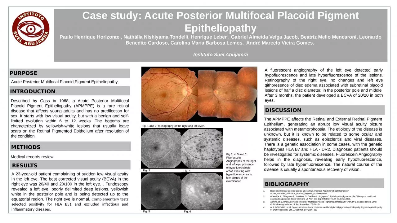
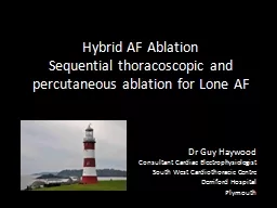
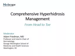
A fluorescent angiography of the left eye detected early hypofluorescence and late hyperfluorescence of the lesions. Retinography of the right eye, no changes and left eye qithpresence of disc edema associated with subretinal placoid lesions of half a disc diameter, in the posterior pole and middle After 3 months, the patient developed a BCVA of 20/20 in both eyes.
Described by Gass in 1968, a Acute Posterior Multifocal Placoid Pigment Epitheliopathy (APMPPE) is a rare retinal disease that affects young adults and has no predilection for sex. It starts with low visual acuity, but with a benign and self-limited evolution within 6 to 12 weeks. The bottoms are characterized by yellowish-white lesions that usually leave scars on the Retinal Pigmented Epithelium after resolution of the condition.
INTRODUCTION
Medical records review
METHODS
RESULTS
DISCUSSION
The APMPPE affects the Retinal and External Retinal Pigment Epithelium, generating an abrupt low visual acuity picture associated with metamorphopsia. The etiology of the disease is unknown, but it is known to be related to some ocular and systemic diseases, such as episcleritis and viral diseases. There is a genetic association in some cases, with the genetic haplotypes HLA B7 and HLA - DR2. Diagnosed patients should be investigated for systemic diseases. Fluorescein Angiography helps in the diagnosis, revealing early hypofluorescence, followed by late hyperfluorescence. The natural course of the disease is usually a spontaneous recovery of vision.
BIBLIOGRAPHY
Basic and Clinical Science Course 2016-2017 American Academy of Ophthalmology -Acute_Posterior_Multifocal_Placoid_Pigment_Epitheliopathy.Gibelalde A., Bidaguren A., Ostolaza JI, Cortázar L., Irigoyen C. Epiteliopatia pigmentar placóide agudo multifocal associado à paralisia do par craniano VI. Arch Soc Esp Oftalmol vol.84 no.3 mar.2009.Xerri O, et al. Untreated Acute Posterior Multifocal Placoid Pigment Epitheliopathy (APMPPE): a case series. BMC Ophthalmology volume 18, Article number: 76 (2018)A. F. DEUTMAN, et al. Communications Acute posterior multifocal placoid pigment epitheliopathy Pigment epitheliopathy or choriocapillaritis. Brit. J. Ophthat. (I972) 56, 863
A 23-year-old patient complaining of sudden low visual acuity in the left eye. The best corrected visual acuity (BCVA) in the right eye was 20/40 and 20/100 in the left eye. . Fundoscopy revealed a left eye, poorly delimited deep lesions, yellowish white in the posterior pole and is being detected up to the equatorial region. The right eye is normal. Complementary tests selected positivity for HLA B51 and excluded infectious and inflammatory diseases.
Fig. 1 and 2: retinography of the right and left eyes.
Fig 3, 4, 5 and 6: Fluorescein Angiography of the right and left eye: presence of hypofluoroscopic areas evolving with hyperfluorescence in late stages of the examination
Fig. 3
Fig. 4
Fig. 5
Fig. 6