
And Salivary Glands Undergraduate Graduate Histology Lecture Series Larry Johnson Professor Veterinary Integrative Biosciences Texas AampM University College Station TX 77843 ID: 920473
Download Presentation The PPT/PDF document "Liver, Gallbladder, Pancreas" is the property of its rightful owner. Permission is granted to download and print the materials on this web site for personal, non-commercial use only, and to display it on your personal computer provided you do not modify the materials and that you retain all copyright notices contained in the materials. By downloading content from our website, you accept the terms of this agreement.


Slide1
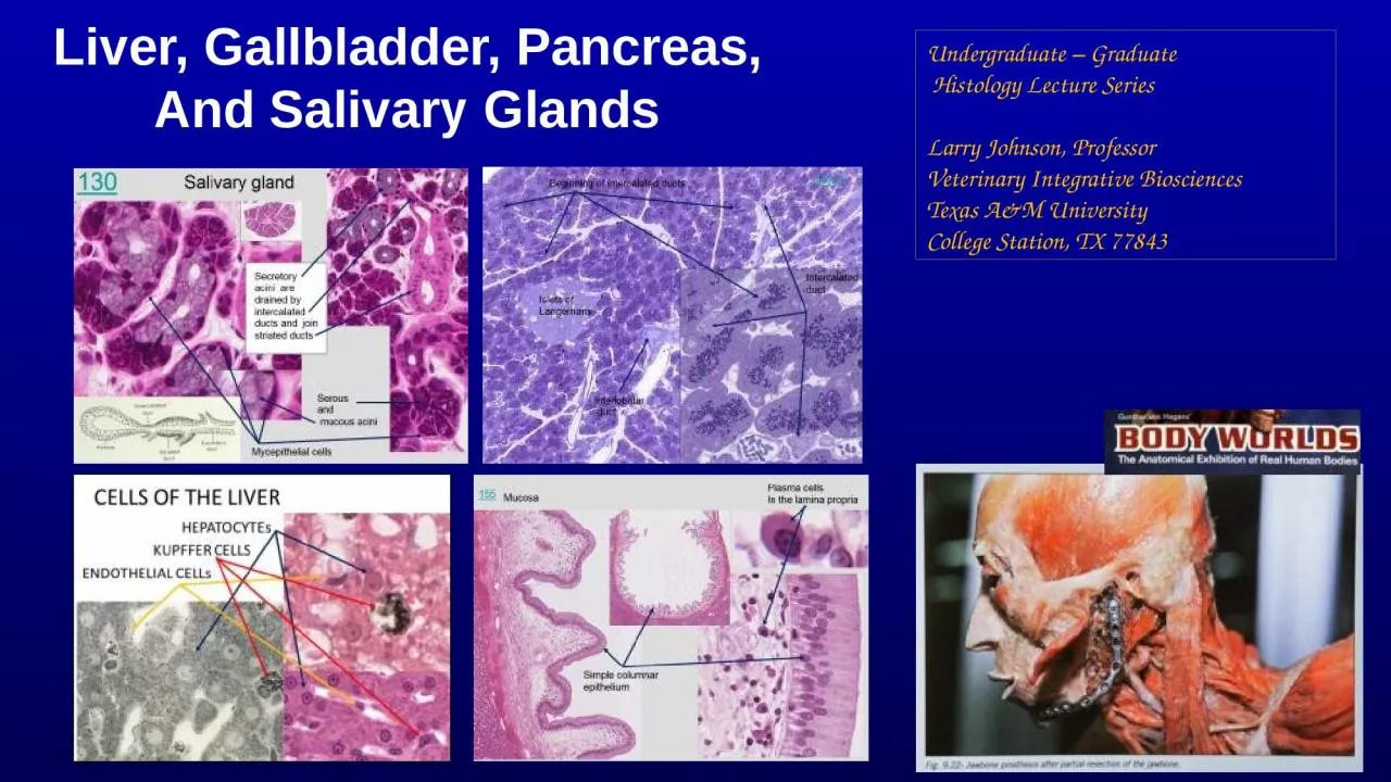
Liver, Gallbladder, Pancreas, And Salivary Glands
Undergraduate – Graduate
Histology Lecture
Series
Larry Johnson, ProfessorVeterinary Integrative BiosciencesTexas A&M UniversityCollege Station, TX 77843
Slide2ObjectivesTo understand the general organization of the accessory organs of the digestive system and how they contribute to obtaining metabolites necessary for growth and energy for the body.
To learn the origin of these glands and how structural features of these glands contribute to their function in digestion and absorption of food stuffs
Slide3Slide4Origin And Distribution Of Epithelium Ectoderm - epidermis of skin and epithelium of cornea together covers the entire surface of the body; sebaceous and mammary glands, oral cavity
Endoderm - alimentary tract, Liver, pancreas, gastric
glands, intestinal glandsEndocrine glands - lose connection with surface
MesodermEndothelium - lining of blood vesselsMesothelium - lining serous cavities
Ectoderm
Endoderm
Mesoderm
Slide5Function of the Digestive SystemMovement of foodSecretion of digestive juices
Absorption of digested foods, water, and electrolytes
Salivary glands and pancreas secretes digestive juices and liver secretes bile
Salivary glands lubricates
Liver stores nutrients and cleans the blood. Also, the accessory digestive organs contribute antibodies and antibacterial/viral growth substances.Role of liver, gall bladder, salivary glands, and pancreas
Slide6ORIGIN AND DISTRIBUTION OF EPITHELIUM con’d
LiverHisto 67
155
Gallbladder
Salivary gland
19758
Pancreas
158
Slide7Slide8Slide9Histo 067 pig liver
118
Connective
tissue capsule
Mesothelium
454
Classical liver lobules
Separated and surrounded with
connective tissue in the pig
Human liver
Monkey liver
Slide10Liver
The hepatocyte functions as an
endocrine-like cell (e.g., secretion of glucose and plasma proteins directly into the blood vascular system) and as an exocrine cell (e.g., secretion of bile into the bile canaliculi). This dual export of secretory products by a single cell requires a unique cellular arrangement in the liver in order to separate and compartmentalize the exocrine and endocrine-like products. Hepatocytes are arranged in fenestrated, anastomosing plates of one cell thick. Also each hepatocyte may have as many as four areas of access to the lumen.
Slide11Landscape of the Hepatocyte – Four Luminal Regions
Slide12HEPATOCYTE
Slide13LIVER FUNCTION - LARGEST GLANDEXOCRINE - BILE ACIDS, BILIRUBINENDOCRINE - ALBUMIN, FIBRINOGEN, ETC.
Slide14LIVER FUNCTIONS
Blood filtration - 1.2 x 10⁷
Kupffer cells/gBlood storage - liver size and sinusoids expandMaintain normal blood glucose concentrationsMetabolism and transport of lipidsSecrete plasma proteins - blood clottingNutritional metabolism and bile secretion
Drug metabolism - drug toleranceExcretion of bilirubin - jaundiceSecrete bile - emulsifying fats
Slide15Slide16Slide17Portal radicles containing:A bile duct Branch of portal vein,Branch of hepatic arteryLymphatic vessel (usually)
Liver
or portal canals
Cords of hepatocytes
155
155
Slide18454
Liver
454
Portal radicles containing:
A bile duct
Branch
of hepatic
artery
Branch of portal
vein
Lymphatic vessel (usually)
Cords of
hepatocytes
Central vein
Slide19Bile Canaliculi
Bile duct
Bile luminal
surfaces
Blood luminal surface
155
Slide20Liver
Slide21Cells of the Liver LobuleHepatocyteKupffer and fat-storing cellsEndothelial cell
Kupffer cells
Endothelial cell
Hepatocyte
Slide22Cells of the Liver LobuleHepatocyteKupffer cells
Endothelial cell
Slide23Triad with bile duct and central vein Liver with colloidal carbon, rat
118
Slide24Liver
Slide25Liver LobulePortal triadBlood supplyCentral vein
Hepatic sinusoidsZonation of
the liver
Slide26Slide27Slide28Acinus with portal vein and artery in center
Zonation of The Liver
2. Portal lobule with triad in center
1. Classical lobule
3. Acinus layers between
two central veins
Slide29Zonation of
the liver
Classical lobule
Slide30Portal Lobulewith Triad in Center
Slide31Acinus
with portal vein and artery in center
Slide32Acinus
with portal vein and artery in center
If liver damage is due to a toxicant, it kills hepatocytes in Zone I first.
If liver damage is due to a oxygen deprivation, it will kill the hepatocytes in Zone III first.
Acinus
Slide33HEPATOCYTE
Slide34Hepatocyte
Slide35Slide36Histological Reaction for Peroxidase
Hepatocyte
Slide37HepatocyteSpace of DisseBile canaliculi
Slide38Space of Disse
Slide39EM 18
Hepatic sinusoid
Hepatic parenchymal
cells with microvilli
Bile canaliculi
with lysosomes close
by the canaliculi
Space of Disse
containing reticular fibers
Endothelial cell
projecting into sinusoid
Liver cells
Platelet
Slide40Sugar and
protein
Slide41Slide42Glycogen in Hepatocytes
Slide43Dietary Differences In Amount Of Glycogen In Hepatocytes2-hour Fast (8.2% Glycogen) 24-hour Fast (0.9% Glycogen)
Slide44Slide45Disease opportunity at each step in a pathway
SER
Slide46Transcytosis to get antibodies
into secretions
Surface Specializations of Epithelia
Slide47Bile canaliculusFour + compounds that are deposited/secreted into this space.a. Cholesterol b. EGF c. insulin d. IgA also bile salts and BILIRUBIN
Slide48Slide49Slide50Slide51Bile Canaliculi
Slide52Bile Duct
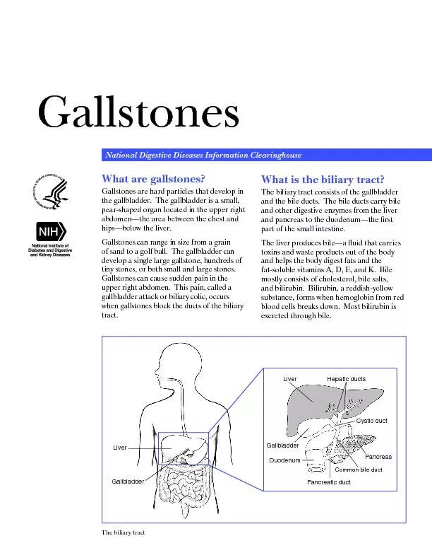
Slide53Slide54Gallbladder & Bile Ducts FunctionBiliary tractOrganization of gallbladder
EpitheliumConnective tissueHistophysiology
Slide55Slide56Slide57Slide58GallbladderThe mucosa is thrown into folds which project into the lumen of the gallbladder.
Lamina
propria
.
Smooth muscle
layer or branching
layers
A thick
perimuscular
layer of connective tissue.
Peritoneal
serosal
layer
Simple columnar epithelium
155
Slide59The gallbladder stores and concentrates the bile elaborated by the liver
Plasma cells
In the lamina
propria
Mucosa
Simple columnar
epithelium
155
Slide60Bile duct with portal vein, monkey
126
Common hepatic duct
Cystic duct
The wall of the cystic duct is convoluted and contains abundant smooth muscle fibers which represent the spiral valve preventing distention or collapse of the cystic duct when the latter is subject to sudden changes of pressure.
Portal
vein
Slide61Slide62Slide63Distinguishing characteristics between the mucosa of the various parts of the stomach, intestines, and gallbladder.
Intestines
148
Cardiac stomach
437Pyloric stomach141
Fundic stomach
145
Gallbladder
155
Mucosa = surface epithelium, lamina
propera
, and
muscularis
mucosa
Slide64Salivary GlandsFunctionHistological organization
Acinus = functional unitSerousMucousMixed
Slide65Origin of Salivary Glands?Ectoderm - oral ectoderm epithelial sheetEndoderm - alimentary tract
Slide66Slide67Saliva Helps Prevents Infections Contains secreted IgAContains Lactoferin - bind up iron needed for bacteria divisionContains lysosome that kills bacteriaConstantly washes mouth to dislodge and sweep bacteria down GI tract
Slide68Salivary Glands
Slide69Salivary Glands
Slide70Ducts of Salivary GlandsIntercalated Striated
Slide7119758
Slide72Slide73Slide74Submandibular gland - intercalated duct runs into Striated duct of salivary gland
130
The salivary gland is a compound,
tubuloacinar
gland.Intercalated ducts
Secretory
acini
Striated
Ducts
These striations reflect vertically arranged mitochondria associated with deep
enfolding
of the basal plasma membrane
Slide75Salivary gland
130
Myoepithelial
cells
Serous
and
mucous
acini
Secretory
acini
are drained by intercalated ducts and join striated ducts
Slide76Salivary gland
130
Individual secretory
acini
are drained by intercalated ducts and join striated ducts
Striated ducts
drain into a series of interlobular ducts
Serous and mucous
acini
Demilune
Vein
Artery
Lobules
Adipose
cells
Slide77Salivary glands 440Lobules
Interlobular ducts
Serous and mucous
acini
Myoepithelial
cells
19758
Striated ducts
Nerve
Nerve
cell bodies
Histo
52
440
Slide78Slide79PancreasFunction1. Exocrine2. Endocrine
Histological organization,Exocrine portion1. Acini
2. DuctsEndocrine portionIslets of LangerhansHistophysiology
Slide80Slide81PANCREASFUNCTION1. EXOCRINE2. ENDOCRINEHISTOLOGICAL ORGANIZATION,EXOCRINE PORTION1. ACINI2. DUCTS
ENDOCRINE PORTIONISLETS OF LANGERHANSHISTOPHYSIOLOGY
Slide82Slide8336723
Beginning of intercalated ducts
Islets of
Langerhans
Interlobular
duct
Intercalated
duct
Slide84Slide85Slide86Slide87156 and 157 Pancreas
157Intercalatedduct
156
Secretory
granules
All
acini
are of the serous type and many contain
centroacinar
cells initiate the duct inside the
acinus
.
36723
157
36723
Slide88Pancreas - Islets of Langerhans
158
Lobes composed
of lobules
Islets of Langerhans
Insulin is secreted by the B cells which are most numerous and centrally located in the islets.
Interlobular duct
Nerve
Blood vessels
Connective tissue septa.
Intercalated
duct
The pancreas is a compound
tubuloalveolar
(
tubuloacinar
) gland which functions in the digestion of food.
Slide89Pancreatic
acinar
cell (EM 1)
Lumen
Zymogen granuleVesiclesCentral acinar cell
EM 1
Slide90Slide91Slide92In summary
Slide93Slide94Slide95Slide96Slide97Slide98Slide99Slide100Slide101Slide102Bruce Alberts, et al. 1983. Molecular Biology of the Cell. Garland Publishing, Inc., New York, NY.Bruce Alberts, et al. 1994. Molecular Biology of the Cell. Garland Publishing, Inc., New York, NY.William J. Banks, 1981. Applied Veterinary Histology. Williams and Wilkins, Los Angeles, CA.Hans Elias, et al. 1978. Histology and Human Microanatomy. John Wiley and Sons, New York, NY.Don W. Fawcett. 1986. Bloom and Fawcett. A textbook of histology. W. B. Saunders Company, Philadelphia, PA.
Don W. Fawcett. 1994. Bloom and Fawcett. A textbook of histology. Chapman and Hall, New York, NY.Arthur W. Ham and David H. Cormack. 1979. Histology. J. S. Lippincott Company, Philadelphia, PA.Luis C. Junqueira, et al. 1983. Basic Histology. Lange Medical Publications, Los Altos, CA.L. Carlos
Junqueira, et al. 1995. Basic Histology. Appleton and Lange, Norwalk, CT.L.L. Langley, et al. 1974. Dynamic Anatomy and Physiology. McGraw-Hill Book Company, New York, NY.W.W. Tuttle and Byron A. Schottelius. 1969. Textbook of Physiology. The C. V. Mosby Company, St. Louis, MO.Leon Weiss. 1977. Histology Cell and Tissue Biology. Elsevier Biomedical, New York, NY.
Leon Weiss and Roy O. Greep. 1977. Histology. McGraw-Hill Book Company, New York, NY.Nature (http://www.nature.com), Vol. 414:88,2001.A.L. Mescher 2013
Junqueira’s Basis Histology text and atlas, 13th ed. McGrawInternet images and videos on biological presentationsMany illustrations in these VIBS Histology YouTube videos were modified from the following books and sources: Many thanks to original sources!
Slide103Questions on the Liver, pancreas, and salivary glands
The humoral activity of the immune system is illustrated by the transfer of IgA immunoglobin by
epithelial
cells into which of the following body fluids?
a. saliva b. milk c. bile d. a and b e. a, b, and c
Which function(s) do the gallbladder and urinary bladder have in common?
a. temporary storage of waste products
b. concentration of their respective luminal contents
c. similar type of luminal epithelium
d. a and b
e. a, b, and c
Characteristics of the pancreas include:
a. a portal blood vascular system
b. endocrine cells of the islets of Langerhans
c. acinar cells and striated ducts
d. a and b
e. a, b, and c
Slide104Slide105Slide106