
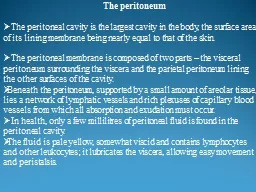
The peritoneal cavity is the largest cavity in the body the surface area of its lining membrane being nearly equal to that of the skin The peritoneal membrane is composed of two ID: 914721
Download Presentation The PPT/PDF document "The peritoneum" is the property of its rightful owner. Permission is granted to download and print the materials on this web site for personal, non-commercial use only, and to display it on your personal computer provided you do not modify the materials and that you retain all copyright notices contained in the materials. By downloading content from our website, you accept the terms of this agreement.


Slide1
The peritoneum
The
peritoneal cavity is the largest cavity in the body
,
the
surface area
of its lining membrane
being nearly equal
to that of the skin
.
The
peritoneal membrane is
composed of two
parts – the visceral
peritoneum surrounding
the viscera and the parietal peritoneum lining
the other
surfaces of the cavity
.
Beneath
the peritoneum,
supported by
a small amount of areolar tissue, lies a network of
lymphatic vessels
and rich plexuses of capillary blood vessels from
which all absorption
and exudation must
occur.
In
health, only a
few millilitres
of peritoneal fluid is found in the peritoneal
cavity.
The
fluid is pale yellow, somewhat viscid and contains
lymphocytes and
other leukocytes
;
it lubricates the viscera,
allowing easy
movement and peristalsis
.
Slide2The parietal portion is richly supplied with nerves and, when irritated, causes severe pain that is accurately localised to the affected area.
The visceral peritoneum, in contrast, is poorly supplied with nerves and its irritation causes pain that is usually poorly localised to the midline
.
The
peritoneum can also produce large volumes of fluid (ascites) and an inflammatory exudate when injured (peritonitis).
The circulation of peritoneal fluids may be responsible for the occurrence of abscesses distant from primary disease
Slide3The peritoneum has the capacity to absorb large volumes of fluid: this ability is used during
peritoneal
dialysis in the treatment of renal failure.
.
When parietal peritoneal defects are created, healing occurs not from the edges but by the development of new mesothelial cells throughout the surface of the defect. In this way, large defects heal as rapidly as small defects.
Functions of the peritoneum
In health
Visceral lubrication
Fluid and particles absorption
In disease
Pain perception (mainly parietal)
Inflammatory and immune responses
Fibrinolytic activity
Slide4PERITONITIS
Peritonitis
is
inflammation of the peritoneum
Localized
Generalised.
Most of cases are
caused by an invasion of the peritoneal cavity by
bacteria, free
fluid spills into the peritoneal cavity and circulates
largely directed
by the normal peritoneal attachments and
gravity.
S
pillage
from a perforated peptic ulcer may
run down
the right paracolic gutter leading to presentation
with pain
in the right iliac fossa (Valentino’s syndrome).
Even in patients
with non-bacterial peritonitis (e.g. acute
pancreatitis, intraperitoneal
rupture of the bladder or haemoperitoneum
), the
peritoneum often becomes infected by transmural spread of
organisms from the bowel
.
Slide5Causes of peritoneal inflammation
Bacterial, gastrointestinal and non-gastrointestinal
Chemical, e.g. bile, barium
Allergic, e.g. starch peritonitis
Traumatic, e.g. operative handling
Ischaemia, e.g. strangulated bowel, vascular occlusion
Miscellaneous, e.g. familial Mediterranean fever
Slide6Although acute bacterial peritonitis most commonly arises from a perforation of a viscus of the alimentary tract, other routes of infection can include the female genital tract and exogenous contamination.
There are also less common forms in which the aetiology is
a primary
‘spontaneous’ peritonitis, in which a pure infection with streptococcal, pneumococcal or haemophilus bacteria occurs.
Slide7Paths to peritoneal infection
Gastrointestinal perforation, e.g. perforated ulcer, appendix, diverticulum
Transmural translocation (no perforation), e.g. pancreatitis, ischaemic bowel
Exogenous contamination, e.g. drains, open surgery,
trauma
Female genital tract infection, e.g. pelvic inflammatory disease
Haematogenous spread (rare), e.g. septicaemia
Slide8Microbiology
Bacteria from the gastrointestinal tract
The number of bacteria within the lumen of the gastrointestinal tract is normally low until the distal small bowel is reached.
Disease leading to stasis and overgrowth (e.g. obstruction, chronic and acute motility disturbances) may increase proximal colonisation.
The biliary and pancreatic tracts are also normally free from bacteria, although they may be infected in disease, e.g. gallstones.
Slide9Peritoneal infection is usually caused by two or more bacterial strains.
Gram-negative bacteria contain endotoxins (lipopolysaccharides) in their cell walls that have multiple toxic effects on the host, primarily by causing the release of tumour necrosis factor (TNF) from host leukocytes
.
Systemic absorption of endotoxin may produce endotoxic shock with hypotension and impaired tissue perfusion.
Clostridium welchii
produce harmful exotoxins
.
Slide10Bacteroides
are commonly found in peritonitis. These anaerobic Gramnegative, non-sporing organisms, although predominant in the lower intestine. These organisms are resistant to penicillin and streptomycin but sensitive to metronidazole, clindamycin, lincomycin and cephalosporin compounds. Since the widespread use of metronidazole (Flagyl), Bacteroides infections have greatly diminished
.
Slide11Non-gastrointestinal causes of peritonitis
Pelvic infection via the Fallopian. The most common organisms are chlamydia and gonococcus.
These organisms lead to a thinning of cervical mucus and allow bacteria from the vagina into the uterus and oviducts.
Perihepatitis which can cause scar tissue to form on Glisson’s capsule, a thin layer of connective tissue surrounding the liver.
Tuberculosis and other mycobacterial strains and those causing primary peritonitis (pneumococcus, staphylococcus and streptococcus spp).
Fungal peritonitis is uncommon but may complicate severely ill patients.
Slide12Localised peritonitis
Anatomical and pathological factors may favour the localisation of peritonitis.
Anatomical
The greater sac of the peritoneum is divided into
The subphrenic spaces
The pelvic space
The peritoneal cavity proper, divided into a ;
Supracolic compartment
Infracolic compartment
Slide13Pathological
Inflamed
peritoneum
loses its glistening appearance and becomes reddened and velvety.
Flakes of fibrin appear and cause loops of intestine to become adherent to one another.
There is an outpouring of serous inflammatory exudate rich in leukocytes and plasma proteins that soon becomes turbid; if localisation occurs, the turbid fluid becomes frank pus.
Peristalsis is retarded in affected bowel and this helps to prevent distribution of the infection.
The greater omentum, by enveloping and becoming adherent to inflamed structures, often forms a substantial barrier to the spread of infection
Slide14Diffuse (generalised) peritonitis
A number of factors may favour the development of diffuse peritonitis:
Speed of peritoneal contamination is a prime factor. If an inflamed appendix or other hollow viscus perforates before localisation has taken place, there will be an efflux of contents into the peritoneal cavity, which may spread over a large area.
Perforation proximal to an obstruction or from sudden anastomotic separation is associated with severe generalised peritonitis and a high mortality rate.
Slide15Stimulation of peristalsis by the ingestion of food or water.
Violent peristalsis occasioned by the administration of a purgative or an enema may cause
the widespread
distribution of an infection.
The virulence of the infecting organism may be so great.
Slide16Young children have a small omentum, which is less effective in localising infection.
Disruption of localised collections may occur with injudicious handling, e.g. appendix mass or pericolic abscess.
Deficient natural resistance (‘immune deficiency’) may result from use of drugs (e.g. steroids), disease (e.g. acquired immune deficiency syndrome (AIDS) or old age.
Slide17With
appropriate treatment, localised peritonitis usually resolves
;
I
n
about 20 per cent of cases, an abscess follows.
Infrequently
, localised peritonitis becomes diffuse.
Conversely
, in favourable circumstances, diffuse peritonitis can become localised, most frequently in the pelvis or at multiple sites
within the
abdominal cavity
.
Slide18Clinical features
Localised peritonitis
The initial symptoms and signs are ( abdominal pain, specific GI symptoms + malaise, anorexia and nausea).
When the peritoneum becomes inflamed, abdominal pain will worsen and, in general, temperature and pulse rate will rise.
The pathognomonic signs are localised guarding (involuntary abdominal wall contraction to protect the viscus from the examining hand), a positive ‘release’ sign (rebound tenderness) and, sometimes, rigidity (involuntary constant contraction of the abdominal wall over the inflamed parietes).
Slide19If
inflammation arises under the diaphragm, shoulder tip (‘phrenic’) pain may be felt as the pain is referred to the C5 dermatome.
In
cases of pelvic peritonitis arising from an inflamed appendix in the pelvic position or from salpingitis, the abdominal signs are often slight; there may be deep tenderness of one or both lower quadrants alone, but a rectal or vaginal examination reveals marked tenderness of the pelvic
peritoneum.
Slide20Diffuse (generalised) peritonitis
Early
Abdominal pain is severe and made worse by moving or breathing.
It is first experienced at the site of the original lesion and spreads outwards from this point.
Tenderness and generalised guarding are found on palpation.
Bowel sounds may still be heard for a few hours but they cease with the onset of paralytic ileus.
Pulse and temperature rise in accord with degree of inflammation and infection.
Slide21Late
The
abdomen will become rigid (generalised rigidity).
Distension
is common and bowel sounds are absent.
Circulatory
failure, with cold, clammy extremities, sunken eyes, dry tongue, thready (irregular) pulse and drawn and anxious face (Hippocratic facies).
The
patient finally lapses into unconsciousness.
With
early diagnosis and adequate treatment, this condition is rarely seen in modern surgical practice
Slide22The Hippocratic facies in terminal diffuse peritonitis.
Slide23Diagnostic aids
Bedside
G
eneral
u
rine examination for urinary tract infection
ECG if diagnostic doubt (as to cause of abdominal pain) or cardiac history.
Bloods
Full blood count for white cell count (WCC)
Serum amylase estimation may establish the diagnosis of acute pancreatitis.
Group and save serum may be taken as an adjunct to impending surgery.
Slide24Imaging
Erect chest radiograph to demonstrate free subdiaphramatic gas.
A supine radiograph of the abdomen may confirm the presence of dilated gas-filled loops of bowel (consistent with a paralytic ileus). In the patient who is too ill for an ‘erect’ film, a lateral decubitus film can show gas beneath the abdominal wall (if CT unavailable).
Multiplanar computed tomography (CT) is increasingly used to identify the cause of peritonitis and may also influence management decisions.
Ultrasound scanning has undoubted value in certain situations such as pelvic peritonitis in females and localised right upper quadrant peritonism.
Slide25Management
General care of the patient
Correction of fluid loss and circulating volume
Patients are frequently hypovolaemic with electrolyte disturbances.
The plasma volume must be restored and electrolyte concentrations corrected.
Special measures may be needed for cardiac, pulmonary and renal support, especially if septic shock is present, including central venous pressure monitoring in patients with concurrent disease.
Slide26Urinary
catheterisation ± gastrointestinal decompression
A urinary catheter will give a guide to central perfusion and will be required if abdominal surgery is to proceed
.
A nasogastric tube is commonly passed to allow drainage ± aspiration
until paralytic
ileus has resolved.
Antibiotic therapy
Administration of parenteral broad-spectrum (aerobic and anaerobic) antibiotics prevents the multiplication of bacteria and the release of endotoxins.
Slide27Analgesia
The patient should be nursed in the sitting-up position and must be relieved of pain before and after operation.
If appropriate expertise is available, epidural infusion may provide excellent analgesia.
Freedom from pain allows early mobilisation and adequate physiotherapy in the postoperative period, which helps to prevent basal pulmonary collapse, deep-vein thrombosis and pulmonary embolism.
Slide28Specific treatment of the cause
In patients where specific treatment has not been guided by CT scanning, early surgical intervention is to be preferred to a ‘wait and see’ policy assuming that the patient is fit for anaesthesia and that resuscitation has resulted in a satisfactory restitution of normal body physiology.
More caution is, of course, required in patients at high operative risk because of
comorbidity or
advanced age.
Slide29In those patients with a preoperative diagnosis, if the cause of
peritonitis
is amenable to surgery, operation must be carried out
as soon
as the patient is fit.
This is usually within a few hours.
In peritonitis caused by pancreatitis or salpingitis, or in cases of primary peritonitis of streptococcal or pneumococcal origin, non-operative treatment is preferred provided the diagnosis can be made with confidence
Slide30In general, surgery is directed to removing (or diverting) the cause and subsequent adequate peritoneal lavage ± drainage.
In operations for generalised peritonitis it is essential that, after the cause has been dealt with, the whole peritoneal cavity is explored with the sucker and, if necessary, mopped dry until all seropurulent exudate is removed.
The use of a large volume of saline (typically 3 litres) containing dissolved antiseptic or antibiotic has been shown to be effective.
Slide31Prognosis
D
iffuse peritonitis carries a mortality rate of about 10 per cent reflecting;
The degree and duration of peritoneal contamination
.
Age
of the
patient.
F
itness of the patient.
T
he nature of the underlying cause.
Slide32Complications of peritonitis
Abdominal complications
Paralytic ileus
Residual or recurrent abscess/inflammatory mass
Portal pyaemia/liver abscess
Adhesional small bowel obstruction
Systemic
complications
Bacteraemic/endotoxic shock
Systemic inflammatory response syndrome
Multiorgan dysfunction syndrome
Death
Slide33SPECIAL FORMS OF PERITONITIS
Bile peritonitis
Causes of bile peritonitis
Perforated cholecystitis
Postcholecystectomy due to;
Cystic duct stump leakage
Leakage from an accessory duct in the gall bladder bed
Bile duct injury
T-tube drain dislodgement (or tract rupture on removal)
Following other operations/procedures
Leaking duodenal stump postgastrectomy
Leaking biliary–enteric anastomosis
Leakage around percutaneous placed biliary drains
Following liver trauma
Slide34Clinical features and treatment
S
evere abdominal pain and signs of diffuse peritonitis.
After a few hours a tinge of jaundice is not unusual.
Laparotomy (or laparoscopy) should be undertaken with evacuation of the bile and peritoneal lavage.
The source of bile leakage should be identified and treated accordingly.
Infected bile is more lethal than sterile bile.
Slide35A
‘blown’ duodenal stump should be drained as it is too oedematous to repair, but sometimes it can be covered by a jejunal patch.
The patient is often jaundiced from absorption of peritoneal bile, but the surgeon must ensure that the abdomen is not closed until any obstruction to a major bile duct has been either excluded or relieved.
Bile leaks after cholecystectomy or liver trauma may be dealt with by percutaneous (ultrasoundguided) drainage and endoscopic biliary stenting to reduce bile duct pressure.
The drain is removed when dry and the stent at 4–6 weeks.
Slide36Primary peritonitis
Primary pneumococcal peritonitis may complicate nephrotic syndrome or cirrhosis in children.
Healthy children, particularly girls between three and nine years of age, may also be affected, and it is likely that the route of infection is sometimes via the vagina and Fallopian tubes.
Always in males, the infection is blood-borne and secondary to respiratory tract or middle ear disease.
The prevalence of pneumococcal peritonitis has declined greatly and the condition is now rare.
Slide37Clinical features
Sudden pain localised to the lower half of the abdomen.
Fever with vomiting.
After 24–48 hours, profuse diarrhoea is characteristic.
There is usually increased frequency of micturition.
The last two symptoms are caused by severe pelvic peritonitis
On examination, peritonism is usually diffuse
Slide38Investigation and treatment
A leukocytosis 30 000 μL with approximately 90 per cent polymorphs suggests pneumococcal peritonitis rather than another cause, e.g. appendicitis.
Antibiotic therapy, correcting dehydration and electrolyte imbalance.
Early surgery is required (Laparotomy or laparoscopy).
In pneumococcal peritonitis the exudate is odourless and sticky, but it is essential to perform a careful exploration to exclude other pathology, some of the exudate is aspirated and sent to the laboratory for microscopy, culture and sensitivity tests
.
Slide39Peritoneal
lavage is carried out and the incision closed.
Antibiotic and fluid replacement therapy.
Other organisms are now known to cause some cases of primary peritonitis in children, including
Haemophilus, other streptococci
and a few Gram-negative bacteria.
Underlying pathology (including an intravaginal foreign body in girls) must always be excluded before primary peritonitis can be diagnosed with certainty.
Idiopathic streptococcal and staphylococcal peritonitis can also occur in adults.
Slide40Slide41Tuberculous peritonitis
Intra-abdominal tuberculosis is very common in the developing world.
The incidence is however also rising in areas of the developed world as a consequence of migration and immunosuppression where
Mycobacterium avium-intracellulare
is becoming increasingly prevalent with the widespread increase in human immunodeficiency virus (HIV) coinfection.
Abdominal tuberculosis (TB) includes intraperitoneal, GI tract and solid organ disease forms with TB peritonitis being a common site-specific variant.
TB peritonitis is resulting in morbidity and mortality.
Slide42Tuberculosis can spread to the peritoneum through the GI tract (typically ileocaecal region) via mesenteric lymph nodes or directly from the blood, usually from the ‘miliary’ but occasionally the ‘cavitating’ form of pulmonary TB, lymph and the Fallopian tubes.
50–83 per cent of patients with abdominal TB can be expected to have peritoneal involvement.
Clinical or subclinical ascites is reported in all patients with TB peritonitis and is frequently a presenting feature.
In the most common form of the disease, wet-type peritonitis, ascites may be localised or generalised throughout the peritoneal cavity.
Multiple tubercle deposits appear on both layers of the peritoneum.
Slide43Diagnosis is via abdominal ultrasound or CT to detect ascites and lymphadenopathy ± diffuse thickening of the peritoneum, mesentery and/or omentum.
Ascitic fluid is typically a straw-coloured exudate (protein >25–30 g/L) with white cells >500 mm3 and lymphocytes >40 per cent.
Diagnostic smears for acid-fast bacilli are diagnostic in <3 per cent of patients and culture may take up to 4–8 weeks with no guarantee of a positive result.
Laparoscopy and peritoneal biopsy may thus be helpful to couple typical appearances with histology.
Slide44Tuberculous peritonitis
Acute (may be clinically indistinguishable from acute bacterial peritonitis)
C
hronic
Abdominal pain, sweats, malaise and weight loss are frequent
Ascites common, may be loculated
Caseating peritoneal nodules are common – distinguish from metastatic carcinoma and fat necrosis of pancreatitis
Intestinal obstruction may respond to antituberculous treatment without surgery
Slide45Management is principally;
Supportive (nutrition and hydration)
Medical (systemic antituberculous therapy)
Surgery may be required for specific complications such as intestinal obstruction.
Slide46Familial Mediterranean fever (periodic peritonitis)
Is characterised by abdominal pain and tenderness, mild pyrexia, polymorphonuclear leukocytosis and, occasionally, pain in the thorax and joints.
The duration of an attack is 24–72 hours, when it is followed by complete remission, but exacerbations recur at regular intervals.
Most of the patients have undergone appendicectomy in childhood.
This disease, often familial, is limited principally to Arab, Armenian and Jewish populations; other races are occasionally affected.
Slide47Mutations in the
MEFV
(Mediterranean fever) gene. This gene produces a protein called pyrin.
Usually, children are affected.
Exceptionally, the disease becomes manifest in patients over 40 years of age.
At operation, which may be necessary to exclude other causes, the peritoneum is inflamed, particularly in the vicinity of the spleen and the gall bladder.
Colchicine therapy is used during attacks and to prevent recurrent attacks
.
Slide48INTRAPERITONEAL ABSCESS
Following intraperitoneal sepsis, abscess development usually occupies one of a number of specific abdominal or pelvic sites.
Larger abscesses will give rise to the picture of swinging pyrexia, pulse and a palpable mass.
Blood tests will reveal elevated inflammatory markers.
Slide49Clinical features of an abdominal/pelvic abscess
Symptoms
Malaise, lethargy – failure to recover from surgery as expected
Anorexia and weight loss
Sweats ± rigors
Abdominal/pelvic pain
Symptoms from local irritation, e.g. shoulder tip/hiccoughs (subphrenic), diarrhoea and mucus (pelvic), nausea and vomiting (any upper abdominal)
Signs
Increased temperature and pulse ± swinging pyrexia
Localised abdominal tenderness ± mass (including on pelvic exam)
Slide50Pelvic abscess
The pelvis is the most common site of abscess formation because the vermiform appendix is often pelvic in position and the Fallopian tubes are also frequent sites of infection.
A pelvic abscess can also occur as a sequel to any case of diffuse peritonitis and is common after anastomotic leakage following colorectal surgery.
Clinical features
The most characteristic symptoms are of pelvic pain, diarrhoea and the passage of mucus in the stools.
Rectal examination reveals a bulging of the anterior rectal wall, which, when the abscess is ripe, becomes softly cystic.
Slide51Investigation and management
The presence of pus should be confirmed by ultrasound or CT scanning
Left to nature, a proportion of these abscesses burst into the rectum.
In women, vaginal drainage through the posterior fornix is often chosen.
When the abscess is definitely pointing into the rectum, rectal drainage is employed.
Laparotomy is almost never necessary and rectal drainage of a pelvic abscess is far preferable to suprapubic drainage, which risks exposing the general peritoneal cavity to infection.
Drainage tubes can also be inserted percutaneously or via the vagina or rectum under ultrasound or CT guidance.
Slide52Intraperitoneal abscess
Anatomy
The complicated arrangement of the peritoneum results in the formation of four intraperitoneal spaces in which pus may commonly collect.
Left subphrenic space
This is bounded above by the diaphragm and behind by the left triangular ligament and the left lobe of the liver, the gastrohepatic omentum and the anterior surface of the stomach.
To the right is the falciform ligament and to the left the spleen, gastrosplenic omentum and diaphragm.
The common cause of an abscess here is an operation on the stomach, the tail of the pancreas, the spleen or the splenic flexure of the colon.
Slide53Right subphrenic space
This space lies between the right lobe of the liver and the diaphragm.
It is limited posteriorly by the anterior layer of the coronary and the right triangular ligaments and to the left by the falciform ligament.
Common causes of abscess here are perforating cholecystitis, a perforated duodenal ulcer, a duodenal cap ‘blow-out’ following gastrectomy and appendicitis.
Left subhepatic space/lesser sac
The most common cause of infection here is complicated acute pancreatitis.
In practice, a perforated gastric ulcer rarely causes a collection here because the potential space is obliterated by adhesions.
Slide54Right subhepatic space
This lies transversely beneath the right lobe of the liver in Rutherford Morison’s pouch.
It is bounded on the right by the right lobe of the liver and the diaphragm.
To the left is situated the foramen of Winslow and below this lies the duodenum.
Slide55In
front are the liver and the gall bladder and behind are the upper part of the right kidney and the diaphragm.
The
space is bounded above by the liver and below by the transverse colon and hepatic flexure.
It
is the deepest space of the four and the most common site of a subphrenic abscess, which usually arises from appendicitis, cholecystitis, a perforated duodenal ulcer or following upper abdominal surgery.
Slide56Clinical features
Sweating and anorexia are present.
There is sometimes epigastric fullness and pain, or pain in the shoulder on the affected side due to irritation of sensory fibres in the phrenic nerve, this being referred along the descending branches of the cervical plexus.
Persistent hiccoughs may be a presenting symptom.
A swinging pyrexia is usually present.
If the abscess is anterior, abdominal examination will reveal some tenderness, rigidity or even a palpable swelling.
Sometimes the liver is displaced downwards but more often it is fixed by adhesions.
Slide57Investigation and management
CXR , as in the majority of cases, collapse of the lung or evidence of basal effusion or even an empyema are evident.
Ultrasound or CT guidance followed by drainage. The same tube can be used to instill antibiotic solutions or irrigate the abscess cavity if necessary.
Radiolabelled white cell scanning may occasionally prove helpful when other imaging techniques have failed.
In most cases, with the aid of percutaneous drainage and antibiotic treatment, the abscess or mass gradually reduces in size until, finally, it is undetectable.
Open drainage of an intraperitoneal collection is thus now uncommon but may be necessary.
Slide58If a swelling can be detected in the subcostal region or in the loin, an incision is made over the site of maximum tenderness or over any area where oedema or redness is discovered.
Cautious blunt finger exploration can then be used to avoid dissemination of pus into the peritoneal or pleural cavities and minimise the risk of an intestinal fistula.
When the cavity is reached, all of the fibrinous loculi must be broken down with the finger and one or two drainage tubes fully inserted.
These drains are withdrawn gradually during the next 10 days and the closure of the cavity can be checked by sinograms or scanning.
Appropriate antibiotics are also given.
Slide59ASCITES
Ascites is defined as an accumulation of excess serous fluid within the peritoneal cavity.
Pathophysiology
The balanced effects of plasma and peritoneal colloid osmotic and hydrostatic pressures determine the exchange of fluid between the capillaries and the peritoneal fluid.
Protein-rich fluid enters the peritoneal cavity when capillary permeability is increased, as in peritonitis and carcinomatosis peritonei.
Capillary pressure may be increased because of generalised water retention, cardiac failure, constrictive pericarditis or vena cava obstruction.
Slide60Capillary pressure is raised selectively in the portal venous system in the Budd–Chiari syndrome, cirrhosis of the liver or extrahepatic portal venous obstruction.
Plasma colloid osmotic pressure may be lowered in patients with reduced nutritional intake, diminished intestinal absorption, abnormal protein losses or defective protein synthesis such as occurs in cirrhosis.
Peritoneal lymphatic drainage may be impaired, resulting in the accumulation of protein-rich fluid.
Slide61Causes of ascites
Transudates (protein <25 g/L)
Low plasma protein concentrations
Malnutrition
Nephrotic syndrome
Protein-losing enteropathy
High central venous pressure
Congestive cardiac failure
Portal hypertension
Portal vein thrombosis
Cirrhosis
Slide62Exudates (protein >25 g/L)
Tuberculous peritonitis
Peritoneal malignancy
Budd–Chiari syndrome (hepatic vein occlusion or thrombosis)
Pancreatic ascites
Chylous ascites
Meigs’ syndrome
Slide63Clinical features
Ascites can usually only be recognised clinically when the amount of fluid present exceeds 1.5 L depending on body habitus: in the obese a greater quantity than this is necessary before there is clear evidence.
The abdomen is distended with fullness of the flanks, which are dull to percussion.
Usually, shifting dullness is present but when there is a very large accumulation of fluid this sign is absent.
In such cases, on flicking the abdominal wall, a characteristic fluid thrill is transmitted from one side to the other.
In women, ascites must be differentiated from an enormous ovarian cyst.
Slide64Congestive heart failure, the most common cause of ascites, results in increased venous pressure in the vena cava and consequent obstruction to the venous outflow from the liver.
The ascitic fluid is light yellow and of low specific gravity, about 1.010, with a low protein concentration (<25 g/L).
Patients with constrictive pericarditis (Pick’s disease) have both peritoneal and pleural effusions because of engorgement of the venae cavae consequent upon the diminished capacity of the right side of the heart.
Ascites occurs with low plasma albumin concentrations, for example in patients with albuminuria or starvation.
The ascites in this instance is caused by alterations in the osmotic pressure of the capillary blood and has a low specific gravity
.
Slide65In cirrhosis, there is obstruction to the portal venous system, which is caused by obliterative fibrosis of the intrahepatic venous bed. In the Budd–Chiari syndrome, thrombosis or obstruction of the hepatic veins is responsible for obstruction to venous outflow from the liver.
The ascites seen in patients with peritoneal metastases is caused by excessive exudation of fluid and lymphatic blockage. The fluid is dark yellow and frequently blood-stained. The specific gravity, 1.020 or over, and the protein content (>25 g/L) are high.
Slide66Microscopic
examination often reveals cancer cells, especially if large quantities of fluid are ‘spun down’ to produce a concentrated deposit for sampling.
Rarely
, ascites and pleural effusion are associated with solid fibromas of the ovary (Meigs’ syndrome).
The
effusions disappear when the tumour is excised
Slide67Investigation
Liver and cardiac function tests.
Ultrasound and/or CT imaging will ;
determine much smaller quantities of ascites.
diagnose aetiology, e.g. carcinomatosis, liver disease.
Ascitic aspiration or tap is now most commonly performed under imaging guidance to minimise the risk of visceral injury,
t
he bladder having been emptied, puncture of the peritoneum is carried out under local anaesthetic using a moderately sized trocar and cannula.
Slide68Alternatively
, a peritoneal drain may be inserted.
In cases where the effusion is caused by cardiac failure, the fluid must be evacuated slowly.
Fluid
is sent for microscopy/cytology, culture, including mycobacteria, and analysis of protein content and amylase.
Unless
other measures are taken the fluid soon reaccumulates, and repeated tappings remove valuable protein.
Slide69Treatment
Treatment of the specific cause;
If portal venous pressure is raised, it may be possible to lower it by treatment of the primary condition.
Dietary sodium restriction to 200 mg/day may be helpful.
Diuretics are usually required.
Slide70In
rare cases in which ascites accumulates rapidly after paracentesis and the patient is otherwise fit, permanent drainage of the ascitic fluid via a peritoneovenous shunt (e.g. LeVeen, Denver) may render the patient more comfortable.
A
catheter (e.g. of silicone) is constructed with a valve so as to allow one-way flow from the peritoneum to a central vein (e.g. internal jugular).
A
chamber placed subcutaneously over the chest wall may be included for manual
compression.
Slide71The complications include;
Overloading the venous system.
Cardiac failure
Disseminated intravascular coagulopathy.
Dissemination of malignant cells
The frequency of these complications may be reduced by evacuating ascitic fluid and partially replacing it with normal saline at the time of shunt insertion.
The procedure may also be used for patients with terminal malignant ascites, giving improved quality of life.
Slide72Special cases
Chylous ascites
The ascitic fluid appears milky because of an excess of chylomicrons (triglycerides).
Most cases are associated with;
Malignancy, usually lymphomas;
Cirrhosis,
Tuberculosis,
Filariasis,
Nephrotic syndrome,
Abdominal trauma
(including surgery),
Constrictive pericarditis,
Sarcoidosis,
Congenital lymphatic abnormality.
The prognosis is poor unless the underlying condition can be cured.
In addition to other measures used to treat ascites, patients should be placed on a fatfree diet with medium-chain triglyceride supplements.
Slide73TUMOURS OF THE PERITONEUM
Primary tumours
Are rare and in most cases take their origin not from the serous layer but from some adjacent structure, e.g
. lipoma
from appendices epiploicae
, fibroma
from connective tissue.
Mesothelioma
of the peritoneum is less frequent than in the pleural cavity but equally lethal. Asbestos is a recognised cause. It has a predilection for the pelvic peritoneum.
Chemocytotoxic agents are the mainstay of treatment.
Desmoid tumours
which have a relationship to the peritoneum are considered under familial adenomatous polyposis.
Slide74Secondary tumours
Carcinomatosis peritonei
This is a common terminal event in many cases of carcinoma of the stomach, colon, ovary or other abdominal organs and also of the breast and bronchus.
The peritoneum, both parietal and visceral, is studded with secondary growths and the peritoneal cavity becomes filled with clear, straw-coloured or blood-stained ascitic fluid.
Slide75The main forms of peritoneal metastases are:
discrete nodules – by far the most common variety;
plaques varying in size and colour;
diffuse adhesions – this form occurs at a late stage of the disease and gives rise, to a ‘frozen pelvis
’.
Gravity probably determines the distribution of free malignant cells within the peritoneal cavity.
The main differential diagnosis is from tuberculous peritonitis.
Treatment;
Are as for underlying malignancy.
Slide76Pseudomyxoma peritonei
This rare condition occurs more frequently in women.
The abdomen is filled with a yellow jelly, large quantities of which are often encysted.
The condition is associated with mucinous cystic tumours of the ovary and appendix.
Recent studies suggest that most cases arise from a primary appendiceal tumour with secondary implantation on to one or both ovaries.
Slide77It is often painless and there is frequently no impairment of general health.
Does not give rise to extraperitoneal metastases.
The diagnosis is more often suggested by ultrasound and CT scanning or made at operation.
At laparotomy, masses of jelly are scooped out.
The appendix, if present, should be excised together with any ovarian tumour.
Slide78Unfortunately
, recurrence is inevitable, but patients may gain symptomatic benefit from repeated ‘debulking’ surgery.
Occasionally, the condition responds to radioactive isotopes or intraperitoneal chemotherapy.
The role of early radical peritoneal excision is uncertain.
Slide79Peritoneal loose bodies (peritoneal mice
)
May be confused with a small tumour but almost never cause symptoms.
One or more may be found in a hernial sac or in the pouch of Douglas.
The loose body may come from an appendix epiploica that has undergone axial rotation followed by necrosis of its pedicle and detachment but they are also found in those who suffer from subacute attacks of pancreatitis.
These hyaline bodies attain the size of a pea or bean and contain saponified fat surrounded by fibrin
.
Slide80Slide81ADHESIONS
Pathophysiology
Are strands of fibrous tissue that form, as a result of surgery, between surgically injured tissues.
After injury, there is bleeding and an increase in vascular permeability with extravasation of fibrinogen-rich fluid from the injured surfaces forming a temporary fibrin matrix.
Slide82An
inflammatory response with cell migration, release of cytokines,
and
activation of
the coagulation cascade.
The
activation of the coagulation system results in thrombin formation, which is necessary for the conversion of fibrinogen to fibrin.
In the absence of fibrinolysis, adhesions will form within 5–7 days as the matrix gradually becomes more organised with collagen secretion by fibroblasts.
The ischaemic tissue loses its ability to break down fibrin and inhibits fibrinolysis in adjacent tissues.
Slide83Complications
Small bowel obstruction (SBO). Adhesions are the most frequent cause of SBO in the developed world and are responsible for 60–70 per cent of SBO.
Major cause of secondary infertility.
Chronic abdominal and pelvic pain.
Unguided division of adhesions has not been shown to reduce chronic abdominal pain.
laparoscopy under local anaesthesia to direct lysis may improve success rates.
Slide84Prevention
Minimising the production of ischaemic tissue by careful operative technique, including meticulous control of bleeding.
laparoscopic bowel surgery has been shown reduce adhesion.
The effect of a number of drugs including anti-inflammatory drugs like aspirin and steroids, some hormones, anticlotting agents, antibiotics, vitamin E and even methylene blue have been investigated in adhesion prevention but have not achieved widespread use either because of side effects or lack of consistent evidence of effectiveness.
Slide85Interceed TC7® is a mesh-like product shown to significantly reduce the number of adhesions.
A
similar barriertype product (hyaluronic acid/carboxymethyl membrane), there was a significant reduction in the incidence,
extent and
severity of adhesions but no reduction in the incidence of intestinal obstruction or operative intervention.
Such barriers when placed around bowel anastomosis also led to a significant increase in the anastomotic leaks.
For
these reasons barrier approaches have not gained popularity.
Slide86Special forms of intraperitoneal fibrosis
Sclerosing encapsulating peritonitis
Is described in patients as a complication of long-term peritoneal dialysis or portovenous shunting.
The peritoneal cavity becomes obliterated as a result of gross subserosal thickening by fibrosis leading to bowel obstruction.
Surgery should be undertaken with trepidation and avoided if possible.
Slide87Diffuse fibromatosis
A rare tumour characterised by an abnormal proliferation of myofibroblasts.
Although non-metastasising
,
benign, it can nevertheless prove widely invasive, compressing and infiltrating surrounding tissues such as the bowel and mesentery with complications thereof.
Intra-abdominal fibromatosis IAF is very rare within the general population but has a recognised association with familial adenomatous polyposis (FAP).
Slide88THE OMENTUM
Rutherford Morison called the greater omentum ‘the abdominal policeman.
It limit intraperitoneal infective and other noxious processes.
An acutely inflamed appendix is often found wrapped in omentum, and this saves many patients from developing diffuse peritonitis.
It often plugs the neck of a hernial sac and prevents a coil of intestine from entering and becoming strangulated.
It can, of course, also be a cause of obstruction (acting as a large adhesion).
The omentum is usually involved in tuberculous peritonitis and carcinomatosis of the peritoneum.
Slide89Torsion of the omentum
Torsion of the omentum is a rare emergency and consequently is seldom diagnosed correctly.
It is usually mistaken for appendicitis with somewhat abnormal signs.
It may be primary, or secondary to adhesion of the omentum to an old focus of infection or hernia.
The patient is most frequently a middle-aged, obese man. A tender lump may be present in the abdomen.
The blood supply having been jeopardised, the twisted mass sometimes becomes gangrenous, in which case bacterial peritonitis may follow.
Treatment is surgical; the pedicle above the twist is ligated securely and the mass removed.
Slide90THE MESENTERY
Mesenteric injury
A wound of the mesentery can follow severe abdominal contusion and is a cause of haemoperitoneum.
More commonly, it is injured by a torsional force, so-called seatbelt syndrome.
Aside from control of any ongoing haemorrhage, associated ischaemic or ruptured gut will require resection.
Ischaemia
Torsion of the mesentery is lead to volvulus of the small intestine.
Embolism and thrombosis of mesenteric vessels leading to intestinal ischaemia
.
Slide91Acute non-specific ileocaecal mesenteric adenitis
Non-specific mesenteric adenitis was so named to distinguish it from specific (tuberculous) mesenteric adenitis.
It is now much more common than the tuberculous variety.
The aetiology often remains unknown, although some cases are associated with
Yersinia infection of the ileum.
In about 25 per cent of cases, a respiratory infection precedes an attack of non-specific mesenteric adenitis.
This self-limiting disease is never fatal but may be recurrent.
Its significance thus mainly lies in its differential diagnosis with
appendicitis in children.
Slide92Diagnosis
A common condition during childhood.
Central abdominal pain lasting from 10 to 30 minutes, commonly associated with vomiting.
More
than the half of the cases the temperature is elevated.
Abdominal tenderness is poorly localised.
The neck, axillae and groins should be palpated for enlarged lymph nodes.
Leukocytosis of 10 000–12 000 μL (10–12 × 109 L) or more on the first day of the attack, but this falls on the second day.
Treatment
Bed rest
Simple analgesia.
Slide93Tuberculosis of the mesenteric lymph nodes
Tubercle bacilli, usually, but not necessarily, bovine, are ingested and enter the mesenteric lymph nodes by way of Peyer’s patches.
The presentation may be with abdominal pain or with general constitutional symptoms (pyrexia, weight loss, etc.).
Misty mesentery
The term ‘misty mesentery’ indicates a pathological increase in mesenteric fat attenuation at CT .
In patients suffering from acute abdominal disease, misty mesentery may be considered a feature of the underlying disease.
Slide94Mesenteric cysts
Cysts may occur in the mesentery of either the small intestine(60 per cent) or the colon (40 per cent) and can be classified as:
• Chylolymphatic;
• Enterogenous;
• Urogenital remnant (actually retroperitoneal but project into peritoneum);
• Dermoid.
Slide95Pathology
Chylolymphatic cyst
This
is the most common variety, probably arising in congenitally misplaced lymphatic tissue that has no efferent communication with the lymphatic system (most frequently in the mesentery of the ileum).
The
thin wall of the cyst, which is composed of connective tissue lined by flat endothelium, is filled with clear lymph or, less frequently, with chyle varying in consistency from watered milk to cream.
Occasionally
, the cyst attains a great size. More often unilocular than multilocular, a chylolymphatic cyst is almost invariably solitary.
A
chylolymphatic cyst has a blood supply that is independent from that of the adjacent intestine and, thus, enucleation is possible without the need for resection of gut.
Slide96Enterogenous cysts
These are believed to be derived either from a diverticulum of the mesenteric border of the intestine that has become sequestrated from the intestinal canal during embryonic life or from a
duplication of the intestine.
An enterogenous cyst has a thicker wall than a chylolymphatic cyst and it is lined by mucous membrane, sometimes ciliated.
The content is mucinous and is either colourless or yellowish brown as a result of past haemorrhage.
The muscle in the wall of an enteric duplication cyst and the bowel with which it is in contact have a common blood supply; consequently, removal of the cyst always entails resection of the related portion of intestine.
Slide97Urogenital remnant
A cyst developing in the retroperitoneal space often attains very large dimensions and has first to be distinguished from a large hydronephrosis.
Even after the latter condition has been eliminated by scanning or urography, a retroperitoneal cyst can seldom be distinguished with certainty from a retroperitoneal tumour until displayed at operation.
The cyst may be unilocular or multilocular.
Many of these cysts are believed to be derived from a remnant of the Wolffian duct, in which case they are filled with clear fluid.
Slide98Mesenteric cysts: clinical features
Cysts occur most commonly in adults with a mean age of 45 years
Twice as common in women as in men
Rare – incidence around 1 per 140 000
Approximately one-third of cases occur in children younger than 15 years
The mean age of children affected is 4-9 years
The most common presentation is of a painless abdominal swelling with characteristic physical signs there is a fluctuant swelling near the umbilicus the swelling moves freely in a plane at right angles to the attachment of the mesentery (Tillaux’s sign) there is a zone of resonance around the cyst
Slide99Other presentations are with recurrent attacks of abdominal pain with or without vomiting (pain resulting from recurring temporary impaction of a food bolus in a segment of bowel narrowed by the cyst or possibly from torsion of the mesentery) and acute abdominal catastrophe, due to torsion of that portion of the mesentery containing the
cyst,
rupture of the cyst, often as a result of a comparatively trivial accident haemorrhage into the
cyst.
Slide100Investigation and treatment
Ultrasound and CT scanning will demonstrate the lesion.
There are no suitable medical therapies.
The goal of surgical therapy is complete excision of the mass.
The
preferred treatment of mesenteric cysts is enucleation, although bowel resection is frequently required to ensure that the remaining bowel is viable.
Slide101Differential diagnosis
S
erosanguinous
cyst
;
Tuberculous abscess of the mesentery;
Hydatid cyst of the mesentery.
Neoplasms of the mesentery
The mesentery is necessarily affected by local lymphatic spread of carcinoma arising from the peritoneal viscera.
Slide102Other
benign and malignant tumours are less common
Tumours
situated in the mesentery give rise to physical signs that are similar to those of a mesenteric cyst, the sole exception being that they sometimes feel solid.
If
indicated, a benign tumour of the mesentery is excised in the same way as an enterogenous mesenteric cyst, i.e. with resection of the adjacent intestine.
A
malignant tumour of the mesentery requires biopsy confirmation and specific, usually non-surgical, treatment, e.g. chemotherapy for lymphoma.
Slide103THE RETROPERITONEAL SPACE
Retroperitoneal chronic
inflammation/ fibrosis
This is a relatively
rare,
diagnosis characterised by the development of a flat grey/white plaque of tissue which is found first in the low lumbar region but then spreads laterally and upwards
to encase
the common iliac vessels, ureters and aorta.
Histological appearances vary from active inflammation with a high cellular content interspersed with bundles of collagen through to
one of
acellularity and mature fibrosis/calcification.
Slide104Its aetiology is
idiopathic
.
In other patients the cause is known .
The
clinical presentation may be one of ill-defined chronic backache or may occur as a result of compromise to involved structures, e.g. lower limb or scrotal oedema secondary to venous occlusion, or chronic renal failure secondary to ureteric obstruction.
Treatment
will be directed to the cause, to the modification of disease activity when appropriate, e.g. immune suppression with steroids and restoration of flow in affected structures,
e.g.
ureteric
stenting.
Slide105Causes of retroperitoneal fibrosis
Benign
Idiopathic (Ormond’s disease)
Chronic inflammation
Extravasation of urine
Retroperitoneal irritation by leakage of blood or intestinal
content
Aortic aneurysm (inflammatory type)
Trauma
Drugs (chemotherapeutic agents and previously
methysergide)
Malignant
Lymphoma
Carcinoid tumours
Secondary deposits (especially from carcinoma of stomach,
colon, breast and prostate)
Slide106Retroperitoneal (psoas) abscess
Psoas abscess is a relatively uncommon.
At the beginning of the twentieth century, psoas abscess was mainly caused by tuberculosis of the spine (Pott’s disease).
With the decline of
Mycobacterium tuberculosis as a major pathogen in developed
countries a psoas abscess was mostly found secondary to direct spread of infection from the inflamed ± perforated digestive or urinary tract.
Slide107In
recent years, a primary psoas abscess due to haematogenous spread from an occult source is more common, especially in immunocompromised and older patients as well as in association with intravenous drug misuse.
Clinical
presentation is back pain, lassitude and fever.
A
swelling may point to the groin as it tracks along ileopsoas Pain may be elicited by passive extension of the hip or a fixed flexion of the hip evident on inspection.
Radiological investigation
CT
scanning.
Treatment
CT-guided drainage
Antibiotic therapy.
Slide108Retroperitoneal tumours
Retroperitoneal tumour is usually confined to primary tumours arising in other tissues in this region, e.g. muscles, fat, lymph nodes and nerves.
Retroperitoneal lipoma
a swelling or abdominal pain.
Women are more often affected.
These swellings sometimes reach an large size.
Slide109Diagnosis
Ultrasound and CT scanning.
A retroperitoneal lipoma is often malignant (liposarcoma) and may increase rapidly in size.
Retroperitoneal sarcoma
Retroperitoneal sarcomas are rare tumours accounting for only 1–2 per cent of all solid malignancies (10–20 per cent of all sarcomas are retroperitoneal).
The peak incidence is in the fifth decade of life, the most frequently encountered cell types are
:
liposarcoma;
leiomyosarcoma;
malignant fibrous histiocytoma (MFH).
Slide110Clinical features
G
row very large without producing symptoms.
Abdominal pain and fullness.
Investigation
(CT + magentic resonance imaging)
Treatment
The definitive treatment of primary retroperitoneal sarcomas is surgical resection.
Chemotherapy and radiotherapy without surgical debulking have rarely been beneficial.
Prognosis
Survival rates are, in general, poor, even after complete resection, these being in the order of 35–50 per cent (excluding low grade liposarcomas, which may frequently be cured by resection).
Slide111