
lise schoonen 141215 1 What is gel electrophoresis Method for separation and analysis of macromolecules DNA RNA proteins Separation based on size andor charge Electric field Marker can be used to determine size of sample ID: 918531
Download Presentation The PPT/PDF document "Gel electrophoresis Tutorial" is the property of its rightful owner. Permission is granted to download and print the materials on this web site for personal, non-commercial use only, and to display it on your personal computer provided you do not modify the materials and that you retain all copyright notices contained in the materials. By downloading content from our website, you accept the terms of this agreement.


Slide1
Gel electrophoresis
Tutoriallise schoonen14-12-’15
1
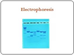
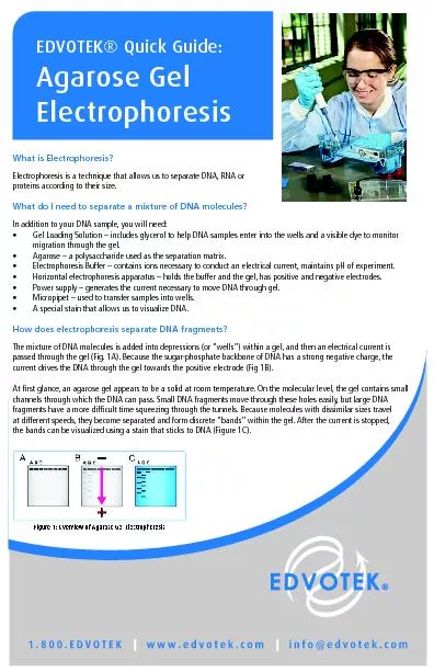
Slide2What is gel electrophoresis?
Method for separation and analysis of macromolecules
DNA, RNA, proteins
Separation based on size and/or charge
Electric fieldMarker can be used to determine size of sample
2
Slide3History
1931 - First report of electrophoresis as a separation technique
1937 - Report on the ‘Tiselius apparatus’ for moving boundary electrophoresis
1955 - Introduction of starch gels
1959 - Introduction of acrylamide gels1969 - Reliable MW determination of proteins using SDS1972 - Agarose gel electrophoresis with ethidium bromide stain
3
Slide4Polyacrylamide gel electrophoresis (PAGE)
Mostly used for protein separation (5-2000 kDa)
Chemical polymerization
Percentage acrylamide
determined pore size, thus separationAdvantage: high resolving power, also for small DNA fragments (5-500 bp’s)
Disadvantage: small range of separation
4
Slide5PAGE gel methods
Denaturing gels
Native
structure of macromolecule is disrupted, mobility depends on linear length
Nucleid acids: urea | Proteins: SDSSDS-PAGENative gels
Analysis
macromolecules
in their folded state, size and shape influence mobility
BN-PAGE: Coomassie Blue provides necessary charges
CN-PAGE
: No
additional charges introduced to proteins
QPNC-PAGE: Preparative variant
5
Slide6SDS-PAGE - Compounds involved
6
Name
Structural formula
FunctionAcrylamidePolymerizes to form the gelTris pH 8.8BufferSDS (sodium dodecyl sulfate)
Anionic detergent to linearize proteins and to impart a negative charge to linearized proteins, leading to a constant mass to charge ratio for every protein
APS (ammonium persulfate)
Persulfate free radicals convert acrylamide monomers to free radicals which react with unactivated monomers to begin the polymerization chain reaction
TEMED (tetramethylethylenediamine)
Accelerates the rate of formation of free radicals from persulfate
Slide7SDS-PAGE - Polymerisation
TEMED accelerates formation of free radicals from persulfate
Persulfate radicals convert acrylamide monomers into radicals
Acrylamide radicals react with unactivated monomers and bis-acrylamide
7
Slide8SDS-PAGE -
Separation mechanism
Power on
Glycine moves towards anode, into stacking gel
Glycine ions become zwitterionic and slow downChlorine ions form ion front ahead of glycine towards anode
Proteins reside between glycine and chlorine ions and become concentrated between the two fronts at high electric field
When the running gel is reached, the pH increases, which
causes deprotonation
of the glycine ions and a high increase in their velocity
The higher acrylamide concentration slows down the proteins according to size
8
Laemmli buffer system
Slide9SDS-PAGE - Separation
Separation depends on:
Size Small proteins move faster
Gel concentration Higher concentration reduces migration speed
Electric field Higher voltage increases migration speed9
Slide10SDS-PAGE -
Visualisation
Coomassie Brilliant Blue
Anionic dye that binds proteins non-specifically
Silver stainingMore sensitive than Coomassie stainingCan also be used to visualize nucleic acids and polysaccharides
Western blot
Antibody-based detection
Very sensitive
10
Slide11SDS-PAGE -
Application
Analysis of proteins in blood serum
Two classes of proteins: serum albumin and globulin
11
Slide12Agarose gel electrophoresis
Mostly used for nucleic acid separation (200-50.000 bp’s)
Material:
natural
polysaccharide polymers extracted from seaweedAgarose is thermally set, no polymerization reaction neededPercentage agarose determines separation
Advantages: large range of separation, easy sample recovery by gel extraction
Disadvantage:
relatively low resolving power
12
Slide13Agarose GE - Separation mechanism
Ogston model
Describes behaviour of DNA smaller than gel pores
Molecules move through pores large enough to accommodate their passage. Movement of large molecules is impeded by collisions with gel matrix.
Reptation modelDescribes behavious of DNA larger than gel pores
DNA crawls through the matrix in a “snake-like” fashion
13
Slide14Agarose GE - Separation
Separation depends on:
Size Small fragments move faster
Conformation Supercoiled DNA moves faster than relaxed DNA
Ethidium bromide concentration Can change the charge and conformation of DNAGel concentration Higher concentration reduces migration speedElectric field Higher voltage increases migration speed
14
Slide15Agarose GE - Visualisation
Ethidium bromide
Intercalates into the major grooves of DNA
Fluoresces under UV light
MutagenicAlternatives: SYBR Green, SYBR Safe15
Slide16Agarose GE - Visualisation
Southern blotting
Detect specific DNA sequence in DNA sample
Method:
Run agarose gelTransfer DNA to nitrocellulose membrane
Expose to hybridization probe, including a radioactive label
Wash away excess probe
Visualize on X-ray film by autoradiography
Northern
blotting
Similar to Southern blotting
Used for RNA detection
16
Slide17Agarose GE - Application
Paternity test
DNA fingerprinting
Restriction enzymes are used to cut DNA into pieces
Pattern of the child should be a combination of the parents’ DNA17
Slide18Conclusions
Gel electrophoresis is a versatile technique for the analysis of proteins and nucleic acids
Different types of gels
Different visualization techniques
It is applied in fields ranging from clinical chemistry to forensic science18
Slide19Moving boundary electrophoresis
Developed by Arna Tiselius
Electrophoresis
free in solution
Nobel prize in Chemistry in 1948The apparatus includes a U-shaped cell filled with buffer solution and electrodes immersed at its ends. The sample applied could be any mixture of charged components like a protein mixture. On applying voltage, the compounds will migrate to the anode or cathode depending on their charges. The change in the refractive index at the boundary of the separated compounds is detected using Schlieren optics at both ends of the solution in the cell.19
Slide20MW determination proteins using SDS
20