
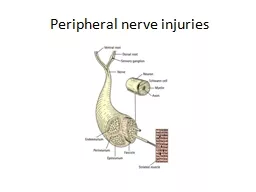
The roots trunks and divisions of the brachial plexus reside in the lower part of the posterior triangle of the neck whereas the cords and most of the branches of the plexus lie in the axilla Complete lesions involving all the roots of the plexus are rare ID: 921284
Download Presentation The PPT/PDF document "Nerve injuries Brachial Plexus Injuries" is the property of its rightful owner. Permission is granted to download and print the materials on this web site for personal, non-commercial use only, and to display it on your personal computer provided you do not modify the materials and that you retain all copyright notices contained in the materials. By downloading content from our website, you accept the terms of this agreement.


Slide1
Nerve injuries
Slide2Brachial Plexus Injuries
The roots, trunks, and divisions of the brachial plexus reside in the lower part of the posterior triangle of the neck
whereas the cords and most of the branches of the plexus lie in the
axilla
Complete lesions involving all the roots of the plexus are rare
Incomplete injuries are common and are usually caused by traction or pressure
individual nerves can be divided by stab wounds.
Slide3Upper Lesions of the Brachial Plexus (
Erb-Duchenne
Palsy)
Upper lesions of the brachial plexus are injuries resulting from excessive displacement of the head to the opposite side and depression of the shoulder on the same side
This causes excessive traction or even tearing of C5 and 6 roots of the plexus
It occurs in infants during a difficult delivery or in adults after a blow to or fall on the shoulder.The suprascapular nerve, the nerve to the subclavius, and the musculocutaneous and axillary nerves all possess nerve fibers derived from C5 and 6 roots and will therefore be functionlessThe following muscles will consequently be paralyzed: the supraspinatus (abductor of the shoulder) and infraspinatus (lateral rotator of the shoulder); the subclavius (depresses the clavicle)
Slide4the biceps
brachii
(
supinator
of the forearm, flexor of the elbow, weak flexor of the shoulder) and the greater part of the
brachialis (flexor of the elbow) and the coracobrachialis (flexes the shoulderand the deltoid (abductor of the shoulder) and the teres minor (lateral rotator of the shoulder)Thus, the limb will hang limply by the side, medially rotated by the unopposed sternocostal part of the pectoralis majorthe forearm will be pronated because of loss of the action of the bicepsThe position of the upper limb in this condition has been likened to that of a porter or waiter hinting for a tip In addition, there will be a loss of sensation down the lateral side of the arm.
Slide5Lower Lesions of the Brachial Plexus (
Klumpke
Palsy)
Lower lesions of the brachial plexus are usually traction injuries caused by excessive abduction of the arm, as occurs in the case of a person falling from a height clutching at an object to save himself or herself
The first thoracic nerve is usually torn.
The nerve fibers from this segment run in the ulnar and median nerves to supply all the small muscles of the handThe hand has a clawed appearance caused by hyperextension of the metacarpophalangeal joints and flexion of the interphalangeal joints.
Slide6The extensor
digitorum
is unopposed by the
lumbricals
and
interossei and extends the metacarpophalangeal jointsthe flexor digitorum superficialis and profundus are unopposed by the lumbricals and interossei and flex the middle and terminal phalanges, respectivelyIn addition, loss of sensation will occur along the medial side of the arm.If the eighth cervical nerve is also damaged, the extent of anesthesia will be greater and will involve the medial side of the forearm, hand, and medial two fingers.Lower lesions of the brachial plexus can also be produced by the presence of a cervical rib or malignant metastases from the lungs in the lower deep cervical lymph nodes.
Slide7Long Thoracic Nerve
The long thoracic nerve, which arises from C5, 6, and 7 and supplies the
serratus
anterior muscle,
can be injured by blows to or pressure on the posterior triangle of the neck or during the surgical procedure of radical mastectomy
Paralysis of the serratus anterior results in the inability to rotate the scapula during the movement of abduction of the arm above a right angleThe patient therefore experiences difficulty in raising the arm above the headThe vertebral border and inferior angle of the scapula will no longer be kept closely applied to the chest wall and will protrude posteriorly, a condition known as winged scapula
Slide8Axillary
Nerve
The
axillary
nerve which arises from the posterior cord of the brachial plexus (C5 and 6), can be injured by the pressure of a badly adjusted crutch pressing upward into the armpit
The passage of the axillary nerve backward from the axilla through the quadrangular space makes it particularly vulnerable here to downward displacement of the humeral head in shoulder dislocations or fractures of the surgical neck of the humerus.Paralysis of the deltoid and teres minor muscles results
Slide9The
cutaneous
branches of the
axillary
nerve, including the upper lateral
cutaneous nerve of the arm, are functionless, and consequently there is a loss of skin sensation over the lower half of the deltoid muscleThe paralyzed deltoid wastes rapidly, and the underlying greater tuberosity can be readily palpatedBecause the supraspinatus is the only other abductor of the shoulder, this movement is much impaired. Paralysis of the teres minor is not recognizable clinically.
Slide10Radial Nerve
The radial nerve which arises from the posterior cord of the brachial plexus, characteristically gives off its branches some distance proximal to the part to be innervated.
In the
axilla
it gives off three branches:
the posterior cutaneous nerve of the arm, which supplies the skin on the back of the arm down to the elbowthe nerve to the long head of the tricepsand the nerve to the medial head of the triceps.
Slide11In the spiral groove of the
humerus
it gives off four branches:
the lower lateral
cutaneous
nerve of the arm, which supplies the lateral surface of the arm down to the elbowthe posterior cutaneous nerve of the forearm, which supplies the skin down the middle of the back of the forearm as far as the wristthe nerve to the lateral head of the tricepsand the nerve to the medial head of the triceps and the anconeus.
Slide12In the anterior compartment of the arm above the lateral
epicondyle
it gives off three branches:
the nerve to a small part of the
brachialis
the nerve to the brachioradialisand the nerve to the extensor carpi radialis longus
Slide13In the
cubital
fossa
it gives off the deep branch of the radial nerve and continues as the superficial radial nerve
The deep branch supplies the extensor carpi radialis brevis and the supinator in the cubital fossa and all the extensor muscles in the posterior compartment of the forearm.The superficial radial nerve is sensory and supplies the skin over the lateral part of the dorsum of the hand and the dorsal surface of the lateral three and a half fingers proximal to the nail beds The radial nerve is commonly damaged in the axilla and in the spiral groove
Slide14Triceps, long head
Triceps, lateral head Triceps, med hd
Brachioradialis
ECRL
ECRB Superficial
Supinator Radial sensExt Digit Abd Pol Longus Post InterosseousExt Pol Longus Ext Pol BrExt Indicies
Slide15Injuries to the Radial Nerve in the
Axilla
In the
axilla
the nerve can be injured by the pressure of the upper end of a badly fitting crutch pressing up into the armpit or by a drunkard falling asleep with one arm over the back of a chair
It can also be badly damaged in the axilla by fractures and dislocations of the proximal end of the humerus.When the humerus is displaced downward in dislocations of the shoulder, the radial nerve, which is wrapped around the back of the shaft of the bone, is pulled downward, stretching the nerve in the axilla excessively.The clinical findings in injury to the radial nerve in the axilla are as follows
Slide16Motor
The triceps, the
anconeus
, and the long extensors of the wrist are paralyzed
The patient is unable to extend the elbow joint, the wrist joint, and the fingers.
Wristdrop, or flexion of the wrist occurs as a result of the action of the unopposed flexor muscles of the wristWristdrop is very disabling because one is unable to flex the fingers strongly for the purpose of firmly gripping an object with the wrist fully flexedIf the wrist and proximal phalanges are passively extended by holding them in position with the opposite hand, the middle and distal phalanges of the fingers can be extended by the action of the lumbricals and interossei, which are inserted into the extensor expansions.The brachioradialis and supinator muscles are also paralyzed, but supination is still performed well by the biceps brachii
Slide17Sensory
A small loss of skin sensation occurs down the posterior surface of the lower part of the arm and down a narrow strip on the back of the forearm.
A variable area of sensory loss is present on the lateral part of the dorsum of the hand and on the dorsal surface of the roots of the lateral three and a half fingers
The area of total anesthesia is relatively small because of the overlap of sensory
innervation
by adjacent nerves.Trophic changes are slight.
Slide18Injuries to the Radial Nerve in the Spiral Groove
In the spiral groove of the
humerus
, the radial nerve can be injured at the time of fracture of the shaft of the
humerus
, or subsequently involved during the formation of the callusThe pressure of the back of the arm on the edge of the operating table in an unconscious patient has also been known to injure the nerve at this site.The prolonged application of a tourniquet to the arm in a person with a slender triceps muscle is often followed by temporary radial palsy.The injury to the radial nerve occurs most commonly in the distal part of the groove, beyond the origin of the nerves to the triceps and the anconeus and beyond the origin of the cutaneous nerves.
Slide19The clinical findings in injury to the radial nerve in the spiral groove are as follows
Motor: The patient is unable to extend the wrist and the fingers, and
wristdrop
occurs
Sensory: A variable small area of anesthesia is present over the dorsal surface of the hand and the dorsal surface of the roots of the lateral three and a half fingers.
Trophic changes: These are very slight or absent
Slide20Injuries to the Deep Branch of the Radial Nerve
The deep branch of the radial nerve is a motor nerve to the extensor muscles in the posterior compartment of the forearm.
It can be damaged in fractures of the proximal end of the radius or during dislocation of the radial head.
The nerve supply to the
supinator
and the extensor carpi radialis longus will be undamagedand because the latter muscle is powerful, it will keep the wrist joint extended, and wristdrop will not occur.No sensory loss occurs because this is a motor nerve
Slide21Injuries to the Superficial Radial Nerve
Division of the superficial radial nerve, which is sensory, as in a stab wound
results in a variable small area of anesthesia over the dorsum of the hand and the dorsal surface of the roots of the lateral three and a half fingers
Slide22Musculocutaneous
Nerve
The
musculocutaneous
nerve is rarely injured because of its protected position beneath the biceps
brachii muscleit is injured high up in the arm, the biceps and coracobrachialis are paralyzed and the brachialis muscle is weakened (the latter muscle is also supplied by the radial nerve). Flexion of the forearm at the elbow joint is then produced by the remainder of the brachialis muscle and the flexors of the forearm.When the forearm is in the prone position, the extensor carpi radialis longus and the brachioradialis muscles assist in flexion of the forearm
Slide23There is also sensory loss along the lateral side of the forearm.
Wounds or cuts of the forearm can sever the lateral
cutaneous
nerve of the forearm, a continuation of the
musculocutaneous
nerve beyond the cubital fossaresulting in sensory loss along the lateral side of the forearm.
Slide24Median Nerve
The median nerve which arises from the medial and lateral cords of the brachial plexus, gives off no
cutaneous
or motor branches in the
axilla
or in the arm.In the proximal third of the front of the forearm, by unnamed branches or by its anterior interosseous branch, it supplies all the muscles of the front of the forearm except the flexor carpi ulnaris and the medial half of the flexor digitorum profundus, which are supplied by the ulnar nerveIn the distal third of the forearm, it gives rise to a palmar cutaneous branch, which crosses in front of the flexor retinaculum and supplies the skin on the lateral half of the palmIn the palm the median nerve supplies the muscles of the thenar eminence and the first two
lumbricals
and gives sensory
innervation
to the skin of the
palmar
aspect of the lateral three and a half fingers, including the nail beds on the dorsum.
Slide25From a clinical standpoint, the median nerve is injured occasionally in the elbow region in
supracondylar
fractures of the
humerus
.
It is most commonly injured by stab wounds or broken glass just proximal to the flexor retinaculumhere it lies in the interval between the tendons of the flexor carpi radialis and flexor digitorum superficialis, overlapped by the palmaris longus.
Slide26Injuries to the Median Nerve at the Elbow
Motor
The
pronator
muscles of the forearm and the long flexor muscles of the wrist and fingers, with the exception of the flexor
carpi ulnaris and the medial half of the flexor digitorum profundus, will be paralyzedAs a result, the forearm is kept in the supine position; wrist flexion is weak and is accompanied by adduction.The latter deviation is caused by the paralysis of the flexor carpi radialis and the strength of the flexor carpi ulnaris and the medial half of the flexor digitorum profundus.No flexion is possible at the interphalangeal joints of the index and middle fingers, although weak flexion of the
metacarpophalangeal
joints of these fingers is attempted by the
interossei
Slide27When the patient tries to make a fist, the index and to a lesser extent the middle fingers tend to remain straight, whereas the ring and little fingers flex
The latter two fingers are, however, weakened by the loss of the flexor
digitorum
superficialis
.Flexion of the terminal phalanx of the thumb is lost because of paralysis of the flexor pollicis longusThe muscles of the thenar eminence are paralyzed and wasted so that the eminence is flattened.The thumb is laterally rotated and adducted. The hand looks flattened and apelike
Slide28Sensory
Skin sensation is lost on the lateral half or less of the palm of the hand and the
palmar
aspect of the lateral three and a half fingers
Sensory loss also occurs on the skin of the distal part of the dorsal surfaces of the lateral three and a half fingers
The area of total anesthesia is considerably less because of the overlap of adjacent nerves.
Slide29Vasomotor Changes
The skin areas involved in sensory loss are warmer and drier than normal because of the arteriolar dilatation and absence of sweating resulting from loss of sympathetic control.
Trophic
Changes
In long-standing cases, changes are found in the hand and fingers
The skin is dry and scaly, the nails crack easily, and atrophy of the pulp of the fingers is present.
Slide30Injuries to the Median Nerve at the Wrist
Motor:
The muscles of the
thenar
eminence are paralyzed and wasted so that the eminence becomes flattened. The thumb is laterally rotated and adducted
The hand looks flattened and apelike.Opposition movement of the thumb is impossibleThe first two lumbricals are paralyzed, which can be recognized clinically when the patient is asked to make a fist slowly, and the index and middle fingers tend to lag behind the ring and little fingers.Sensory, vasomotor, and trophic changes: These changes are identical to those found in the elbow lesions.Perhaps the most serious disability of all in median nerve injuries is the loss of the ability to oppose the thumb to the other fingers and the loss of sensation over the lateral fingers. The delicate pincerlike action of the hand is no longer possible.
Slide31Carpal Tunnel Syndrome
The carpal tunnel, formed by the concave anterior surface of the carpal bones and closed by the flexor
retinaculum
, is tightly packed with the long flexor tendons of the fingers, with their surrounding synovial sheaths, and the median nerve
Clinically, the syndrome consists of a burning pain or pins and needles along the distribution of the median nerve to the lateral three and a half fingers and weakness of the
thenar musclesIt is produced by compression of the median nerve within the tunnelThe exact cause of the compression is difficult to determine, but thickening of the synovial sheaths of the flexor tendons or arthritic changes in the carpal bones are thought to be responsible in many casesno paresthesia occurs over the thenar eminence because this area of skin is supplied by the palmar cutaneous branch of the median nerve, which passes superficially to the flexor retinaculum.The condition is dramatically relieved by decompressing the tunnel by making a longitudinal incision through the flexor
retinaculum
.
Slide32Ulnar
Nerve
The
ulnar
nerve which arises from the medial cord of the brachial plexus (C8 and T1), gives off no
cutaneous or motor branches in the axilla or in the armAs it enters the forearm from behind the medial epicondyle, it supplies the flexor carpi ulnaris and the medial half of the flexor digitorum profundusIn the distal third of the forearm, it gives off its palmar and posterior cutaneous branches.The palmar cutaneous branch supplies the skin over the hypothenar eminence; the posterior branch supplies the skin over the medial third of the dorsum of the hand and the medial one and a half fingers
Not uncommonly, the posterior branch supplies two and a half instead of one and a half fingers
It does not supply the skin over the distal part of the dorsum of these fingers.
Slide33Having entered the palm by passing in front of the flexor
retinaculum
, the superficial branch of the
ulnar
nerve supplies the skin of the
palmar surface of the medial one and a half fingers including their nail beds; it also supplies the palmaris brevis muscleThe deep branch supplies all the small muscles of the hand except the muscles of the thenar eminence and the first two lumbricals, which are supplied by the median nerveThe ulnar nerve is most commonly injured at the elbow, where it lies behind the medial epicondyle, and at the wrist, where it lies with the ulnar artery in front of the flexor retinaculumThe injuries at the elbow are usually associated with fractures of the medial epicondyle
.
The superficial position of the nerve at the wrist makes it vulnerable to damage from cuts and stab wounds.
Slide34Ulnar
nerve
Elbow
Flexor
carpi ulnaris Flex Dig Prof III/IV Dorsal uln cut WristAdductor Pollicus AbductorFlex Pollicus Br Opponens Digiti Minimi FlexorDorsal/palmar
Interosseous
3rd/4th
lumbricals
Slide35Injuries to the
Ulnar
Nerve at the Elbow
Motor
The flexor
carpi ulnaris and the medial half of the flexor digitorum profundus muscles are paralyzedThe paralysis of the flexor carpi ulnaris can be observed by asking the patient to make a tightly clenched fistNormally, the synergistic action of the flexor carpi ulnaris tendon can be observed as it passes to the pisiform bonethe tightening of the tendon will be absent if the muscle is paralyzedThe profundus tendons to the ring and little fingers will be functionless, and the terminal phalanges of these fingers are therefore not capable of being markedly flexed
Flexion of the wrist joint will result in abduction, owing to paralysis of the flexor
carpi
ulnaris
The medial border of the front of the forearm will show flattening owing to the wasting of the underlying
ulnaris
and
profundus
muscles
Slide36The small muscles of the hand will be paralyzed, except the muscles of the
thenar
eminence and the first two
lumbricals
, which are supplied by the median nerve.
The patient is unable to adduct and abduct the fingers and consequently is unable to grip a piece of paper placed between the fingersthe extensor digitorum can abduct the fingers to a small extent, but only when the metacarpophalangeal joints are hyperextended.It is impossible to adduct the thumb because the adductor pollicis muscle is paralyzedIf the patient is asked to grip a piece of paper between the thumb and the index finger, he or she does so by strongly contracting the flexor pollicis longus and flexing the terminal phalanx (Froment's sign).
Slide37The
metacarpophalangeal
joints become
hyperextended
because of the paralysis of the
lumbrical and interosseous muscles, which normally flex these jointsBecause the first and second lumbricals are not paralyzed (they are supplied by the median nerve), the hyperextension of the metacarpophalangeal joints is most prominent in the fourth and fifth fingersThe interphalangeal joints are flexed, owing again to the paralysis of the lumbrical and interosseous muscles, which normally extend these joints through the extensor expansion.
Slide38The flexion deformity at the
interphalangeal
joints of the fourth and fifth fingers is obvious because the first and second
lumbrical
muscles of the index and middle fingers are not paralyzed
In long-standing cases the hand assumes the characteristic claw deformity (main en griffe). Wasting of the paralyzed muscles results in flattening of the hypothenar eminence and loss of the convex curve to the medial border of the hand.Examination of the dorsum of the hand will show hollowing between the metacarpal bones caused by wasting of the dorsal interosseous muscles
Slide39Sensory
Loss of skin sensation will be observed over the anterior and posterior surfaces of the medial third of the hand and the medial one and a half fingers.
Vasomotor Changes
The skin areas involved in sensory loss are warmer and drier than normal because of the arteriolar dilatation and absence of sweating resulting from loss of sympathetic control.
Slide40Injuries to the
Ulnar
Nerve at the Wrist
Motor:
The small muscles of the hand will be paralyzed and show wasting, except for the muscles of the
thenar eminence and the first two lumbricals The clawhand is much more obvious in wrist lesions because the flexor digitorum profundus muscle is not paralyzed, and marked flexion of the terminal phalanges occurs.Sensory:The main ulnar nerve and its palmar cutaneous branch are usually severedthe posterior cutaneous branch, which arises from the ulnar nerve trunk about 2.5 in. (6.25 cm) above the
pisiform
bone, is usually unaffected
The sensory loss will therefore be confined to the
palmar
surface of the medial third of the hand and the medial one and a half fingers and to the dorsal aspects of the middle and distal phalanges of the same fingers.
Vasomotor and
trophic
changes:
These are the same as those described for injuries at the elbow.
It is important to remember that with
ulnar
nerve injuries, the higher the lesion, the less obvious the clawing deformity of the hand
Slide41Unlike median nerve injuries, lesions of the
ulnar
nerve leave a relatively efficient hand
The sensation over the lateral part of the hand is intact, and the
pincerlike
action of the thumb and index finger is reasonably goodalthough there is some weakness owing to loss of the adductor pollicis.