

Gout Acute inflammatory arthritis caused by accumulation of monosodium urate Typical Features Monoarticular Rapid onset wI 24 hrs Incapacitating pain Pathophysiology Uric Acid product of purine degradation accumulates to the point of precipitating into the joint ID: 775431
Download Presentation The PPT/PDF document " Gout Scott Smith PGY-1 1/11/2018" is the property of its rightful owner. Permission is granted to download and print the materials on this web site for personal, non-commercial use only, and to display it on your personal computer provided you do not modify the materials and that you retain all copyright notices contained in the materials. By downloading content from our website, you accept the terms of this agreement.


Slide1
Gout
Scott Smith PGY-1
1/11/2018
Slide2Gout
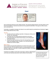
Acute inflammatory arthritis caused by accumulation of monosodium urate
Slide3Typical Features
Monoarticular
Rapid onset w/I 24
hrs
Incapacitating pain
Slide4Pathophysiology
Uric Acid (product of purine degradation) accumulates to the point of precipitating into the joint
Precipitation of uric acid can be affected by variety of factors, including body pH and temperature
Hyperuricemia (serum urate >7mg/
dL
in men >6mg/
dL
in women) is the major risk factor
Precipitated urate crystals phagocytosed by macrophages
Crystals bins to cellular NLRP3
inflammasome
Secretion of IL-1
IL-1 binds IL-1 receptor on synovial cells, secreting
proinflammatory
factors
Arrival of neutrophils and secretion of additional inflammatory factors causes inflammation and pain
Slide5Pathophysiology
Understanding of pathophysiology presents targets
for control of disease
Slide6Clinical Manifestations
Early gout is typically
monoarticular
, most frequently the MTP joint of 1
st
toe (
Podagra
)
Develops rapidly from asymptomatic to incapacitating pain in <25hrs
Joint is red, warm, swollen and exquisitely tender
Common sites 1
st
toe > arch of foot > ankle > knee
Wrist, elbow and shoulder involvement is more characteristic of more advanced gout
Slide7Progression of Disease
After having years of disease a patient will have more frequent and more severe diseasePolyarticular GoutSoft tissue deposits of monosodium urate (tophi) esp. on auricle, extensor surface of hand/fingers, olecranon bursa and achilles tendonCan develop chronic arthritis as opposed to flares of acute goutErosive Arthropathy
Slide8Gout Stages
Asymptomatic
hyperuricemia:
majority never develop
This usually lasts for years-decades before developing acute gouty
arthritis
Acute gouty arthritis
:
monoarticular
pain (
podagra
, ankle, etc.); attacks last hours-2
week
Intercritical
gout
: completely asymptomatic between acute attacks
~65% will have another acute attack within 1-2 years
Presence of asymptomatic periods between
monoarthritic
attacks is almost unique to gout and used as a diagnostic
criterion
Chronic
tophaceous
gout
:
intercritical
periods are now symptomatic (chronic swelling and worsening pain) & develop subcutaneous
tophaceous
deposits of MSU; usually after >10y of acute intermittent gout
Slide9Differential Diagnosis of Monoarthropathy
Gout
Pseudogout
Septic Arthritis
Hemorrhagic
Arthropathy
Reactive Arthritis
Traumatic
Arthropathy
Cellulitis
Slide10Risk Factors - Demographics
Gender
Gout is more prevalent in men by a factor of 3 (6% vs 2%)
However Gap is narrowed in post-menopausal women
Slide11Risk Factors - Demographics
Age
Increasing age brings heavier burden of disease to age cohort
1% prevalence in 20’s
3.3% prevalence in 40’s
12.6% prevalence in 80’s
Slide12Risk Factors - Demographics
Ethnicity
More common in African Americans >
Causasians
> Mexican Americans
5% vs 4% vs 1.5%
Slide13Risk Factors - Diet
Intuitively, a diet high in purines increases risk
Alcohol is also associated with higher risk, especially beer
Red meats and seafood
Caffeinated Coffee, low-fat dairy, Cherries are associated with lower risk
Slide14Risk Factors - Diet
Slide15Risk Factors - Clinical
Slide16Diagnosis
Gold standard is arthrocentesis with microscopy
Aspirate in disease will be yellow vs. normal clear
Microscopy under polarized light shows needle-shaped, negatively birefringent crystals
Leukocyte count of 15,000
–
80,000 with neutrophil predominance
Soft Tissue Tophi
Shown to contain monosodium urate crystals under polarized microscopy
Slide17Slide18Diagnosis
Arthrocentesis is not always feasibleClinical Criteria for diagnosisPut forth by American College of Rheumatology in 19776 or more is sufficient for diagnosis of gout
Slide19Treatment of Acute Gout
Corticosteroids
Can be administered orally, IV or
intraarticularly
Rapid relief of symptoms 1-5 hours
Methylprednisolone dose pack (starting 24mg daily, reducing 4mg daily)
Prednisone starting 20-30mg daily, tapering in 1-2 weeks
Intra-articular injection of triamcinolone in
monoarticular
gout attack
Slide20Treatment of Acute Gout
NSAID’s
Indomethacin most commonly used
Limited in many patients
PUD
CKD
Anticoagulation
Slide21Treatment of Acute Gout
Colchicine
Commonly prescribed as 0.6mg tablets
Old Therapy included 1 tablet per hour until gout attack is improved
or until diarrhea develops
New Standard of 1.2mg dose followed by 0.6mg dose after an hour shown to be equal and less toxic
Can Impair Hematopoiesis, induce
neuromyotoxicity
, and is contraindicated in CKD
Slide22Prevention of Gout Attacks
Eliminating precipitants
Certain Medications can increase serum Uric Acid
HCTZ
Immunosuppressant medications (Tacrolimus, Cyclosporine)
Low Dose aspirin
Slide23Gout Prophylaxis
Criteria for prophylaxis
Based on clinical picture, not lab values
M
ore than one gout flare per year
Presence of
tophaceous
gout
Slide24Gout Prophylaxis – Xanthine Oxidase Inhibitors
Allopurinol
Typically started at 300mg daily but can be lower at 50-100mg daily
Maximum dose of 600-800mg daily
Patients can rarely develop allopurinol hypersensitivity syndrome, more common in patients with impaired renal
function
Feboxistat
Relative indication is impaired renal function or other situation where allopurinol cannot be used
Metabolized by liver instead of kidney
Starting dose of 40mg daily maximum dose of 80mg daily
Should treat to goal serum uric acid level of <6mg/
dL
Both can precipitate new gout attack, so treatment should be started in conjunction with colchicine 0.6mg daily or naproxen 250mg 2x daily until steady dose is achieved
Slide25Gout Prophylaxis – Colchicine
Can be used for prophylaxis at different dosage
0.6 or 1.2mg daily
Lower dosage should be considered in those with impaired renal function
Must monitor for drug-induced myopathy and neuropathy
Slide26Gout Prophylaxis – Probenecid
Uricosuric
Agent
Inhibits reabsorption of uric acid in proximal renal tubule
Starting dose of 250mg 2x daily increased to 500mg 2x daily in one week
Ineffective in impaired renal function
Contraindicated in patient with prior urate nephrolithiasis
Slide27Thank you!