
Study Histology Often called Microscopic Anatomy Greek word Histos tissue Logia sciencestudy Cells work together in functionally related groups called tissues 4 basic type of tissues ID: 912306
Download Presentation The PPT/PDF document "Histology Study of cells, tissues and or..." is the property of its rightful owner. Permission is granted to download and print the materials on this web site for personal, non-commercial use only, and to display it on your personal computer provided you do not modify the materials and that you retain all copyright notices contained in the materials. By downloading content from our website, you accept the terms of this agreement.


Slide1
Histology
Study of cells, tissues and organs as seen with the help of microscope
Study
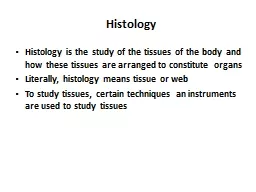
Slide2Histology
Often called Microscopic AnatomyGreek word – Histos
= tissue
Logia=science/studyCells work together in functionally related groups called tissues
4 basic type of tissues:
Epithelial
– lining and covering
Connective
– support
Muscle
– movement
Nervous
– control
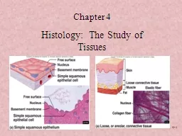
Slide3Epithelial Tissue
or Epithelium
Consist of sheets of cells
Covers
a external surface of the bodyMay line the internal cavities and the organsForms most organs & glands
Slide4Special Characteristics of Epithelia
Cellularitycells are in close contact with each other with little or no intercellular space between them
Specialized
contacts:
Junctional Complexesmay have junctions for both attachment and communication
Slide5Special Characteristics of Epithelia
Polarityepithelial tissues always have an apical and basal surface
Support by connective tissue
at the basal surface, both the epithelial tissue and the connective tissue contribute to the basement membrane
Avascular..No blood vessel, no lymphaticsnutrients must diffuse
Slide6Special Characteristics of
EpitInnervated
Regeneration
epithelial tissues have a high capacity for regeneration
Invaginates and grows in the underlying CT..specializing as glandsApical surface shows modification: presence of cilia or
microvilli
Nuclear shape corresponds
to the cell shape: oval in
columnar,round
in
cuboidal
or polyhedral and flat in
squamous
cells
Slide7Functions
Functions of epitheliumProtectionAbsorption,
May secrete material and ion transport
Filtration
Forms slippery surfacesFunction as sensory surfaces
Slide8Embryologically
Epithelia are derived from all the 3 germ layers:Ectoderm Epithelium of skinEndoderm Epithelium of gutMesoderm Epithelium of pericardial, peritoneal and pleural cavities
Slide9Basement Membrane
All cells rest on it.Thin, non-cellularSeparates epithelium from underlying connective tissueEasily seen with light microscope
Made up of:
Basal Lamina-Amorphous, product of epithelium Reticular Lamina-Reticular fibres, product of CT
Slide10Basement Membrane
The epithelial cells lie on the reticular lamina
(collagen – CT)
Reticular lamina is bound to another CT called
areolar CT. Together this structure is called the "basement
membrane
”
Slide11Basal Feature: The Basal Lamina
Noncellular supporting sheet between the epithelium and the connective tissue deep to it Consists of proteins secreted by the epithelial cells
Functions:
Acts as a selective filter, determining which molecules from capillaries enter the epithelium
Acts as scaffolding along which regenerating epithelial cells can migrate Basal lamina and reticular layers of the underlying connective tissue deep to it form the basement membrane
Slide12Intercellular Junctions OR
Junctional ComplexesZonula
occludens
(Tight Junctions)Zonula adherensMacula adherens (Desmosome) and hemidesmosome
Gap junction (Nexus)
Slide13Lateral Surface Features
Factors holding epithelial cells togetherAdhesion proteins link plasma membranes of adjacent cellsContours of adjacent cell membranes Special cell junctions
Slide14Tight junctions (
zona occludens)
– close off intercellular space
Found at apical region of most epithelial typesSome proteins in plasma membrane of adjacent cells are fusedPrevent molecules from passing between cells of epithelial tissue…..Serves as a SELECTIVE BARRIER, giving it a sealing effect.
Example- Intestine and urinary bladder
Slide15Adherens
junctions (zonula adherens) – anchoring junction
Transmembrane
linker proteins attach to actin microfilaments of the cytoskeleton and bind adjacent
cells
Along with tight junctions, form the tight
junctional
complex around apical lateral borders of epithelial
tissues
Present just below the tight junctions
Provides Rigidity to the apex of the cell.
Presence of dense plaque – like material on the
cytoplasmic
surface of the plasma membranes of the junction.
Slide16Slide17Desmosomes
(Macula Adherens) Hemidesmosomes
Gap of 30nm
Transmembrane
ProteinsElectron dense plaqueAttachment to Intermediate
Filaments
FIRM ADHESION between cells
Subjected to friction,
Epidermis of skin.
Slide18Desmosomes
Desmosomes – two disc-like plaques connected across intercellular spacePlaques of adjoining cells are joined by proteins called cadherins Proteins interdigitate
into extracellular space
Intermediate filaments insert into plaques from
cytoplasmic side
Slide19Desmosomes
Slide20Gap junctions (Nexus)
passageway between two adjacent cellsLet small molecules move directly between neighboring cells
Cells are connected by hollow cylinders of
protein
Passage of inorganic ionsExchange of chemical messengers in cell recognition and differentiation.
Slide21Gap Junction
Slide22Slide23Slide24Tight Junctions
In the apical Band or beltBarrier device
Slide25Surface Modifications
Glycocalyx-rich in polysaccharides Concentrates ions prior to absorption
Act as receptor sites for hormones and enzymes.
Microvilli
- minute finger like projectionsIncrease absorptive surface Stereocilia –
Long thick
Microvilli
, Non motile, may show branching, Increase surface area(
Epididimis
), helps perception of stimuli (Internal Ear)
Cilia-
long, hair like projections of plasma membrane
Slide26Microvilli and Cilia
NonmotileContain MicrofilamentsFunction-AbsorptionIntestinal epithelium, proximal convoluted tubules of the kidney
Motile
Contain 9+2 pattern of microtubules
Driving the entangled particles, transport in one dcirectionExamples: Respirastory
tract,uterine
tube and
ependyma
Slide27Epithelial Tissues
Slide28According to the number of cell layers
First name of tissue indicates number of layersSimple – one layer of cells
Stratified – more than one
layer of cells
Classifications & Naming of Epithelia
Slide29Classification & Naming of Epithelia
Last name of tissue describes shape of cellsSquamous – cells wider than tall (plate or “scale” like)
Cuboidal – cells are as wide
as tall, as in cubes
Columnar – cells are taller than they are wide, like columns
Slide30Naming Epithelia
Naming the epithelia includes both the layers (first) and the shape of the cells (second)i.e. stratified cuboidal epitheliumThe name may also include any accessory structures
Goblet cells
Cilia
KeratinSpecial epithelial tissues (don’t follow naming convention)PsuedostratifiedTransitional
Slide31Simple Squamous
EpitheliumDescription single layer of flat cells with disc-shaped nucleiSpecial types Endothelium (inner covering)
slick lining of hollow organs
Mesothelium (middle covering)
Lines peritoneal, pleural, and pericardial cavities Covers visceral organs of those cavities
Slide32Simple Squamous
EpitheliumFunction Passage of materials by passive diffusion and filtration
Secretes lubricating substances in serosae
Location
Renal corpusclesAlveoli of lungs Lining of heart, blood and lymphatic vessels
Lining of ventral body cavity (serosae)
Slide33Simple Squamous Epithelium
Simple squamous lining the walls of the capillary
Slide34Simple Cuboidal Epithelium
Descriptionsingle layer of cube-like cells with large, spherical central nucleiFunction secretion and absorption
Location
kidney tubules, secretory
portions of small glands, ovary & thyroid follicles
Slide35Simple Columnar Epithelium
Description single layer of column-shaped (rectangular) cells with oval nucleiSome bear cilia at their apical surface
May contain goblet cells
Function
Absorption; secretion of mucus, enzymes, and other substancesCiliated type propels mucus or reproductive cells by ciliary action
Slide36Simple Columnar Epithelium
Location Non-ciliated form Lines digestive tract, gallbladder, ducts of some glandsCiliated form
Lines small bronchi,
uterine tubes, uterus
Slide37Slide38Pseudostratified Columnar Epithelium
DescriptionAll cells originate at basement membraneOnly tall cells reach the apical surfaceMay contain goblet cells and bear ciliaNuclei lie at varying heights within cells
Gives false impression of stratification
Function
secretion of mucus; propulsion of mucus by cilia
Slide39Pseudostratified Columnar Epithelium
LocationsNon-ciliated type Ducts of male reproductive tubes
Ducts of large glands
Ciliated variety
Lines trachea and most of upper respiratory tract
Slide40Stratified Epithelia
Contain two or more layers of cellsRegenerate from belowMajor role is protectionAre named according to the shape of cells at apical layer
Slide41Stratified Squamous Epithelium
DescriptionMany layers of cells – squamous in shapeDeeper layers of cells appear cuboidal or columnar Thickest epithelial tissue – adapted for protection
Slide42Stratified Squamous
EpitheliumSpecific types Keratinized – contain the protective protein keratinSurface cells are dead and full of keratin
Non-keratinized – forms moist lining of body openings
Function
Protects underlying tissues in areas subject to abrasionLocation Keratinized – forms epidermisNon-keratinized – forms lining of esophagus, mouth, and vagina
Slide43Transitional Epithelium
Description Basal cells usually cuboidal or columnarSuperficial cells dome-shaped or squamousFunctionstretches and permits distension of urinary bladder
Location
Lines ureters, urinary bladder and part of urethra
Slide44Introduction
Histology
There are (4) types of tissue:
1. Epithelial
2. Connective
3. Muscle
4. Nervous
Similarities
between tissue types:
1. All contain cells
2. Cells that make up tissues have similar functions
Slide45Epithelial Structure
Apical
Basement Membrane
Apical
Slide46Basement Membrane
The epithelial cells lie on the reticular lamina
(collagen – CT)
Reticular lamina is bound to another CT called
areolar CT. Together this structure is called the "basement
membrane
”
Slide47Classification and Examples
1. Simple Epithelium
Single layer
All cells anchored to basement membrane
2. Simple SquamousKidney – filtration
3.
Simple
Cuboidal
Kidney tubules
Filtration; secretion, absorption
Slide48Simple Epithelia
4. Simple Columnar
Tall, thin cells
Absorptive cells (small intestine)
Goblet Cells 5. Pseudostratified ‘Ciliated
’
Columnar
Epithelium
“
Pseudostratified
” ?
Trachea
Goblet Cells and Mucus
Slide49Stratified Epithelium
Characteristics2+ layers
Stratified
SquamousSkin – outer layer hardened by ‘keratin’ 4 to 5 layers thick
3.
Stratified
Cuboidal
Ducts of sweat glands
This type + stratified columnar are rare!
Slide50MCQ
Transitional epithelium is found inUterusUreterGall bladder
vagina
Slide51MCQ
Stomach is lined bySimple columnar epitheliumStraified squamous epithelium
Simple
cuboidal
epitheliumPseudostratified columnar epithelium
Slide52MCQ
Simple Squamous epithelium is seen inAlveoli of lungsStomach
Urinary bladder
Tongue
Slide53MCG
Glycocalyx coat present in the absorptive surface of small intestine1.Increases the surface area2.Transports the absorbed material3.Concentrates ions prior to absorption
4.Participates in the digestion of carbohydrates
Slide54MCQ
Pseudostratified Epithelium is seen in1. Ureter2.Skin3.Trachea
4.Kidney
Slide55Simple
squamous epithelium lining of serous membrane is called………..Basement membrane is made up of how many layers….What are Microvilli?Desmosomes
?