
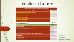
Asst Professor Department of Medicine AIIMS Rishikesh 5 th Semester Classes on Infectious Diseases 89AM Thursdays LT4 Topics Date Approach to Infectious Diseases and their prevention ID: 777001
Download The PPT/PDF document "Infectious diseases Dr. P. K. Panda," is the property of its rightful owner. Permission is granted to download and print the materials on this web site for personal, non-commercial use only, and to display it on your personal computer provided you do not modify the materials and that you retain all copyright notices contained in the materials. By downloading content from our website, you accept the terms of this agreement.


Slide1
Infectious diseases
Dr. P. K. Panda, Asst. ProfessorDepartment of MedicineAIIMS, Rishikesh
5
th
Semester Classes on Infectious
Diseases, 8-9AM, Thursdays (LT-4)
Topics
Date
Approach to Infectious Diseases and their prevention
04/Jul/17
Community-Acquired Infections
27/
Jul /17
Health Care–Associated Infections
03/ Aug/17
Gram-Positive
Bacteria (part-1)
10/ Aug/17
Gram-Positive Bacteria (part-2)
17/Aug/17
Gram-Negative
Bacteria
(part-1)
24/
Aug /17
Class bunked due to strike
31/ Aug /17
Gram-Negative Bacteria (part-2)
07/ Sep/17
Spirochetal Diseases
14/Sep/17
Diseases Caused by Atypical Bacterial Infections
21/Sep/17
Infections Due to DNA Viruses
28/Sep/17
Infections Due to RNA Viruses
05/Oct/17
Human Immunodeficiency Virus Disease:
AIDS and
Related Disorders
12/Oct/17
Fungal Infections
26/Oct/2017
Protozoal Infections
02/Nov/2017
Helminthic Infections
09/Nov/2017
Slide2ENTEROBACTERIACEAE
(E. coli, Klebsiella, Proteus
,
Enterobacter)
In healthy humans, E. coli is the predominant species of gram-negative bacilli (GNB) in the colonic flora; Klebsiella and Proteus are less prevalentMultiple bacterial virulence factors are required for the pathogenesis
Slide3Certain strains of
E. coli are capable of causing diarrheal diseaseExPEC strains are the most common enteric GNB to cause community-acquired and health care–associated bacterial infections (All age groups, all types of hosts, and nearly
all organs
and anatomic sites)Humans are the major reservoir [except
for STEC/EHEC]
Slide4Transmission occurs predominantly
via contaminated food and water for ETEC, STEC/EHEC/STEAEC, EIEC, and EAEC and by person-to-person spread for EPEC (and occasionally STEC/EHEC/STEAEC)
Except
in the cases of EHEC and EAEC, disease occurs primarily in developing countriesDistinguish
noninflammatory (mainly by ETEC, EPEC, and DAEC) from inflammatory diarrhea (suggested by grossly bloody or mucoid stool or a positive test for fecal leukocytes)Definitive diagnosis generally is not necessary except for STECThe
mainstay of treatment for all diarrheal
syndromes is
replacement of water and
electrolytes, especially for
STEC/EHEC/STEAEC
infection where antibiotics may
increase the incidence of
HUS
If
diarrhea persists for >10
days
despite
treatment,
Giardia
or
Cryptosporidium
should
be sought
Slide5Person-to-person spread is the predominant
mode of acquisition of KlebsiellacKP Causes pneumonia, UTI, abdominal infection, intravascular device infection, surgical site infection, soft tissue infection, and
subsequent bacteremia
hvKP (of Asian origin) infection distinguished from traditional infections due to
cKP by (1) presentation as community-acquired pyogenic liver abscess (2) occurrence in patients lacking a history of hepatobiliary disease, and (3) a propensity for metastatic spread to distant sitesUrine samples with unexplained
alkalinity should be cultured for
Proteus
, and
identification of
a
Proteus
species in urine should prompt consideration of
an evaluation
for
urolithiasis
Enterobacter
/
citrobacter
/
serratia
/
morganella
/
edwardsiella
causes a spectrum of
extraintestinal
infections
similar to other GNB
Slide6Two species: Salmonella
enterica and Salmonella bongoriSerotyping is based on the somatic O antigen (lipopolysaccharide cellwall components), the surface Vi antigen
(restricted to
S. typhi and S. paratyphi C), and the
flagellar H antigenThe growth of serotypes Salmonella typhi and Salmonella paratyphi is restricted to human hosts, remaining serotypes (
nontyphoidal
Salmonella
, or
NTS
)
can colonize the gastrointestinal tracts of a broad range of animals, reptiles, birds, and insectsIngestion in contaminated food or water with the ingested dose as determinant of incubation period and disease severity Conditions that decrease either stomach acidity or intestinal integrity increase susceptibility to infectionOnce reach the small intestine, they penetrate the mucus layer of the gut, traverse the intestinal layer through phagocytic microfold (M) cells that reside within Peyer’s patches, phagocytosed by macrophages but in a protective manner, and then via the lymphatics colonize reticuloendothelial tissuesIn contrast to enteric fever, NTS gastroenteritis is characterized by massive polymorphonuclear leukocyte infiltration into both the large- and small-bowel mucosa
SALMONELLA
Slide7Enteric (typhoid
) fever is a systemic disease characterized by fever and abdominal pain and caused by dissemination of S. typhi or S. paratyphi
Most
commonly, food-borne or waterborne transmission results from fecal contamination; Sexual transmission between male partners has been
described; Health care workers occasionally acquire tooIP; 10–14 days but ranges from 5 to 21 daysRisk factors include contaminated water or ice, flooding,
food
and drinks purchased from
street vendors
,
raw
fruits and vegetables grown
in fields fertilized with sewage, ill household contacts, lack of hand washing and toilet access, and evidence of prior Helicobacter pylori infectionIt is estimated that there is one case of paratyphoid fever for every four cases of typhoid fever
Slide8SYMPTOMS
SIGNS
Fever
(>75%)
headache (80%)anorexia (55%)chills (35–45%)
Abdominal pain
(30-40%)
cough
(30%)
sweating (20–25%)
myalgias
(20%),nausea (18–24%),vomiting (18%), diarrhea (22–28%) Constipation (13–16%)malaise (10%)
arthralgia
(2–4%).
coated tongue (51–56%),relative bradycardia at the peak of high fever (<50%)rose spots (30%), splenomegaly (5–6%), abdominal tenderness (4–5%)hepatosplenomegaly (3–6%), epistaxis,
The
development of severe disease
(which occurs in ~10–15%
of patients
) depends on host
factors,
strain virulence and
inoculum, and
choice of antibiotic
therapy
Gastrointestinal
bleeding (10–20%)
Intestinal
perforation (1–3
%)
Neurologic
manifestations (2-40%)
Slide9Up to 10% of untreated patients excrete
S. typhi in the feces for up to 3 months, and 1–4% develop chronic asymptomatic carriage, shedding S. typhi in either urine or stool
for >1 year
The definitive diagnosis of enteric fever requires the isolation of S. typhi or
S. paratyphi from blood, bone marrow, intestinal secretions,(THESE 3 IN COMBINATION POSITIVE >90%) other sterile sites, rose spots, and stoolSerologic tests, including the classic Widal test for “febrile agglutinins,” and rapid tests to detect antibodies to outermembrane
proteins
have
lower positive
predictive
values
than blood
cultureTwo typhoid vaccines: Ty21a, (given on days 1, 3, 5, and 7, with a booster every 5 years); Vi CPS, (given in a single dose, with a booster every 2 years)cumulative efficacy was 48% for Ty21a at 2.5–3.5 years and 55% for Vi CPS at 3 years
Slide10Shigella cannot be
distinguished from Escherichia coli by DNA hybridization and remains a separate species only on hstorical and clinical groundsUnlike E
. coli
, is nonmotile and does not produce gas from sugarsHuman intestinal tract represents the major reservoirBacteria are
transmitted most efficiently by the fecal-oral route via hand carriage, rarely by flies and sexuallyHighest prevalences in the most impoverished areasShigellosis typically evolves through four phases:
Incubation (
1–4
days),
Watery diarrhea,
Dysentery -
dysentery—a
clinical syndrome of fever, intestinal cramps, and frequent passage of small, bloody, mucopurulent stoolsPostinfectious phase – Reactive arthritis, toxic megacolon, and HUS (in developing countries)Ciprofloxacin is recommended as first-line treatment; others like ceftriaxone, azithromycin, pivmecillinam, and some fifth-generation quinolonesSHIGELLA
Slide11Cholera now refers to
disease caused by V. cholerae serogroup O1 or O139—i.e., the serogroups with epidemic potentialResponsible for seven
global
pandemics and much suffering over the past two centuriesIn nature, vibrios most commonly reside in tidal rivers and bays under conditions of
moderate salinity; They proliferate in the summer monthsCholera is predominantly a pediatric disease in endemic areas, but it affects adults and children equally when newly introduced into a populationCholera toxin, toxin-coregulated pilus,
and several other virulence factors are
coordinately regulated
by
ToxR
With
IP
24- to 48-hSome individuals are asymptomatic or have only mild diarrhea; others present with the sudden onset of explosive and life-threatening diarrhea (cholera gravis); “rice-water” stool WITH absent fever; Complications derive exclusively from the effects of volume and electrolyte depletionClinical suspicion of cholera can be confirmed by the identification in stoolTreatment; first and foremost requires fluid resuscitation with macrolides (DOC)VIBRIO
Slide12Pseudomonas, Burkholderia
, and StenotrophomonasThe pathogenicity is based on opportunism with the exceptions (melioidosis
by
Burkholderia pseudomallei and glanders by B.
mallei)P. aeruginosa remains the most common contributing factor to respiratory failure in Cystic FibrosisB. cepacia gained notoriety as the cause of a rapidly fatal syndrome of respiratory distress and septicemia (the “cepacia syndrome”) in CF patients
Cytotoxic
chemotherapy,
mechanical ventilation
, and broad-spectrum antibiotic therapy probably
paved the
way for colonization and
infectionP. aeruginosa is found in most moist environments; infection Often occurs concomitantly with host defense compromiseOf the common gram-negative bacteria, no other species produces such a large number of putative virulence factorsAmong gram-negative bacteria, it probably produces the largest number of substances that are toxic to cells and thus may injure tissuesPSEUDOMONADS (an inability to ferment lactose)
Slide13P. aeruginosa causes infections at almost all sites in the body but
shows a rather strong predilection for the lungsBacteremia; only point differentiating this entity from gram-negative sepsis of other causes may be ecthyma
gangrenosum
, which occur almost exclusively in markedly neutropenic patients and patients with AIDSCombination therapy became the standard of
care, recently newer antipseudomonal drugs (colistin, tigecycline, cefepime) can be used as monotherapy
Slide14Acinetobacter baumannii
is particularly formidable because of its propensity to acquire antibiotic resistance determinantsContrary to previous thought of nonmotile characteristic it demonstrate motility under certain growth conditions
Widely distributed in nature
, like water, soil, on vegetables, a component of the skin flora,
and sometimes a contaminant in blood samplesColonizes patients exposed to heavily contaminated hospital environments or to the hands of health care workersIt must be considered in the differential diagnosis of hospital-acquired pneumonia, central line–associated bloodstream infection, posttraumatic wound infection in military personnel, and
postneurosurgical
meningitis
It should
be suspected
when plump
coccobacilli
are seen in Gram’s-stained samplesOnly sulbabctam, cotrimoxazole, carbapenams, amikacin, tigecycline, colistin are possible treatmentACINETOBACTER
Slide15It colonizes the stomach in ~50%
of the world’s human population, essentially for life unless eradicated by antibiotic treatmentHumans are the only important reservoir
Lifelong colonization
may offer some protection against complications of gastroesophageal reflux disease (GERD), including esophageal adenocarcinoma
Treatment against the organism prevent/treat PUD and low-grade gastric MALT lymphoma, however, no benefit in the treatment of gastric adenocarcinomaNongastric (intestinal) Helicobacter species can cause clinical features resembling those of Campylobacter
infections
Prevalence varies
with age:
H
. pylori
is usually acquired in
childhood, The age association is due mostly to a birth-cohort effectCombination of factors lead to disease state: bacterial strain differences, host susceptibility to disease, and environmental factorsHELICOBACTER
Slide16Slide17Slide18Whether or not the ulcers are currently active, H. pylori
should be eradicated in patients with documented ulcer disease to prevent RelapseOverall most treatment of asymptomatic H. pylori carriage is given without a firm evidence base
Test-and-treat
has emerged as a common clinical practice
Slide19Campylobacter,
Arcobacter, and HelicobacterIt is more common than that due to Salmonella and Shigella combined
Although
acute diarrheal illnesses are most common, these organisms may cause infections in virtually all parts of the body, especially in compromised hosts
, and these infections may have late nonsuppurative sequelae (Reactive A, GBS)The human pathogens fall into two major groups: those that primarily cause diarrheal disease (C. jejuni mainly)those that cause
extraintestinal
infection
(
C.
fetus mainly
)
Transmitted to humans in raw or undercooked food products or through direct contact with infected animalsThe symptoms of Campylobacter enteritis are not sufficiently unusual to distinguish this illness from that due to Salmonella, Shigella, Yersinia, and clostridium (inflammatory bacterial diarrhea)Diagnosis of inflammatory bowel disease should not be made until Campylobacter infection has been ruled outIndications for therapy include high fever, bloody diarrhea, severe diarrhea, persistence for >1 week, and worsening of symptomsA 5- to 7-day course of erythromycin (250 mg orally four times daily) is DOCCAMPYLOBACTER
Slide20Thank you
Next Class
Spirochetal Diseases
14/
Sep/17