
M Fahud Khurram MS MCh DNB Plastic Surgery MNAMS Former fellow Stanford University USA Department of Plastic Surgery ORAL CAVITYBOUNDARIES Anteriorly vermilion border of upper amp lower lips ID: 999972
Download Presentation The PPT/PDF document "Tumours of the ORAL CAVITY" is the property of its rightful owner. Permission is granted to download and print the materials on this web site for personal, non-commercial use only, and to display it on your personal computer provided you do not modify the materials and that you retain all copyright notices contained in the materials. By downloading content from our website, you accept the terms of this agreement.


1. Tumours of the ORAL CAVITY M Fahud KhurramMS, MCh, DNB (Plastic Surgery) , MNAMSFormer fellow Stanford University, USA Department of Plastic Surgery
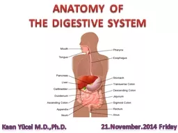
2. ORAL CAVITY-BOUNDARIESAnteriorly - vermilion border of upper & lower lipsPosteriorly – junction between the hard & soft palate superiorly and junction of anterior 2/3 & posterior 1/3 of tongue inferiorly
3. STRUCTURES OF ORAL CAVITYLipsGingivobuccal sulciGingiva & teethBuccal mucosaFloor of the mouthTongue – dorsal & ventral surface (ant.2/3)Retromolar trigoneHard palate
4. Retromolar trigone – Mucosa behind the last mandibular molar tooth extending superiorly to the maxillary tuberosity .
5. Neoplasms of retromolar trigone have important peculiarities due to their spatial relationships with the surrounding structures. Tumours that involve this area can extend to nearby muscles; adipose spaces; and other anatomic structures, such as the soft palate, the tonsillar fossa, the parapharyngeal space, and the floor of the mouth.
6. Tumors and growths in the oral cavity
7. Tumors and growths in the oral cavityThey fit into 3 general categories: Benign or non-cancerous growths that do not invade other tissues and do not spread to other parts of the body. Harmless growths that can later develop into cancer. These are known as precancerous conditions. Cancerous/ malignant tumors that can grow into surrounding tissues and spread to other parts of the body
8. Benign (non-cancerous) tumors Eosinophilic granuloma Fibroma Granular cell tumor Keratoacanthoma Leiomyoma Osteochondroma Lipoma Schwannoma Neurofibroma Papilloma Condyloma acuminatum Verruciform xanthoma Pyogenic granuloma Rhabdomyoma Odontogenic tumors (tumors that start in tooth-forming tissues)
9. Pre-cancerous conditions/lesions (Potentially malignant disorders)
10. The World Health Organization classifies oral precancerous/potentially malignant disorders into 2 general groups, as follows:A precancerous lesion is “a morphologically altered tissue in which oral cancer is more likely to occur than its apparently normal counterpart.” These precancerous lesions include leukoplakia, erythroplakia, and the palatal lesions of reverse smokers. A precancerous condition is “a generalized state associated with significantly increased risk of cancer.” The precancerous conditions include submucous fibrosis, lichen planus, epidermolysis bullosa, and discoid lupus erythematous.
11. PREMALIGNANT LESIONS /CONDITIONS Morphologically altered tissue in which cancer is more likely to occur than its normal counter part. Leukoplakia: white patch, unrelated to any other disease; malignant - 5% cases Erythroplakia: Slightly raised, red area that bleeds easily if scraped; malignant - 90% Oral Submucous fibrosis : Mucosa tough, leathery with vertical fibrous bands. Lichen planus: rarely malignant, erosive form needs regular follow up.
12.
13. Leukoplakia The term leukoplakia was first used by Schwimmer in1877 to describe a white lesion of the tongue, which probably represented a syphilitic glossitis.It is the most common premalignant lesion (85%)WHO working group defines leukoplakia as ‘A white patch or plaque that cannot be characterized clinically or pathologically as any other disease’.In Indian studies the rate of malignant transformation ranges from 0.13 to 2.2% per year.
14.
15. Erythroplakia Any lesion of the oral mucosa that presents as bright red velvety plaques which cannot be characterized clinically or pathologically as any other recognizable condition.All erythroplakia cases showed some degree of epithelial dysplasia; 51 percent showed invasive squamous cell carcinoma, 40 percent were carcinoma in situ or severe epithelial dysplasia, and the remaining 9 percent demonstrated mild-to-moderate dysplasia.
16. Erythroplakia
17. Oral submucous fibrosis (OSF) It is a chronic disorder characterized by fibrosis of the lining mucosa of the upper digestive tract involving the oral cavity, oropharynx and frequently the upper third of the esophagus.OSF is particularly associated with areca nut chewing, the main component of betel quid
18. Oral Submucous fibrosis
19. Oral cavity cancers SCCVerrucuous carcinomaMelanoma AdenocarcinomaSarcoma
20. Risk factors:Tobacco use Drinking alcohol Drinking and smoking together : 15 times riskBetel quid and gutka Human papilloma virus (HPV) infection Gender AgeUltraviolet (UV) light Poor nutrition Weakened immune system Genetic syndromes :Fanconi anemia, Dyskeratosis congenitaLichen planus Irritation from dentures
21. Besides smoking, use of smokeless tobacco is widely prevalent. The use of Betel quid (pan) - consisting of pieces of areca nut, processed or unprocessed tobacco, aqueous calcium hydroxide (slaked lime) and some spices wrapped in the leaf of piper betel vine leaf - is very common Additionally, gutka, zarda, kharra, mawa and khainni are all dry mixtures of lime, areca nut flakes and powdered tobacco custom mixed by vendors. Risk factors
22. In recent years, commercially available sachets of premixed areca nut, lime, catechu, condiments with or without powdered tobacco have become very popular, particularly among younger Indians. Typically, the pan or gutka is kept in the cheek and chewed or sucked for 10-15 minutes, with some users keeping it in overnight.Risk factors
23. FIELD CANCERISATIONCarcinogens like tobacco & alcohol affect all the mucosal surfaces of the aerodigestive tract with which they come in contact. The primary tumour thus arises in a field of abnormal mucosa, in which a second primary tumour may arise in a geographically separate locationSynchronous primaries- If primary tumour & second primary tumour are present at the same time.Metachronous primaries- If primary tumour & second primary tumour present more than 6 months apart. Oral cavity tumours have the highest rate of second primaries
24. Signs and symptoms of oral cavity cancer A sore in the mouth that does not heal (most common symptom) Pain in the mouth that doesn’t go away (also very common) A lump or thickening in the cheek A white or red patch on the gums, tongue, tonsil, or lining of the mouth A sore throat or a feeling that something is caught in the throat that doesn’t go away Trouble chewing or swallowing Numbness of the tongue or other area of the mouth Swelling of the jaw that causes dentures to fit poorly or become uncomfortable Loosening of the teeth or pain around the teeth or jaw Voice changes A lump or mass in the neck Weight loss Constant bad breath Trouble moving the jaw or tongue
25. Aphthous ulcer
26.
27. INCIDENCEIndia – highest incidence of oral cancers in the world India - 40% of all cancers UK - 4% of all cancers 80% - of head & neck cancersMale:Female :: 2:1
28. The age-adjusted rates of oral cancer vary from over 20 per 100,000 population in India, to 10 per 100,000 in the U.S., and less than 2 per 100,000 in the Middle East.
29. Histology of Oral Cancers More than 90% - squamous cell carcinomas .Invasive squamous cell cancer - the cancer cells have spread into deeper layers of the oral cavity Other types - adenocarcinoma, lymphoma, sarcoma, melanoma.
30. Verrucous CarcinomaLow grade variant of sqamous cell carcinoma.Buccal mucosa of older women.No history of smoking & alcohol. Exophytic & nonaggressiveNever metastasize to lymph nodes or other sites Surgery only treatment – radioresistant.Recurrence – not as a local failure but as a second or even a third primary tumor.Follow-up – as with more invasive disease
31. Local ExtensionSuperficial exophytic – spreads along mucosal surface.Infiltrating –involves deeper structures.Ulcerating / fungating – metastasize earlier.
32. Regional MetastasisZone I :Submental & Submandibular Zone II :Upper jugular (jugulodigastric)Zone III :Middle jugular (jugulomohyoid)Zone IV :Lower jugular (supraclavicular)Zone V : Posterior triangle
33. Distant MetastasisUntreated patient : Lung, liver and bone
34. Evaluation (diagnosis)HistoryGeneral evaluation – assessment of overall health & fitness for GAExamination of oral cavityCervical lymph nodes Primary lesion - Biopsy (HPE)Lymph nodes - FNAC (fine needle aspiration cytology)
35. Diagnostic Studies Haemogram – Hb%, TLC, DLC Chemistry – Bl.sugar, Bl.urea, S.creatinineCoagulation profileLiver function tests Radiograph chest ECGCT scan of the head & neckExtent of tumour involvement (resectable)Cervical lymph nodes for metastasis MRI
36. Vital tissue staining with Tolonium chloride (TB). It is a metachromatic vital dye that may bind preferentially to tissues undergoing rapid cell division (such as inflammatory, regenerative and neoplastic tissue), to sites of DNA change associated with oral PMD or both.
37. TNM Classification & Staging
38.
39.
40.
41.
42. Treatment goalsTo eradicate primary tumour and lymph node metastasisTo maintain the function and aesthetics
43. Factors affecting treatment
44. Management modalitiesDepends upon stage of tumourSurgery alone – mainstay of treatmentSurgery + post op. radiotherapy Surgery + post op. radiotherapy +chemotherapyRadiotherapy alone.Brachytherapy
45. Resectable tumourResectable – Complete excision of primary tumour with negative pathologic margins (frozen section). A cuff of 1cm of normal tissue around the visible and palpable tumour Frozen section cannot be used to assess bone because it must be decalcified.Stage1,2(T1,2 N0)– Excision of tumour + ipsilateral supraomohyoid neck dissectionStage 3(T3,N0) – Excision of tumour +ipsilateral modified radical neck dissectionStage 4(T4,N0/T1-4,N2,3) – Excision of tumour + ipsilateral radical neck dissection
46. Unresectable tumour Unresectable - all gross tumour cannot be removed local control will not be achieved after an operation even with the addition of radiotherapyTreatment - Radiotherapy
47. Neck dissectionRadical neck dissection- Removes LN level 1-5, sternomastoid muscle, internal jugular V, & spinal accessory nerve(CN 11)Modified radical neck dissection – Removes LN levels 1-5.Supraomohyoid neck dissection – Removes LN levels 1-3.
48. Post op radiotherapy-indicationsT4 (stage 4) lesion Close/positive margins Perineural/Vascular invasion Extracapsular extension of LN diseaseMore than 4 positive nodes
49. Reconstruction“Like tissue with like tissue” Soft tissue -skin graft -local flaps -regional flaps -free flapBone -rib or iliac crest graft -fibula free flap
50. Primary reconstructionImproves quality of life by:Shortening recovery timePreserving useful speaking and swallowingMinimising cosmetic deformityLimiting complications
51. ComplicationsWound infection Seroma & HaematomaFlap loss - ischemic necrosisNeck flap necrosisFistulaDonor site complicationsNeed for revision
52. PrognosisStage Approx. 5 yrs survival rate 190-92 %275-85 %350 %425-35 %
53. Local Recurrence A tumor that occurs in the previously treated anatomic area within 2 years of the initial definitive treatment.If it occur more than 2 years after then it is second primary tumor. Confirmation by biopsy Treatment - Surgical excision
54. Follow upEssentialEven after small cancers are treated & fully cured – 40% develop second primary tumour (field cancerisation)Oral cavity tumours have the highest rate of second primaries which occur mostly in the head & neck
55. Follow upYear 1Every 1-3 monthsYear 2Every 2-4 monthsYear 3-5Every 4-6 monthsMore than 5 yearsEvery 6-12 months X-ray chest annually TSH every 6-12 months if neck initiated
56. RehabilitationFrom the time of diagnosisCounselling – Psycho socialDietary adviseProsthesis to replace missing jaw & teethSpeech therapist
57. PreventionQuit tobacco – all forms Replace with nicotine gum or patch if required Avoid / minimise alcohol useMaintain good oral hygienePromptly treat sharp teeth Replace ill fitting denturesLip cancer- avoid sun /sunscreen
58. Gutkha, Tobacco, Pan Masala, Cigarette can causeORAL CANCER
59. Squamous cell carcinoma of the oral cavityKey facts95% of the malignancies of the oral cavity are SCCsIncidence is 6 per 100000 (20/lac in India )Accounts for 0.6% - 5% of all cancers in the US, and upto 45% in IndiaMale to female ratio 2.2:1, seventh decade of lifePredisposing factors: alcohol, tobacco use, and betel nut chewing (India). HPV Most important prognostic factor– cervical lymph nodes6% of patients have a synchronous SCC present in aerodigestive tractSummary
60. Squamous cell carcinoma Clinical featuresExophytic lesion or non healing ulcer, may bleed easily on touch, painsymptoms depend on the site of the tumorCancer of tongue may cause referred pain to the ears, slurring, difficulty in eatingCancer of floor : may invade mandible, causing loosening of lower ant teethOther symptoms: odynophagia, bleeding, dyphagia, oral discomfort, reduced movement of oral structures, and trismus
61. Squamous cell carcinoma Diagnosis and investigationsExamination of mouth and teethNeck for LNBiopsyCT or MRI: for stagingEndoscopyEvaluation of distant metastasis
62. Squamous cell carcinoma TreatmentT1 and T2 lesionsSURGICAL EXCISIONLN dissection: SOH, MRNDT3 and T4 lesionsCombination therapy: surgical resection with adjuvant or neoadjuvant radiotherapyMRNDReconstruction STSG for small defects which cannot be closed primarily and donot require radiotherapyFlaps for large reconstructionRegional: PMMC, DPFree flaps: RFFF Free fibula, iliac crest, scapula
63. Squamous cell carcinoma PrognosisEarly stage: 75% 5-yr survivalLate or advanced stage: 35%
64.