
Pettit AC Jahangir AA Wright PW Mycobacterium doricum Osteomyelitis and Soft Tissue Infection Emerg Infect Dis 2011171120752077 httpsdoiorg103201eid1711110460 ID: 1010619
Download Presentation The PPT/PDF document "Figure A1 Figure A1. Computed t..." is the property of its rightful owner. Permission is granted to download and print the materials on this web site for personal, non-commercial use only, and to display it on your personal computer provided you do not modify the materials and that you retain all copyright notices contained in the materials. By downloading content from our website, you accept the terms of this agreement.


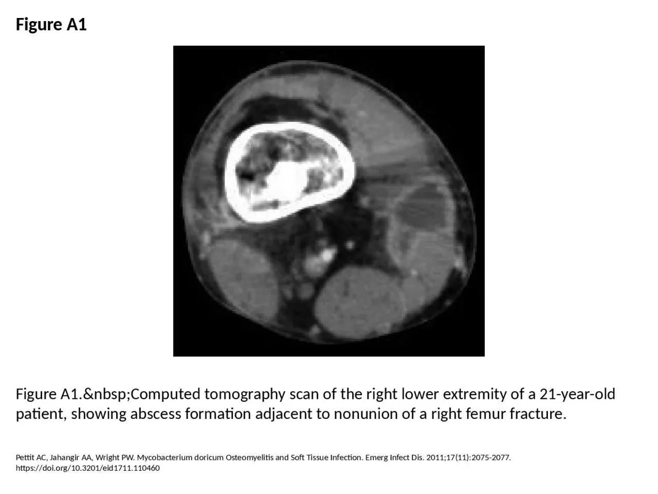
1. Figure A1Figure A1. Computed tomography scan of the right lower extremity of a 21-year-old patient, showing abscess formation adjacent to nonunion of a right femur fracture.Pettit AC, Jahangir AA, Wright PW. Mycobacterium doricum Osteomyelitis and Soft Tissue Infection. Emerg Infect Dis. 2011;17(11):2075-2077. https://doi.org/10.3201/eid1711.110460