
Corman VM Nagy P Ostermann S Arloth J Liljander A Barua R et al Hepatitis E Virus Genotype 7 RNA and Antibody Kinetics in Naturally Infected Dromedary Calves United Arab Emirates Emerg Infect Dis 202026922142217 httpsdoiorg103201eid2609191758 ID: 999165
Download Presentation The PPT/PDF document "Figure 1 Figure 1. HEV RNA and HEV antib..." is the property of its rightful owner. Permission is granted to download and print the materials on this web site for personal, non-commercial use only, and to display it on your personal computer provided you do not modify the materials and that you retain all copyright notices contained in the materials. By downloading content from our website, you accept the terms of this agreement.


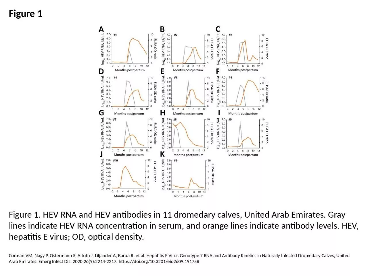
1. Figure 1Figure 1. HEV RNA and HEV antibodies in 11 dromedary calves, United Arab Emirates. Gray lines indicate HEV RNA concentration in serum, and orange lines indicate antibody levels. HEV, hepatitis E virus; OD, optical density.Corman VM, Nagy P, Ostermann S, Arloth J, Liljander A, Barua R, et al. Hepatitis E Virus Genotype 7 RNA and Antibody Kinetics in Naturally Infected Dromedary Calves, United Arab Emirates. Emerg Infect Dis. 2020;26(9):2214-2217. https://doi.org/10.3201/eid2609.191758