
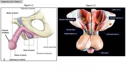
Bulbospongiosus m Perineal body Levator ani m Figure 11 Figure 12 Urogenital Lab 2 Station 1 Oburator internus fascia Glans penis Glans penis Corpus cavarnosum Corpus spongiosum ID: 908456
Download Presentation The PPT/PDF document "Oburator internus m. Ischiocavernosus m..." is the property of its rightful owner. Permission is granted to download and print the materials on this web site for personal, non-commercial use only, and to display it on your personal computer provided you do not modify the materials and that you retain all copyright notices contained in the materials. By downloading content from our website, you accept the terms of this agreement.

Slide1
Oburator
internus m.
Ischiocavernosus m.
Bulbospongiosus m.
Perineal body
Levator
ani
m.
Figure 1.1
Figure 1.2
Urogenital, Lab 2: Station 1
Oburator
internus fascia
Glans penis
Slide2Glans
penis
Corpus
cavarnosum
Corpus
spongiosum
Bulb
of penis
Deep Dorsal vein
Deep (Buck’s
) Fascia
Figure 1.3
Figure 1.4
Tunica albuginea
Urogenital, Lab 2: Station 1
Slide3Figure 2.1
Figure 2.2
Inguinal canal
Superficial (external) ring
Testicular v.
Testicular a.
Ductus deferens
Ureter
Epididymis: Head
Epididymis: Body
Epididymis: Tail
Testis
Pampiniform plexus
Testicular a.
Ductus deferens
Cremaster m.
Conjoint tendon (internal and transversus aponeurosis)
Deep (internal) ring
Superficial (external) ring
Conjoint tendon (internal and transversus aponeurosis)
Ext Abdominal oblique aponeurosis
Ext Abdominal oblique aponeurosis
Deep (internal) ring
Figure 2.3
Urogenital, Lab 2: Station 1
Slide4Figure 2.4
Ureter
Ampulla
ductus
deferens
Seminal Gland
Prostate Gland
Ejaculatory duct
Prostate gland
Figure 3.2
Figure 3.1
Urogenital, Lab 2: Station 1
Figure 3.3
Slide5Bulbourethral gland
Perineal membrane
Bulbourethral gland: Note that the model incorrectly shows the gland in the superficial space. It should be shown superior to the perineal membrane.
Perineal membrane
Fat
in A.
Recess of
Ischioanal
fossa
A Recess of
Ischioanal
fossa
Figure 4.1
Figure 4.2
Fibromuscular “urogenital diaphragm”
Spongy (penile) urethra
External urethral orifice
External urethral sphincter
Membranous
urethra
Urogenital, Lab 2: Station 1