

Dr AlSaghbini M S MD PhD Pathology Cyto Histopathology Consultant Assistant Prof Autophagy Is a process in which a cell eats its own contents It is a survival mechanism when the starved cell lives by cannibalizing itself and recycling the digested contents ID: 743190
Download Presentation The PPT/PDF document "General Pathology Autophagy, Intracellul..." is the property of its rightful owner. Permission is granted to download and print the materials on this web site for personal, non-commercial use only, and to display it on your personal computer provided you do not modify the materials and that you retain all copyright notices contained in the materials. By downloading content from our website, you accept the terms of this agreement.


Slide1
General PathologyAutophagy, Intracellular Accumulations, Pathologic Calcification and Cellular Aging
Dr. Al-Saghbini M. S.
MD. PhD. Pathology
Cyto
/Histopathology Consultant
Assistant Prof.Slide2
AutophagyIs a process in which a cell eats its own contents. It is a survival mechanism, when the starved cell lives by cannibalizing itself and recycling the digested contents.
Cellular stresses, such as nutrient deprivation, activate autophagy genes that create vacuoles in which cellular organelles are sequestered and then degraded following fusion of the vesicles with lysosomes.
The digested materials are recycled to provide nutrients for the cell. Slide3
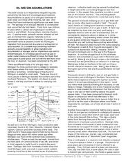
Intracellular AccumulationsOne of the manifestations of metabolic derangements in cells is the intracellular accumulation of abnormal amounts of various substances. The stockpiled substances fall into two categories: (1) a normal cellular constituent, such as water, lipids, proteins, and carbohydrates, that accumulates in excess.(2) an abnormal substance, either exogenous, such as a mineral or products of infectious agents, or endogenous, such as a product of abnormal synthesis or metabolism.Slide4
1. A normal endogenous substance is produced at a normal or increased rate, but the rate of metabolism is inadequate to remove it. Most accumulations are attributable to four types of abnormalities.
Examples
of this type of process are
fatty change
in the liver and reabsorption protein droplets in the tubules of the kidneys Slide5
2. An abnormal endogenous substance, typically the product of a mutated gene, accumulates because of defects in protein folding and transport and an inability to degrade the abnormal protein efficiently. Examples include the accumulation of
mutated α1-antitrypsin
in liver cells and various mutated proteins in degenerative disorders of the central nervous systemSlide6
3. A normal endogenous substance accumulates because of defects, usually inherited, in enzymes that are required for the metabolism of the substance. Examples include diseases caused by genetic defects in enzymes
involved in the metabolism of
lipid and carbohydrates
, resulting in intracellular deposition of these substances, largely in lysosomes.Slide7
4. An abnormal exogenous substance is deposited and accumulates because the cell has neither the enzymatic machinery to degrade the substance nor the ability to transport it to other sites. Accumulations of carbon particles
and nonmetabolizable chemicals such as
silica
are examples of this type of alteration.Slide8
All major classes of lipids can accumulate in cells: triglycerides, cholesterol/cholesterol esters, and phospholipids. Phospholipids are components of the myelin figures found in necrotic cells.Abnormal complexes of lipids and carbohydrates accumulate in the lysosomal storage diseases.
LIPIDS
The terms
steatosis and fatty change
describe abnormal accumulations of triglycerides within
parenchymal
cells.
Fatty change is often seen in the liver because it is the major organ involved in fat metabolism, but it also occurs in heart, muscle, and kidney.
myelin figure in the parietal cellSlide9
The causes of steatosis include toxins, protein malnutrition, diabetes mellitus, obesity, and anoxia. In developed nations the most common causes of significant fatty change in the liver (fatty liver) are alcohol abuse and nonalcoholic fatty liver disease, which is often associated with diabetes and obesity.
Free fatty acids (FFA) from adipose tissue or ingested food are normally transported into hepatocytes.
This EM photo shows numerous membrane bound myelin figures. These myelin figures are composed of whorls of membranes enclosed within the lysosomes.Slide10
In the liver the FFA are esterified to triglycerides, converted into cholesterol or phospholipids, or oxidized to ketone bodies. Release of triglycerides from the hepatocytes requires association with apoproteins to form
lipoproteins
, which may then be transported from the blood into the tissues
Excess accumulation of triglycerides within the liver may result from excessive entry or defective metabolism and export of lipids Slide11
Fatty change is most often seen in the liver and heart. In all organs fatty change appears as clear vacuoles within parenchymal cells. Intracellular accumulations of water or polysaccharides (e.g., glycogen) may also produce clear vacuoles.Morphology
Fatty change begins with the development of minute, membrane-bound inclusions (
liposomes
) closely applied to the ER. Slide12
Accumulation of fat is first seen by light microscopy as small vacuoles in the cytoplasm around the nucleus. As the process progresses the vacuoles coalesce, creating cleared spaces that displace the nucleus to the periphery of the cell.Slide13
Accumulations manifested histologically by intracellular vacuoles are seen in several pathologic processes.Cholesterol and Cholesterol Esters
1-
Atherosclerosis.
smooth muscle cells and macrophages within the intimal layer of the aorta and large arteries are filled with lipid vacuoles, most of which are made up of cholesterol and cholesterol esters.
Here is a coronary artery with atherosclerotic plaques. There is recent hemorrhage into the plaque.Slide14
3- Cholesterolosis. This refers to the focal accumulations of cholesterol-laden macrophages in the lamina propria of the gallbladder . The mechanism of accumulation is unknown.
Cholesterol-laden macrophages (foam cells,
arrow
) in a focus of gallbladder
cholesterolosis.
2-
Niemann
-Pick disease, type C
. This lysosomal storage disease is caused by mutations affecting an enzyme involved in cholesterol trafficking, resulting in cholesterol accumulation in multiple organs.Slide15
4- Xanthomas. Intracellular accumulation of cholesterol within macrophages is also characteristic of acquired and hereditary hyperlipidemic states. Clusters of foamy cells are found in the subepithelial connective tissue of the skin and in tendons, producing tumorous masses known as xanthomas.
Histology picture of
xanthoma
showing lipid-laden foam cells with large areas of cholesterol clefts.
10 × magnification, H & E stain
The term
xanthoma
denotes any accumulation of fat anywhere in the skinSlide16
Intracellular accumulations of proteins usually appear as rounded, eosinophilic droplets, vacuoles, or aggregates in the cytoplasm. By electron microscopy they can be amorphous, fibrillar, or crystalline in appearance. In some disorders, such as certain forms of
amyloidosis
, abnormal proteins deposit primarily in extracellular spaces.
PROTEINS
[CARDIAC (HEART) AMYLOIDOSIS]. .
Amyloidosis
is an insoluble extracellular deposition of abnormal
fibrillar
substance composed of specific protein fragments.Slide17
1- Reabsorption droplets in proximal renal tubules are seen in renal diseases associated with protein loss in the urine (proteinuria).
Excesses of proteins within the cells sufficient to cause morphologically visible accumulation have diverse causes.
In disorders with heavy protein leakage across the glomerular filter there is increased reabsorption of the protein into vesicles, and the
protein appears as pink
hyaline droplets within
the cytoplasm of the
tubular cell.
(Courtesy of Dr. Helmut
Rennke
, Department of Pathology, Brigham and Women's Hospital, Boston, MA.) Slide18
2- The proteins that accumulate may be normal secreted proteins that are produced in excessive amounts, as occurs in certain plasma cells engaged in active synthesis of immunoglobulins. The ER becomes hugely distended, producing large, homogeneous eosinophilic inclusions called Russell bodies
.
3-
Defective intracellular transport and secretion of critical proteins. In
α
1
-antitrypsin
deficiency, mutations in the protein significantly slow folding, resulting in the buildup of partially folded intermediates, which aggregate in the ER of the liver and are not secreted. The resultant deficiency of the circulating enzyme causes emphysema.Slide19
4- Accumulation of cytoskeletal proteins.Intermediate filaments, which provide a flexible intracellular scaffold that organizes the cytoplasm and resists forces applied to the cell, are divided into five classes:
1-
Keratin filaments
(characteristic of epithelial cells).
2- Neurofilaments (neurons).
3-
Desmin
filaments
(muscle cells).
4-
Vimentin
filaments
(connective tissue cells),
and
5-
Glial filaments
(astrocytes).Slide20
5- Aggregation of abnormal proteins ( which can be intracellular, extracellular, or both), and the aggregates may either directly or indirectly cause the pathologic changes. These disorders are sometimes called proteinopathies or protein-aggregation diseases.Slide21
The term hyaline refers to an alteration within cells or in the extracellular space that gives a homogeneous, glassy, pink appearance in routine histologic sections stained with H & E. Intracellular accumulations of protein, are examples of intracellular hyaline deposits. Hyaline Change
Glycogen
Is a readily available energy source stored in the cytoplasm of healthy cells.
Excessive intracellular deposits of glycogen are seen in patients with an abnormality in either glucose or glycogen metabolism.Slide22
Glycogen accumulates within the cells in a group of related genetic disorders that are collectively referred to as the glycogen storage diseases, or glycogenoses. In these diseases enzymatic defects in the synthesis or breakdown of glycogen result in massive accumulation, causing cell injury and cell death.Slide23
Pigments are colored substances, some of which are normal constituents of cells (e.g., melanin), whereas others are abnormal and accumulate in cells only under special circumstances. Pigments can be exogenous, or endogenous.PigmentsSlide24
Exogenous PigmentsThe most common exogenous pigment is carbon (coal dust). When inhaled it is picked up by macrophages within the alveoli and is then transported through lymphatic channels to the regional lymph nodes in the tracheobronchial region.
Accumulations of this pigment blacken the tissues of the lungs (
anthracosis
) and the involved lymph nodes.
Tattooing is a form of localized, exogenous pigmentation of the skin. The pigments inoculated are phagocytosed
by dermal macrophages.
The pigments do not usually evoke any inflammatory response.Slide25
Lipofuscin is an insoluble pigment, also known as lipochrome or wear-and-tear pigment. Lipofuscin is composed of polymers of lipids and phospholipids in complex with protein, suggesting that it is derived through lipid peroxidation of polyunsaturated lipids of subcellular membranes.
Lipofuscin
is not injurious to the cell or its functions.
Endogenous Pigments
Its importance lies in its being a telltale sign of
free radical injury and lipid peroxidation.
Slide26
Lipofuscin granules in a cardiac myocyte shown by (A) light microscopy (deposits indicated by arrows), and
(B) electron microscopy (note the
perinuclear
,
intralysosomal location). In tissue sections it appears as a yellow-brown, finely granular cytoplasmic, often
perinuclear
, pigment.
It is seen in cells undergoing slow, regressive changes and is particularly prominent in the liver and heart of aging patients or patients with severe malnutrition and cancer
cachexia
.Slide27
Melanin: is an endogenous, non-hemoglobin-derived, brown-black pigment formed when the enzyme tyrosinase catalyzes the oxidation of tyrosine to dihydroxyphenylalanine in melanocytes. Melanin is the only endogenous brown-black pigment.
Hemosiderin
:
Is a hemoglobin-derived, golden yellow-to-brown, granular or crystalline pigment that serves as one of the major storage forms of iron.
Slide28
When there is a local or systemic excess of iron, ferritin forms hemosiderin granules, which are easily seen with the light microscope. Hemosiderin pigment represents aggregates of ferritin micelles.
Hemosiderin granules in liver cells.
A-
H&E stain showing golden-brown, finely granular pigment.
B- Prussian blue stain, specific for iron (seen as blue granules). Slide29
When there is systemic overload of iron, hemosiderin may be deposited in many organs and tissues, a condition called hemosiderosis. The main causes of hemosiderosis are (1) Increased absorption of dietary iron.(2)
Hemolytic anemias, in which abnormal quantities of iron are released from erythrocytes, and
(3)
Repeated blood transfusions because the transfused red cells constitute an exogenous load of iron.
Bilirubin is the normal major pigment found in bile.
It is derived from hemoglobin but contains no iron.
Its normal formation and excretion are vital to health, and jaundice is a common clinical disorder caused by excesses of this pigment within cells and tissues.Slide30
Pathologic CalcificationIs the abnormal tissue deposition of calcium salts, together with smaller amounts of iron, magnesium, and other mineral salts.
There are two forms of pathologic calcification.
1-
Deposition
occurs locally in dying tissues it is known as
dystrophic calcification
.
2-
Deposition of calcium salts in otherwise normal tissues is known as
metastatic calcification
, and it almost always results from hypercalcemia secondary to some disturbance in calcium metabolism.Slide31
Is encountered in areas of necrosis, whether they are of coagulative, caseous, or liquefactive type, and in foci of enzymatic necrosis of fat. Calcification is almost always present in the atheromas of advanced atherosclerosis. It also commonly develops in aging or damaged heart valves, further hampering their functionDystrophic Calcification
View looking down onto the unopened aortic valve in a heart with
calcific
aortic
stenosis
. It is markedly narrowed (
stenosis
).
The
semilunar
cusps are thickened and fibrotic, and behind each cusp are irregular masses of piled-up
dystrophic calcification
. Slide32
Histologically, with the usual H & E stain, calcium salts have a basophilic, amorphous granular, sometimes clumped appearance. They can be intracellular, extracellular, or in both locations. In the course of time, heterotopic bone may be formed in the focus of calcification. The progressive acquisition of outer layers may create lamellated configurations, called
psammoma
bodies
because of their resemblance to grains of sand.
Morphology. Slide33
Pathogenesis.It is thought that calcium is concentrated in membrane-bound vesicles in cells by a process that is initiated by membrane damage and has several steps: (1) calcium ion binds to the phospholipids present in the vesicle membrane; (2) phosphatases
associated with the membrane generate phosphate groups, which bind to the calcium; Slide34
(3) the cycle of calcium and phosphate binding is repeated, raising the local concentrations and producing a deposit near the membrane; and (4) a structural change occurs in the arrangement of calcium and phosphate groups, generating a microcrystal, which can then propagate and lead to more calcium deposition.Slide35
Metastatic calcification may occur in normal tissues whenever there is hypercalcemia. Hypercalcemia also accentuates dystrophic calcification. There are four principal causes of hypercalcemia:Metastatic Calcification(1)
Increased secretion
of
parathyroid hormone (PTH)
with subsequent bone resorption, as in hyperparathyroidism.
(2)
Destruction of bone tissue
, secondary to primary tumors of bone marrow
(e.g., multiple myeloma, leukemia)
or diffuse skeletal metastasis
(e.g., breast cancer),
accelerated bone turnover
(e.g., Paget disease),
or immobilization.Slide36
(4) Renal failure, which causes retention of phosphate, leading to secondary hyperparathyroidism.Less common causes include aluminum intoxication
,
which occurs in patients on chronic renal dialysis, and
milk-alkali syndrome
, which is due to excessive ingestion of calcium and absorbable antacids such as milk or calcium carbonate.(3)
Vitamin D–related disorders
,
including vitamin D intoxication,
sarcoidosis
(in which macrophages activate a vitamin D precursor),
and idiopathic hypercalcemia of infancy
(Williams syndrome),
characterized by abnormal sensitivity to vitamin D.Slide37
Metastatic calcification may occur widely throughout the body but principally affects the interstitial tissues of the gastric mucosa, kidneys, lungs, systemic arteries, and pulmonary veins. Though quite different in location, all of these tissues excrete acid and therefore have an internal alkaline compartment that predisposes them to metastatic calcification.Slide38
Cellular aging is the result of a progressive decline in cellular function and viability caused by genetic abnormalities and the accumulation of cellular and molecular damage due to the effects of exposure to exogenous influences.How calorie restrictions prolong life span is net established.
Cellular AgingSlide39
The known changes that contribute to cellular aging include the following:1- Decreased cellular replication. After a fixed number of divisions all somatic cells become arrested in a terminally non-dividing state, known as senescence.With each cell division there is incomplete replication of chromosome ends (telomere shortening),
thus
the ends of chromosomes cannot be protected and are seen as broken DNA, which activates the DNA damage response and signals cell cycle arrest.
Telomere length is normally maintained by nucleotide addition mediated by an enzyme called telomerase, which is a
specialized
RNA-protein complex
that uses its own RNA as a template for adding nucleotides to the ends of chromosomes.Slide40
Germ cells and stem cells both contain active telomerase, but only the germ cells have sufficient levels of the enzyme to stabilize telomere length completely. In normal somatic cells there is no telomerase activity, and telomeres progressively shorten with successive cell divisions until growth arrest, or senescence, occurs. Telomerase activation in cancer cells counteracts the telomere shortening that limits the proliferative capacity of normal somatic cells.Telomere-telomerase hypothesis and proliferative capacity of cells. Telomere length is plotted against the number of cell divisions. Slide41
2- Accumulation of metabolic and genetic damage.One group of potentially toxic products of normal metabolism are reactive oxygen species. These by-products of oxidative phosphorylation cause covalent modifications of proteins, lipids, and nucleic acids.
The amount of oxidative damage, which increases as an organism ages, may be an important cause of senescence.
Free radicals
may have deleterious
effects on DNA, leading to breaks and genome instability, thus affecting all cellular functions.
Although most
DNA
damage is repaired by
endogenous DNA repair enzymes,
some persists and accumulates as cells age. Slide42
Thus, the balance between cumulative metabolic damage and the response to that damage could determine the rate at which we age. In this scenario aging can be delayed by decreasing the accumulation of damage or by increasing the response to that damage.Not only damaged DNA but damaged cellular organelles also accumulate as cells age. In part this may be the result of declining function of the proteasome
, the
proteolytic
machine that serves to eliminate abnormal and unwanted intracellular proteins.Slide43
Studies in model organisms, from yeast to mammals, have shown that the most effective way of prolonging life span is calorie restriction. How this works is still not established, but the effect of calorie restriction on longevity appears to be mediated by a family of proteins called sirtuins.
Sirtuins
have
histone
deacetylase activity, and are thought to promote the expression of several genes whose products increase longevity.Slide44
These products include proteins that increase metabolic activity, reduce apoptosis, stimulate protein folding, and inhibit the harmful effects of oxygen free radicals.Sirtuins also increase insulin sensitivity and glucose metabolism, and may be targets for the treatment of diabetes.Slide45
Next lectureChapter 2 Acute and Chronic InflammationEnd of Chapter One