
Dr Rehma Dar Assistant professor Pathology Introduction to Immune System Cell mediated vs Humoral Immunity Hypersensitivity reactions Autoimmune diseases Transplant Rejection Immunodeficiency states ID: 774730
Download Presentation The PPT/PDF document " Diseases of Immune System" is the property of its rightful owner. Permission is granted to download and print the materials on this web site for personal, non-commercial use only, and to display it on your personal computer provided you do not modify the materials and that you retain all copyright notices contained in the materials. By downloading content from our website, you accept the terms of this agreement.


Slide1
Diseases of Immune System
Dr Rehma Dar
Assistant professor Pathology
Slide2Introduction to Immune SystemCell mediated vs Humoral ImmunityHypersensitivity reactionsAutoimmune diseasesTransplant RejectionImmunodeficiency states
Learning Objectives
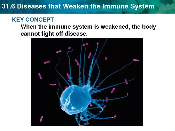
Slide3The immune system is vital for survival- protects us from infectious pathogens.
The Normal Immune Response
Slide4The mechanisms of protection against infections fall into two broad categories.
Innate immunity
Adaptive immunity
Slide5Slide6natural, or native immunity refers to defense mechanisms - present even before infection and protect individuals against infections. first line of defense, because it is always ready to prevent and eradicate infections.
Innate immunity
Slide7epithelial barriers, phagocytic cells (mainly neutrophils and macrophages), dendritic cells, natural killer (NK) cells, andseveral plasma proteins, including complement system.
Components of innate immunity
Slide8The two most important cellular reactions of
innate immunity
are:
inflammation
, in which phagocytic leukocytes are recruited and activated to kill microbes, and
anti-viral defense
, via dendritic cells and NK cells.
Slide9Leukocytes and epithelial cells recognize components of microbes (
called
pathogen associated molecular patterns)
via cellular receptors.
The recognition receptors are a family of proteins called
Toll-like receptors
(TLRs)
that are homologous to the
Drosophila
protein Toll.
Different TLRs are specific for components of different bacteria and viruses.
TLRs are located on the cell surface and in endosomes-recognize and initiate cellular responses to extracellular and ingested microbes.
Slide10Other microbial sensors are located in the cytoplasm -recognize bacteria and viruses that may have colonized cells.
Upon recognition of microbes, the TLRs and other sensors signal by a common pathway - the activation of transcription factors, NF-κB (nuclear factor κB).
NF-κB turns on the production of cytokines and proteins that stimulate the microbicidal activities of phagocytes.
Slide11Epithelia
of
the skin and
GITand
respiratory tracts
-
mechanical barriers to the entry of
microbes
also
produce anti-microbial molecules such as defensins, and lymphocytes located in the epithelia combat microbes at these sites.
Monocytes
and
neutrophils
–
phagocytes -rapidly
be recruited to any site of
infection
Dendritic
cells
produce type I interferons, anti-viral cytokines that inhibit viral infection and
replication
Natural killer cells
-
protection against many viruses and intracellular bacteria
Slide12Complement system
is activated by microbes using the alternative and lectin pathways; in adaptive immunity it is activated by antibodies using the classical pathway.
Other proteins of innate immunity are mannose-binding lectin and C-reactive protein - coat microbes for phagocytosis.
Lung surfactant - provide protection against inhaled microbes.
Slide13Acquired or specific immunity consists of mechanisms that are stimulated by (“adapt to”) microbes and are capable of recognizing microbial and nonmicrobial substances. develops later, and is even more powerful than innate immunity in combating infections. By convention, the term “immune response” refers to adaptive immunity
Adaptive immunity
Slide14The adaptive immune system consists of lymphocytes and their products, including antibodies.
The
receptors of lymphocytes are much more diverse
- not inherently specific for microbes, and they are capable of recognizing a vast array of foreign substances.
Slide15There are two types of adaptive immunity:
Humoral immunity
Cell-mediated
immunity
Slide16Humoral immunity
-
protects
against
extracellular
microbes and their toxins,
mediated by
B
(bone marrow–derived) lymphocytes and their secreted products, antibodies (also called immunoglobulins, Ig)
Cell-mediated
immunity
- responsible for defense against
intracellular
microbes,
mediated by
T
(thymus-derived) lymphocytes.
Both classes of lymphocytes express highly specific receptors for a wide variety of substances, called
antigens.
Slide17Slide18appear morphologically similar – heterogeneous Mature lymphocytes that have not encountered specific antigen - naive (immunologically inexperienced). activated by recognition of antigens - differentiate into effector cells, which perform the function of eliminating microbes, and memory cells, better able to combat the microbe in case it returns
Lymphocytes
Slide19T LymphocytesB LymphocytesNK cells
Lymphocytes
Slide20develop from precursors in the thymus. constitute 60% to 70% of blood lymphocytes and in T-cell zones of peripheral lymphoid organs (paracortical & medullary) Each T cell recognizes a specific cell-bound antigen by means of an antigen-specific T-cell receptor (TCR).
T lymphocytes
Slide21Slide22TCR recognizes peptide antigens that are displayed by major histocompatibility complex (MHC) molecules on the surfaces of antigen-presenting cells (APCs)
.
CD4+ T cells (Helper) :
60% of T lymphocytes
CD8+ T cells (Cytotoxic)
: 30% of T lymphocytes
Normal 2:1 (CD4+ : CD8+)
Slide23Slide24TCR disulfide-linked α and a β polypeptide chain having a variable (antigen-binding) region and a constant region.
TCR diversity - somatic rearrangement of the genes that encode the TCR α and β chains
Each TCR linked to CD3 complex -transduction of signals into the T cell after the TCR has bound the antigen
T cells express CD4, CD8, CD2, integrins, and CD28 -assist the functional responses.
Slide25develop from precursors in the bone marrow. constitute 10% to 20% of the circulating lymphocyte and also present in peripheral lymphoid tissues like lymph nodes(germinal centers), spleen, and mucosa-associated lymphoid tissues.recognize antigen via the B-cell antigen receptor complex.
B Lymphocytes
Slide26Membrane-bound antibodies called IgM and IgD, present on the surface of all mature, naive B cells, are the antigen-binding component of the B-cell receptor complex
After stimulation - B cells -
plasma cells
that secrete antibodies, the mediators of humoral immunity.
B cells also express complement receptors, Fc receptors, and CD40- essential for their responses.
Slide27Slide2810% to 15% of peripheral blood lymphocytes.do not express TCRs or Ig. contain abundant azurophilic granules; because of these characteristics, they are also called large granular lymphocytes.early line of defense against viral infections and some tumorsNeither prior sensitization nor Ab is involved in killingsecrete cytokines, such as interferon-γ (IFN-activates macrophages to destroy ingested microbes- intracellular microbial infections.
Natural Killer Cells
Slide29Slide30numerous fine cytoplasmic processes that resemble dendritesthe most important antigen-presenting cells (APCs) for initiating primary T-cell responsesLocated under epithelia, the common site of entry of microbes and foreign antigens, and in the interstitia of all tissues, where antigens may be produced. Immature dendritic cells within the epidermis are called Langerhans cells. express many receptors for capturing and responding to microbes
Dendritic Cells
Slide31Slide32part of the mononuclear phagocyte phagocytosed microbes and protein antigens process the antigens and present peptide fragments to T cells. - function as APCs in T-cell activation.( MHC II to CD4+)key effector cells in certain forms of cell-mediated immunity, the reaction that serves to eliminate intracellular microbes. also participate in the effector phase of humoral immunity-phagocytose and destroy microbes opsonized (coated) by IgG or C3b.
Macrophages
Slide33Generative Lymphoid Organs also called primary, or central Peripheral Lymphoid Organs or secondary
Tissues of the Immune System
Slide34Generative Lymphoid Organs
in which T and B lymphocytes mature and become competent to respond to antigens
thymus, where T cells develop, and the bone marrow, where B lymphocytes mature.
Peripheral Lymphoid Organs
in which adaptive immune responses to microbes are initiated.
lymph nodes, spleen, and the mucosal and cutaneous lymphoid tissues.
Slide35HLA system – group of related proteins called as HLA antigens.Genes that code for HLA are called histocompatibility genes. responsible for tissue compatibility between individuals. The physiologic function of MHC molecules is to display peptide fragments of proteins for recognition by antigen-specific T cells
Human Leukocyte Antigen(HLA)
system &
Major Histocompatibility Complex(MHC)
Slide36genes encoding the major
histocompatibility
molecules are clustered on a small segment of chromosome 6, the
major
histocompatibility
complex
, or the
human leukocyte antigen
(HLA) complex
Slide37Slide38Slide39Slide40fundamental to the
recognition of antigens
by T cells and are linked to many autoimmune diseases
evoke rejection of
transplanted organs
,
are responsible for
tissue compatibility
between individuals.
highly polymorphic
, - many alleles of each MHC gene in the population and each individual inherits one set of these alleles that is different from the alleles in most other individuals.
Slide41On the basis of their structure, cellular distribution, and function, MHC gene products are classified into three groups.
•
Class I MHC molecules
are expressed on all nucleated cells and platelets except RBC
They are encoded by three closely linked loci, designated
HLA-A
,
HLA-B
, and
HLA-C
Slide42Since
CD8+ T cells
recognize peptides only if presented as a complex with self–class I MHC molecules, CD8+ T cells are said to be
class I MHC–restricted
.
Because one of the important functions of CD8+ CTLs is to eliminate viruses, which may infect any nucleated cell- nucleated cells express class I HLA molecules and can be surveyed by CD8+ T cells.
Slide43Class II MHC molecules
are encoded in a region called
HLA-D
, which has three subregions:
HLA-DP
,
HLA-DQ
, and
HLA-DR
.
The class II - binding site for CD4, and , the class II–peptide complex is recognized by
CD4+ T cells
, which function
as helper cells-
they are referred to as
class II MHC–restricted
.
mainly expressed on cells that present ingested antigens and respond to T-cell help (macrophages, B lymphocytes, and dendritic cells).
Slide44Class III MHC molecules
-
MHC locus also contains genes - encode some complement components and the cytokines tumor necrosis factor (TNF) and lymphotoxin
contains genes that encode many proteins involved in antigen processing and presentation
Slide45Slide46Antigen processing and display by major histocompatibility complex (MHC) molecules
Slide47MHC molecules play key roles in regulating T cell–mediated immune responses
MHC molecules ensure that the correct immune response is mounted against different microbes—
CD8+ T against
cytoplasmic
microbes, and
antibodies and macrophages (both of which are activated by CD4+helper T cells) against
extracellular
microbes
.
Slide48A variety of diseases are associated with the inheritance of certain HLA alleles The most striking is the association between ankylosing spondylitis and HLA-B27; individuals who inherit this class I HLA allele have a 90-fold greater chance (relative risk) of developing the disease as compared with those who do not carry HLA-B27.
HLA and Disease Association
Slide49The diseases that show association with the HLA locus can be broadly grouped into the following categories:
1.
Inflammatory diseases
, including ankylosing spondylitis and several post-infectious arthropathies, all associated with
HLA-B27
2.
Autoimmune diseases
, including autoimmune endocrinopathies, associated mainly with alleles at the
DR
locus
3.
Inherited errors of metabolism
, such as 21-hydroxylase deficiency (
HLA-BW47
) and hereditary hemochromatosis (
HLA-A
)
Slide50Slide51The induction and regulation of immune responses involve multiple interactions among lymphocytes, dendritic cells, macrophages, other inflammatory cells (e.g., neutrophils), and endothelial cellsmediated by short-acting secreted mediators called cytokines
Cytokines: Messenger Molecules of the Immune System
Slide52Cytokines of innate immunity
produced by macrophages, dendritic cells, and NK cells, and mediate inflammation and anti-viral defense; these include TNF, IL-1, IL-12, type I IFNs, IFN-γ, and chemokines
Cytokines of adaptive immune
responses
are made principally by CD4+ T lymphocytes - promote lymphocyte proliferation and differentiation and to activate effector cells. The main ones in this group are IL-2, IL-4, IL-5, IL-17, and IFN-γ
Slide53Activation of T Lymphocytes and Elimination of Intracellular MicrobesNaive T lymphocytes are activated by antigen proliferate and differentiate into effector cells that migrate to any site where the antigen (microbe) is present CD4+ helper T cells - cytokine IL-2 and expression of high-affinity receptors for IL-2. Activated CD8+ lymphocytes differentiate into CTLs - kill cells harboring microbes in the cytoplasm.
Cell-Mediated
Immunity
Slide54Slide55Slide56Activation of B Lymphocytes and Elimination of Extracellular Microbes Upon activation, B lymphocytes proliferate and then differentiate into plasma cells that secrete different classes of antibodies with distinct functions Cytokines that stimulate the production of antibodies with high affinities for the antigen. This process, called affinity maturation,
Humoral Immunity
Slide57The humoral immune response combats microbes in many ways
Antibodies bind to microbes and prevent them from infecting cells, thus
“neutralizing
” the microbes.
IgG antibodies coat
(“opsonize”)
microbes and target them for phagocytosis, since phagocytes (neutrophils and macrophages) express receptors for the Fc tails of IgG.
IgG and IgM activate the
complement system
by the classical pathway- phagocytosis and destruction of microbes.
Slide58Some antibodies serve special roles at particular anatomic sites.
IgA -mucosal epithelia
and neutralizes microbes in the lumens of the respiratory and gastrointestinal tracts
IgG
is actively transported across the placenta and protects the newborn until the immune system becomes mature.
I
gE
and eosinophils cooperate to kill parasites
IgG antibodies have half-lives of about 3 weeks.
Some plasma cells migrate to the bone marrow - continuing to produce low levels of antibodies.
Slide59Slide60effector lymphocytes induced by an infectious pathogen die by apoptosis after the microbe is eliminated, - immune system to its basal resting state, called homeostasis. The activation of lymphocytes also generates long-lived memory cellsMemory cells - antigen-specific lymphocytes (more numerous than the naive cells specific for any antigen that are present before), respond faster and more effectively when re-exposed to the antigen than do naive cells- generation of memory cells is an important goal of vaccination.
Decline of Immune Responses and Immunological Memory
Slide61Hypersensitivity Reaction
Slide62Slide63Individuals who have been previously exposed to an antigen are said to be sensitized. repeat exposures to the same antigen trigger a pathologic reaction; -hypersensitivity, - excessive response to antigen. Both exogenous and endogenous antigens may elicit hypersensitivity reactions.
MECHANISMS OF HYPERSENSITIVITY REACTIONS
Slide64Exogenous
antigens include those in dust, pollens, foods, drugs, microbes, chemicals, blood products that are used in clinical practice.
The immune responses ranging from annoying discomforts, such as itching of the skin, to potentially fatal diseases, such as bronchial asthma and anaphylaxis
Slide65Injurious immune reactions -evoked by
endogenous
tissue antigens.
Immune responses against self- , cause the
autoimmune diseases
.
hypersensitivity diseases - associated with the inheritance of particular susceptibility genes( HLA genes and many non-HLA genes)
Slide66Hypersensitivity diseases -associated with the inheritance of susceptibility genes(HLA genes and many non-HLA genes)
imbalance between the
effector
and the
control
mechanisms of immune responses
Slide67classified on the basis of the immunologic mechanism that mediates the disease In immediate hypersensitivity (type I hypersensitivity), mediated by TH2 cells, IgE antibodies, and mast cells-release of mediators that act on vessels and smooth muscle and of pro-inflammatory cytokines that recruit inflammatory cells.
TYPES
OF HYPERSENSITIVITY REACTIONS
Slide68Slide69Slide70rapid immunologic reaction occurring within minutes after the combination of an antigen with antibody bound to mast cells in individuals previously sensitized to the antigen.These reactions are often called allergy, and the antigens that elicit them are allergens.
Immediate (Type I) Hypersensitivity
Slide71Slide72Immediate hypersensitivity may occur as a
systemic or local reaction
.
The systemic reaction
usually follows injection of an antigen into a sensitized individual.
Sometimes, within minutes the patient goes into a state of shock, which may be fatal.
Slide73Local reactions
are diverse and vary depending on the portal of entry of the allergen.
localized
cutaneous
swellings (skin allergy, hives), nasal and
conjunctival
discharge (allergic rhinitis and conjunctivitis), hay fever, bronchial asthma, or allergic gastroenteritis (food allergy).
Slide74Slide75Slide76Slide77Slide78Slide79Slide80Many local type I hypersensitivity reactions have two well-defined phases
The
immediate
or
initial reaction
characterized by vasodilation, vascular leakage, and depending on the location, smooth muscle spasm or glandular secretions.
evident within
5 to 30 minutes
after exposure to an allergen and tend to subside in
60 minutes.
Slide81a
second,
late-phase reaction
sets in
2 to 24 hours
later without additional exposure to antigen and may last for several days.
This late-phase reaction is characterized by infiltration of tissues with
eosinophils
,
neutrophils
,
basophils
,
monocytes
, and CD4+ T cells as well as tissue destruction, typically in the form of mucosal epithelial cell damage.
Slide82Slide83Slide84T
H
2 cells
play a central role in the initiation and propagation of - by stimulating IgE production and promoting inflammation
.
The first step is the presentation of the antigen to naive
CD4+ helper T cells
, by dendritic cells - cytokines such as
IL-4
- T cells differentiate into
T
H
2 cells
.
T
H
2
cells produce a cytokines
IL-4, IL-5, and IL-13. IL-4
acts
on B cells
to stimulate class
switching to IgE
,
Mast cells and basophils express a high-affinity receptor, called
FcεRI
-
specific for the
Fc
portion of
IgE
Slide85The bridging of the
Fcε
receptors -
signal transduction
- mast cell
degranulation
with the discharge of preformed (primary) mediators and de novo synthesis and release of secondary mediators, including lipid products and cytokines -
mediators responsible for the clinical expression of immediate hypersensitivity reactions.
Slide86Slide87Slide88Mast cells
are bone marrow–derived cells that are widely distributed in the tissues.
abundant near blood vessels and nerves and in subepithelial tissues, -
why local immediate hypersensitivity reactions often occur at these sites.
Mast cells have cytoplasmic membrane-bound granules that contain a variety of biologically active mediators.
Slide89Basophils
are similar to mast cells in many respects, including the presence of cell surface
IgE
Fc
receptors as well as
cytoplasmic
granules.
But not normally present in tissues but rather circulate in the blood in extremely small numbers. -
basophils
can be recruited to inflammatory sites.
Slide90• Vasoactive amines. Histamine- causes intense smooth muscle contraction, increased vascular permeability, and increased mucus secretion by nasal, bronchial, and gastric glands. • Enzymes. include neutral proteases (chymase, tryptase) and several acid hydrolases. - tissue damage and lead to the generation of kinins and activated components of complement (e.g., C3a)• Proteoglycans. include heparin, and chondroitin sulfate- pack and store the amines in the granules.
Preformed Mediators
Slide91Synthesized in the mast cell membranes that lead to activation of phospholipase A2- acts on membrane phospholipids to yield arachidonic acid. leukotrienes and prostaglandins are derived by the 5-lipoxygenase and cyclooxygenase pathways Leukotrienes. Leukotrienes C4 and D. Prostaglandin D2. (cyclooxygenase pathway) Platelet-activating factor - production of PAF is also triggered by the activation of phospholipase A2, - not a product of arachidonic acid metabolism.
Lipid Mediators.
Slide92TNF, IL-1, and chemokines, - promote leukocyte recruitment (typical of the late-phase reaction); IL-4, which amplifies the TH2 response; and numerous others.
Cytokines
.
Slide93Slide94Slide95Atopic Urticaria ("Wheal and Flare" Reaction)
Edematous - Swollen, Fluid-Influx
"Wheal"
Erythrematous
- Reddened,
Vasodilated
, Blood-cell Influx
"Flare"
Manifestation of Type I Hypersensitivity in Skin:
"Hives"
Slide96Slide97genetically determined. atopy refers to a predisposition to develop localized immediate hypersensitivity reactions to a variety of inhaled and ingested allergenshigher serum IgE levels, and more IL-4–producing TH2 cellstriggered by temperature extremes and exercise, and do not involve TH2 cells or IgE; such reactions are sometimes called “non-atopic allergy.”
Localized immediate hypersensitivity
Slide98characterized by vascular shock, widespread edema, and difficulty in breathing. It may occur in sensitized individuals after administration of foreign proteins (e.g., antisera), hormones, enzymes, polysaccharides, and drugs (such as the antibiotic penicillin), exposure to food allergens (e.g. peanuts, shellfish) or insect toxins.
Systemic Anaphylaxis
Slide99(type I) hypersensitivity is a complex disorder -IgE-mediated - mast cells and inflammatory cells cells (particularly eosinophils), - TH2 helper T - release of mast cell mediators and eosinophil-rich inflammation.
Key words
Slide100In antibody-mediated disorders (type II hypersensitivity), secreted IgG and IgM antibodies participate directly in injury to cells by -phagocytosis or lysis and injury to tissues Antibodies may also interfere with cellular functions and cause disease without tissue injury.
Type II Hypersensitivity
Slide101that react with antigens present on cell surfaces or in the extracellular matrix. The antigenic determinants may be intrinsic to the cell membrane or matrix orexogenous antigen, such as a drug metabolite, that is adsorbed on a cell surface or matrix. In either case the hypersensitivity reaction results from the binding of antibodies to normal or altered cell surface antigens
Antibody-Mediated (Type II) Hypersensitivity)
Slide102The antibody-dependent mechanisms that cause tissue injury and disease
Slide103Slide104Cells opsonized by IgG antibodies are recognized by phagocyte Fc receptors activate the complement system by the classical pathway -generates by-products, mainly C3b and C4b, deposited on the surfaces of the cells and recognized by phagocytes
Opsonization
and Phagocytosis
Slide105Antibody-mediated destruction of cells by another process called
antibody-dependent cellular
cytotoxicity
(ADCC)
Cells coated with low concentrations of
IgG
antibody are killed by
effector
cells,
bind to the target cells by their receptors for the
Fc
fragment of
IgG
- cell
lysis
mediated by
monocytes
,
neutrophils
,
eosinophils
, and NK cells.
Slide106Slide107Transfusion reactions Hemolytic disease of the newborn (erythroblastosis fetalis) Autoimmune hemolytic anemia, agranulocytosis, and thrombocytopenia certain drug reactions, in which a drug acts as a “hapten” by attaching to surface molecules of red cells and antibodies -against the drug–membrane protein complex.
Examples
Slide108Transfusion reactions
Produced by mismatched blood types
Destroys foreign RBC by complement-mediated
lysis
triggered by
IgG
Produces fever, intravascular clots, lower back pain,
Hgb
in urine
Free
Hgb
produced has 2 fates:
passes to the kidneys –
hemoglobinuria
Breaks down to
bilirubin
..can be toxic
Slide109Slide110When antibodies deposit in fixed tissues, such as basement membranes and extracellular matrixantibodies activate complement, generating chemotactic agents (C5a), - migration of polymorphonuclear leukocytes and monocytes, and anaphylatoxins (C3a and C5a), - increase vascular permeability The leukocytes - release or generation of a variety of pro-inflammatory substances- prostaglandins, and chemotactic substances.
Inflammation
Slide111Leukocyte activation - substances that damage tissues, such as
lysosomal
enzymes, including proteases capable of digesting basement membrane, collagen,
elastin
, and cartilage, and reactive oxygen species
EXAMPLES
some forms of
glomerulonephritis
,
vascular rejection
in organ grafts
Slide112antibodies directed against cell surface receptors impair or dysregulate function without causing cell injury or inflammation Examplein myasthenia gravis, antibodies - acetylcholine receptors in the motor end plates of skeletal muscles block neuromuscular transmission -cause muscle weakness.
Cellular Dysfunction
Slide113antibody-mediated stimulation of cell function) -
Graves disease
.
antibodies against the thyroid stimulating hormone(TSH) receptor on thyroid epithelial cells
stimulate
the cells, resulting in hyperthyroidism
Slide114Slide115Immune complex–mediated disorders (type III hypersensitivity), IgG and IgM antibodies bind antigens usually in the circulation, and the antigen-antibody complexes deposit in tissues and induce inflammation- tissue damage by inflammatory cellswithin the circulation (circulating immune complexes), and deposited typically in vessel walls Sometimes at extravascular sites where antigen may have been “planted
Type III
Hypersensitivity
Slide116The antigens may be
exogenous
, such as a foreign protein that is injected or produced by an infectious microbe, or
endogenous
, if the individual produces antibody against self-components (autoimmunity).
Slide117Diseases can be
systemic
, if immune complexes are formed in the circulation and are deposited in many organs, or
localized
to particular organs, such as the kidney (
glomerulonephritis
), joints (arthritis), or the small blood vessels of the skin if the complexes are deposited or formed in these tissues
Slide118Slide119Acute serum sickness - sequela to the administration of large amounts of foreign serum (e.g., serum from immunized horses used for protection against diphtheria.
Systemic Immune Complex Disease
Slide120The
pathogenesis
of systemic immune complex disease can be divided into three phases:
(1) formation of antigen-antibody complexes in the circulation;
(2) deposition of the immune complexes in various tissues, thus initiating
(3) an inflammatory reaction at the sites of immune complex deposition
Slide121Slide122Antigen - antibodies, typically about a week after the injection of the protein. These antibodies are secreted into the blood, where they react with the antigen still present in the circulation and form antigen-antibody complexes.
Formation of Immune Complexes.
Slide123Deposition of Immune Complexes.
circulating antigen-antibody complexes are deposited in various tissues.
In general, complexes that are of medium size, are the most pathogenic.
Organs where blood is filtered at high pressure, like urine and synovial fluid, are
favored
; hence, immune complexes frequently deposit in
glomeruli
and joints
Slide124Once deposited in the tissues - acute inflammatory reactionDuring this phase (approximately 10 days after antigen administration), clinical features such as fever, urticaria, joint pains (arthralgias), lymph node enlargement, and proteinuria appear. Vasculitis - blood vessels, glomerulonephritis -renal glomeruli, arthritis -jointsIt is clear that complement-fixing antibodies (i.e., IgG and IgM induce the pathologic lesions of immune complex disorders.
Tissue Injury Caused by Immune Complexes
Slide125Immune complex
vasculitis
. The necrotic vessel wall is replaced by smudgy, pink “
fibrinoid
” material.
Slide126localized area of tissue necrosis resulting from acute immune complex vasculitis, usually elicited in the skin.intracutaneous injection of antigen in a previously immunized animal that contains circulating antibodies against the antigen- and large immune complexes are formed locally. These complexes precipitate in the vessel walls and cause fibrinoid necrosis, and superimposed thrombosis worsens the ischemic injury.
Local Immune Complex Disease
(Arthus Reaction)
Slide127Slide128antigen-activated (sensitized) T lymphocytes, including CD4+ and CD8+ T cells (TH1 and TH17 cells and CTLs)Cause cellular and tissue injury
T Cell–Mediated
(Type IV) Hypersensitivity
Slide129CD4+ T cell–mediated hypersensitivity induced by environmental and self-antigens - chronic inflammatory disease. Many autoimmune diseases - inflammatory reactions by CD4+ T cells some of T cell–mediated autoimmune diseases, CD8+ cells may also be involved. In viral infections, CD8+ cells may be the dominant effector cells.
(Type IV) Hypersensitivity
Slide130Slide131Mechanisms of T cell–mediated (type IV) hypersensitivity reactions
Slide132exogenously administered antigenschronic inflammatory reactions against self-tissues. Both TH1 and TH17 cells The inflammatory reaction associated with TH1 cells is dominated by activated macrophages, and that triggered by TH17 cells has a greater neutrophil component.
Delayed-Type Hypersensitivity and Immune Inflammation
Slide133The cellular events in T cell–mediated hypersensitivity consist of a series of reactions -
cytokine
s play important roles. The reactions can be divided into the following stages.
Proliferation and Differentiation of CD4+ T Cells.
Responses of Differentiated Effector T Cells.
Slide134CD4+ T cells recognize peptides displayed by dendritic cells - IL-2- autocrine growth factor to stimulate proliferation of the antigen-responsive T cells. The differentiation of antigen-stimulated T cells to TH1 or TH17 cells - cytokines produced by APCs (dendritic cells and macrophages) at the time of T-cell activation IFN-γ by these effector cells - TH1 development- amplifying the reaction. IL-1, IL-6, and IL-12 , IL-23, TGF-β (made by many cell - differentiation of T cells to the TH17 subset.
Proliferation and Differentiation of CD4+ T Cells
Slide135Some of the differentiated
effector
cells enter the circulation remain in the memory pool of T cells for long periods
Slide136Upon repeat exposure previously activated T cells recognize to an antigenTH1 cells IFN-γ, - manifestations of delayed-type hypersensitivity. IFN-γ–activated macrophages are altered in several ways: their ability to phagocytose and kill microorganisms express more class II MHC - antigen presentationTNF, IL-1, and chemokines- inflammation Produce IL-12- amplifying the TH1 response.
Responses of Differentiated Effector T Cells.
Slide137Thus, activated macrophages serve to eliminate the offending antigen; if the activation is sustained- tissue injury result.
Slide138T
H
17
cells activated by e microbial antigens and by self-antigens in
autoimmune diseases
.
secrete IL-17, IL-22,
chemokines
- recruit neutrophils and monocytes to the reaction, -inflammation.
IL-21, which amplifies the T
H
17 response.
Slide139The classic example of
DTH is the
tuberculin reaction
,
intracutaneous
injection of purified protein derivative (
PPD, also called tuberculin
), a protein-containing antigen of the tubercle bacillus.
In a previously sensitized individual, reddening and
induration
of the site appear in 8 to 12 hours, reach a peak in 24 to 72 hours, and thereafter slowly subside.
Slide140Slide141Slide142the accumulation of mononuclear cells, mainly CD4+ T cells and macrophages, around
venules
, producing
perivascular
“cuffing”
venules
show marked endothelial hypertrophy, cytokine-mediated endothelial activation
Slide143With certain
persistent or nondegradable antigens
, such as tubercle bacilli colonizing the lungs or other tissues, infiltrate is dominated by
macrophages
over a period of 2 or 3 weeks.
The activated macrophages - morphologic transformation into -
epithelioid
cells.
A microscopic aggregation of epithelioid cells, usually surrounded by a collar of lymphocytes, is referred to as
a
granuloma
This pattern called
granulomatous inflammation
Slide144Slide145Contact dermatitis
is a common example of tissue injury resulting from DTH reactions.
It may be evoked by contact with urushiol, the antigenic component of poison ivy or poison oak- vesicular dermatitis
Slide146Slide147Slide148CD8+ CTLs kill antigen-bearing target cells. Tissue destruction by CTLs - type 1 diabetes. CTLs directed against cell surface histocompatibility - graft rejectionvirus-infected cell, viral peptides displayed by class I MHC molecules - TCR of CD8+ T lymphocytes- killing of infected cells , cell damage (e.g., in viral hepatitis). Tumor-associated antigens are also presented on the cell surface- tumor rejection
Reactions of CD8+ T Cells: Cell-Mediated Cytotoxicity
Slide149The T cell–mediated killing of targets involves
perforins
and
granzymes
-
preformed mediators contained in granules of CTLs
Granzymes
are proteases that cleave and activate
caspases
, -
apoptosis
of the target cells
CD8+ T cells also produce
IFN-γ
-
iinflammatory
reactions following virus infections and exposure to some contact sensitizing agents.
Slide150Slide151Type I
IgE Mediated
Classic Allergy
Type II
IgG/IgM Mediatedrbc lysis
Type III IgG MediatedImmune complexDisease
Type IV T cellDelayed Type Hypersensitivity
Slide152Immune reactions against self-antigens—autoimmunity—1% to 2% of the US population. Autoantibodies can be found in the serum of apparently normal individuals, particularly in older age groupsautoantibodies are also formed after damage to tissue – serve to remove tissue breakdown products.
AUTOIMMUNE DISEASES
Slide153Also called as
immune-mediated inflammatory diseases
-
uncertainty about the target antigens
contribution of chronic inflammation to the pathogenesis of these diseases.
Slide154Criteria :
(1) the presence of an
immune reaction
specific for some
self-antigen
or self-tissue;
(2) evidence that such a reaction is not secondary to tissue damage but is of
primary pathogenic significance
; and
(3) the absence of another well-defined cause of the disease
Slide155DISEASES MEDIATED BY ANTIBODIES AND IMMUNE COMPLEXESOrgan-specific autoimmune diseasesAutoimmune hemolytic anemiaMyasthenia gravisGraves diseaseGoodpasture syndromeSystemic autoimmune diseasesSystemic lupus erythematosus (SLE)Diseases caused by autoimmunity or by reactions to microbial antigensPolyarteritis nodosa
Immune-Mediated Inflammatory Diseases
Slide156DISEASES MEDIATED BY T CELLS
Organ-specific autoimmune diseases
Type 1 diabetes mellitus
Multiple sclerosis
Systemic autoimmune diseases
Rheumatoid arthritis
Systemic sclerosis
Sjogren syndrome
Diseases caused by autoimmunity or by reactions to microbial antigens
Inflammatory bowel disease (Crohn disease, ulcerative colitis)
Inflammatory myopathies
Slide157The
systemic diseases
tend to involve blood vessels and connective tissues, and therefore, they are often classified as
collagen vascular diseases
.
Slide158BASIC FACTS ABOUT TOLERANCE
Tolerance – a state of unresponsiveness specific for a given antigen
It is specific (negative) immune response
It is induced by prior exposure to that antigen
Slide159BASIC FACTS ABOUT TOLERANCE-2
Self tolerance – prevents the body to elicit an immune attack against its own tissues
Slide160Features of self-tolerance
Self-non-self discrimination is learned during development
Tolerance is NOT genetically programmed
The time of first encounter is critical in determining responsiveness
Slide161Immunological tolerance is unresponsiveness to an antigenSelf-tolerance refers to lack of responsiveness to an individual's own antigens-live in harmony with our cells and tissues. Lymphocytes capable of recognizing self-antigens are being generated constantly, and these cells have to be eliminated or inactivated as soon as they recognize the antigens, to prevent them from causing harm.
Immunological Tolerance
Slide162IMMUNOLOGICALLY PRIVILEGED SITES
Sites in the body where foreign antigens or tissue grafts do not elicit immune responses
Immunosuppressive cytokines such as
TGF-beta
seem to be resposible for such unusual response
The sites include: brain, eye, testis, uterus (fetus)
Slide163The mechanisms of self-tolerance can be broadly classified into two groups:
central tolerance and
peripheral tolerance
Slide164DIVISION OF TOLERANCE
Central
The site for T cells is the thymus
The site for B cells is the bone marrow
The mechanism – clonal deletion
Peripheral The site – everywhere in the bodyCells – both T and BMechanisms – anergy, cell death, immune deviation
Slide165immature self-reactive T- and B-lymphocyte clones that recognize self-antigens during their maturation in lymphoid organs are killed or rendered harmlessT lymphocytes - Negative selection or deletionB lymphocytes - Receptor editing;
Central Tolerance
Slide166developing T cells
, random somatic gene rearrangements – TCRs - produces many lymphocytes that express high-affinity receptors for self-antigens.
When immature lymphocytes encounter the antigens + self-MHC by
thymic
antigen-presenting cells -cells die by apoptosis.
This process, called
negative selection
or
deletion
,
Slide167some of the T-cell lineage cells that see self antigens in the thymus do not die but develop into
regulatory T cells
A protein called
AIRE
(autoimmune regulator) is critical for deletion of immature T cells specific for self antigens
Mutations in the
AIRE
gene are the cause of an
autoimmune polyendocrinopathy
Slide168developing B cells
recognize self-antigens in the bone marrow,- antigen receptor gene rearrangement
to express new antigen receptors, not specific for self-antigens. This process is called
receptor editing
;
If receptor editing does not occur, the self-reactive cells undergo apoptosis,
Slide169NOT 100 % perfect.
Not all self-antigens may be present in the thymus,
and hence T cells bearing receptors for such autoantigens escape into the periphery.
There is similar “slippage” in the B-cell system.
Self-reactive lymphocytes that escape negative selection - tissue injury unless they are deleted in the peripheral tissues.
Slide170Several mechanisms silence potentially autoreactive T and B cells in peripheral tissues Anergy Suppression by regulatory T cellsDeletion by activation-induced cell death
Peripheral Tolerance.
Slide171POSSIBLE WAYS OF PREVENTION OF SELF-REACTIVITY
Clonal deletion
– physical elimination of
cells during
their lifespan
Clonal anergy
– downregulating the intrinsic mechanism of the immune response such as lack of costimulatory molecules or insufficient second signal for cell activation
Suppression
– inhibition of cellular activation by interaction with other cells:
(Treg – CD4+, CD25+ T lymphocytes
)
Slide172Anergy: prolonged or irreversible functional inactivation of lymphocytes, induced by encounter with antigens under certain conditions. activation of antigen-specific T cells requires two signals: recognition of peptide antigen + self-MHC molecules on the surface of APCs and costimulatory signals (“second signals”) from APCs.
Slide173These second signals are provided by certain T cell–associated molecules, such as
CD28
, that bind to their ligands (the costimulators
B7-1 and B7-2
) on APCs.
If APC do not bear the costimulators a negative signal is delivered, and the cell becomes anergic
Anergy also affects mature
B cells
in peripheral tissues.
if B cells encounter self-antigen in the absence of
specific helper T cells
, - unable to respond to subsequent antigenic stimulation and may be excluded from lymphoid follicles, resulting in their death.
Slide174Suppression by regulatory T cells:
develop mainly in the thymus, as a result of recognition of self-antigens
CD4+ cells that express CD25, the α chain of the IL-2 receptor, and a transcription factor of the forkhead family, called Foxp3.
The mechanisms to suppress immune responses are not fully defined.
inhibitory activity -mediated by the secretion of immunosuppressive cytokines such as IL-10 and TGF-β, - inhibit lymphocyte activation
Slide175Deletion by activation-induced cell death:
CD4+ T cells that recognize self-antigens receive signals that promote their death by apoptosis.
T cells recognize self-antigens, they may express a
pro-apoptotic
member of the Bcl family, called
Bim
, without anti-apoptotic members of the family like
Bcl-2 and Bcl-x
Unopposed
Bim
triggers apoptosis
Slide176Slide177A second mechanism involves the
Fas-Fas ligand system
.
Lymphocytes as well as many other cells express
Fas
(CD95), a member of the TNF-receptor family.
FasL
, a membrane protein that is structurally homologous to the cytokine TNF, is expressed mainly on activated T lymphocytes.
The engagement of Fas by FasL induces apoptosis of activated T
Self-reactive
B cells
may also be deleted by FasL on T cells engaging Fas on the B cells
Slide178Some antigens are hidden (sequestered) from the immune system, - these antigens are located do not communicate with the blood and lymph.
self-antigens in these tissues do not induce tolerance
e.g testis, eye, and brain, - called
immune-privileged sites
If the antigens of these tissues are released, for example, as a consequence of trauma or infection, - leads to prolonged tissue inflammation and injury.
Slide179Autoimmunity arises from a combination of the inheritance of susceptibility genes, which may contribute to the breakdown of self-tolerance, and environmental triggers, such as infections and tissue damage, which promote the activation of self-reactive lymphocytes create an imbalance between control mechanisms that normally function to prevent self-reactivity
Mechanisms of Autoimmunity:
Slide180Slide181Role of Susceptibility Genes
The incidence is greater in twins of affected individuals than in the general population, and greater in monozygotic than in dizygotic twins.
Most autoimmune diseases are complex multigenic disorders.
The greatest contribution is that of
HLA genes
.
particular MHC alleles affects the negative selection of T cells in the thymus or the development of regulatory T cells
Slide182multiple
non-MHC genes
are associated with various autoimmune diseases.
Some of these genes are disease-specific, but many of the associations are seen in multiple disorders, suggesting
the products of these genes affect general mechanisms of immune regulation and self-tolerance.
Three recently described genetic associations are.
Slide183Polymorphisms in a gene called
PTPN-22
, - encodes a protein
tyrosine phosphatase
, are associated with rheumatoid arthritis, type 1 diabetes, and several other autoimmune diseases.
phosphatase is functionally defective and is unable to fully control the activity of
tyrosine kinases
, which are involved in many lymphocyte responses. The net result is excessive lymphocyte activation.
Slide184Polymorphisms in the gene for
NOD-2
are associated with Crohn disease,
NOD-2 - cytoplasmic sensor of microbes, expressed in epithelial and many other cells.
the disease-associated variant is ineffective at sensing intestinal microbes, -chronic inflammatory responses against normally well-tolerated commensal bacteria.
Slide185The genes encoding the
IL-2 receptor (CD25)
and
IL-7 receptor
α chains
are associated with multiple sclerosis and other autoimmune diseases.
These cytokines may control the maintenance of regulatory T cells.
Slide186Many autoimmune diseases and clinical flare-ups are associated with infections, Two mechanismsExpression of costimulators on APCsinfections may up-regulate the expression of costimulators on APCs. If these cells are presenting self-antigens, the result may be a breakdown of anergy and activation of T cells specific for the self-antigens
Role of Infections
Slide187Molecular mimicry
some microbes may express antigens that have the same amino acid sequences as self-antigens.
Immune responses against the microbial antigens may result in the activation of self-reactive lymphocytes.
Example is
rheumatic heart disease
, in which antibodies against
streptococcal proteins
cross-react with myocardial proteins and cause myocarditis
Slide188Slide189Some viruses, such as Epstein-Barr virus (EBV) and HIV, cause
polyclonal B-cell activation
, which may result in production of autoantibodies.
The tissue injury that is common in infections may release
self-antigens and structurally alter self-antigens
to activate T cells -not tolerant to new, modified antigens.
Infections may induce the production of
cytokines
that recruit lymphocytes, including potentially self-reactive lymphocytes, to sites of self-antigens.
Slide190common disease, with a prevalence that may be as high as 1 in 2500 in certain populations
predominantly affects women, with a frequency of 1 in 700 among women of childbearing age and a female-to-male ratio of 9 : 1
2–3 fold higher in blacks and Hispanics than in whites.
Slide191Chronic, remitting and relapsing, often febrile multisystem disease of autoimmune origin- vast array of autoantibodies, particularly antinuclear antibodies (ANAs).characterized principally by injury to the skin, joints, kidney, and serosal membranes.
SYSTEMIC LUPUS ERYTHEMATOSUS (SLE)
Slide1921997 Revised Criteria for Classification of Systemic Lupus Erythematosus
Criterion
Definition
1.
Malar rash
Fixed erythema, flat or raised, over the
malar
eminences
2.
Discoid rash
Erythematous raised patches with adherent keratotic scaling and follicular plugging;
3.
Photosensitivity
Rash as a result of unusual reaction to sunlight,
4.
Oral ulcers
Oral or nasopharyngeal painless ulceration
5.
Arthritis
Nonerosive arthritis involving two or more peripheral joints, characterized by tenderness, swelling, or effusion
6.
Serositis
Pleuritis
or pleural
effusion,
or
Pericarditis
or pericardial effusion
7.
Renal disorder
Persistent
proteinuria
or Cellular casts—may be red blood cell, hemoglobin, granular, tubular, or mixed
Slide1938.
Neurologic disorder
Seizures or Psychosis—in the absence of offending drugs or known metabolic derangements (e.g., uremia, ketoacidosis, or electrolyte imbalance
)
9.
Hematologic disorder
Hemolytic anemia—with reticulocytosis,
or
Leukopenia
or
Lymphopenia
10.
Immunological disorder
Anti-DNA antibody, or Anti-Sm—presence of antibody to Sm nuclear antigen, or Positive finding of antiphospholipid antibodies based on (1) an abnormal serum level of IgG or IgM anticardiolipin antibodies, (2) a positive test for lupus anticoagulant or (3) a false-positive serologic test for syphilis known to be positive for at least 6 months and confirmed by negative
Treponema pallidum
immobilization or fluorescent treponemal antibody absorption test
11.
Antinuclear antibody
An abnormal titer of antinuclear antibody by
immunofluorescence
in the absence of drugs known to be associated with drug-induced lupus syndrome
Slide194Slide195A person is said to have systemic lupus erythematosus if any 4 or more of the 11 criteria are present, serially or simultaneously, during any period of observation.
Slide196Some antibodies are against nuclear and cytoplasmic components of the cell that are neither organ- nor species-specific, and others are directed against cell surface antigens of blood cells. Responsible for the immune complex–mediated glomerulonephritis -typical of this diseaseAntinuclear antibodies (ANAs) are directed against nuclear antigens and can be grouped into four categories: (1) antibodies to DNA, (2) antibodies to histones, (3) antibodies to nonhistone proteins bound to RNA, and (4) antibodies to nucleolar antigens.
Spectrum of Autoantibodies in SLE
Slide197The most widely used method for detecting ANAs is
indirect immunofluorescence
,
The pattern of
nuclear fluorescence
suggests the type of antibody present in the patient's serum.
Four basic patterns are recognized:
Homogeneous or diffuse nuclear staining
-
antibodies to chromatin, histones, and, occasionally, double-stranded DNA.
•
Rim or peripheral staining
patterns -
antibodies to double-stranded DNA.
Slide198Speckled pattern
one of the most commonly observed patterns of fluorescence - the presence of antibodies to non-DNA nuclear constituents. Examples include
Sm
antigen,
ribonucleoprotein
, and SS-A and SS-B reactive antigens
Nucleolar
pattern
few discrete spots of fluorescence within the nucleus and represents antibodies to RNA
Slide199Slide200The immunofluorescence test is sensitive because it is positive in every patient with SLE, but not
specific because patients with other autoimmune diseases also frequently score positive
Antibodies to double -stranded DNA and the so-called Smith (Sm) antigen are virtually diagnostic of SLE.
Slide201Antiphospholipid antibodies
-
present in 40% to 50% of lupus patients.
directed against epitopes of plasma proteins -complex with phospholipids.
prothrombin
, annexin V, β
2
-glycoprotein I, protein S, and protein C.
Some of these antibodies interfere with in vitro clotting tests, such as partial thromboplastin time. -referred to as
lupus anticoagulant
.
Slide202Despite having a circulating anticoagulant -have complications associated with a
hypercoagulable state
venous and arterial
thrombosesassociated
with recurrent spontaneous miscarriages and focal cerebral or ocular ischemia.
Antibodies against the phospholipid
–β
2
-
glycoprotein complex also bind to cardiolipin antigen, used in syphilis serology, - have a
false-positive test
result for syphilis
.
Slide203the fundamental defect in SLE is a failure of the mechanisms that maintain self-tolerance.
Etiology and Pathogenesis of SLE
Slide204Genetic Factors
contributions from MHC and multiple non-MHC genes.
Family history
higher rate (>20%) in monozygotic twins
vs
dizygotic
twins (1% to 3%).
Specific alleles of the
HLA-DQ
locus - production of anti–double-stranded DNA, anti-
Sm
, and
antiphospholipid
antibodies,
inherited deficiencies of early complement components, such as C2, C4, or C1q.
Slide205Immunological Factors
Failure of self-tolerance in B cells & CD4
+
helper T cells
-
specific for
nucleosomal
antigens
Nuclear DNA and RNA in immune complexes
activate B lymphocytes -
function normally to sense microbial products
Other cytokines that may play a role in unregulated
B-cell activation
include the TNF family member, BAFF
Slide206Environmental Factors
Exposure to
ultraviolet (UV
) light
exacerbates the disease - induce apoptosis in cells - alter the DNA that it becomes immunogenic,
Sex hormones
- reproductive years the frequency of SLE is 10 times greater in women than in men, and exacerbation during normal menses and pregnancy.
Drugs
such as hydralazine, procainamide, and
d
-penicillamine can induce an SLE-like response in humans.
Slide207Slide208Most of the visceral lesions are caused by immune complexes (type III hypersensitivity). immune complexes- glomeruli and small blood vessels.Autoantibodies specific for red cells, white cells, and platelets opsonize these cells -phagocytosis and lysis
Mechanisms of Tissue Injury
Slide209LE bodies or
hematoxylin
bodies
.
phagocytic
leukocyte that has engulfed the denatured nucleus of an injured cell.
used in the past as a test for SLE.
Slide210The morphologic changes in SLE are extremely variable. The most characteristic lesions result from immune complexes depositing in blood vessels, kidneys, connective tissue, and skin.An acute necrotizing vasculitis - capillaries, small arteries and arterioles - characterized by fibrinoid deposits in the vessel walls. In chronic stages, vessels undergo fibrous thickening with luminal narrowing.
Morphology
Slide211Kidney:
lupus nephritis- immune complex deposition in the in the mesangium or along the entire basement membrane and sometimes throughout the glomerulus, tubular or peritubular capillary basement membranes, or larger blood vessels.
thrombi in glomerular capillaries, arterioles, or arteries
Slide212Five morphological patterns of lupus nephritis are recognized:
minimal mesangial (class I)
mesangial proliferative (class II)
focal proliferative (class III)
diffuse proliferative (class IV)
membranous (class V)
Slide213Mesangial
lupus
glomerulonephritis
:
10% to 25% of patients
characterized by
mesangial
cell proliferation and immune complex deposition without involvement of
glomerular
capillaries.
There is no or slight (class I) to moderate (class II) increase in both
mesangial
matrix and number of
mesangial
cells
Slide214Focal proliferative glomerulonephritis
(class III) 20% to 35% of patients,
<50% of glomeruli involved
may be segmental (affecting only a portion of the glomerulus) or global (involving the entire glomerulus).
Affected glomeruli may exhibit crescent formation, fibrinoid necrosis, proliferation of endothelial and mesangial cells, infiltrating leukocytes, and eosinophilic deposits or intracapillary thrombi - lead to chronic global or segmental glomerular scarring.
Slide215Focal proliferative type
Slide216Diffuse proliferative glomerulonephritis (class IV) -
35% to 60% of patients.
proliferation of endothelial, mesangial and, sometimes, epithelial cells latter producing cellular crescents that fill Bowman's space
>50% glomeruli involved
Membranous glomerulonephritis (class V)
-10% to 15%
diffuse thickening of the capillary walls
may occur concurrently with focal or diffuse lupus nephritis.
Slide217Diffuse proliferative type
Slide218Slide219variable amounts of mesangial deposits.
In
membranous lupus nephritis
, the deposits -
subepithelia
l (between the basement membrane and visceral epithelial cells).
proliferative types (classes III and IV)
–
subendothelial deposits
(between the endothelium and the basement membrane)
subendothelial deposits create a homogeneous thickening of the capillary wall, seen by light microscopy as a
“wire-loop”
lesion - in both focal and diffuse proliferative (class III or IV) lupus nephritis
Slide220Skin
.
Urticaria
, bullae, maculopapular lesions, and ulcerations also occur.
Histologically vacuolar degeneration of the basal layer of the epidermis
Vasculitis with fibrinoid necrosis may be prominent.
deposition of immunoglobulin and complement along the dermoepidermal junction skin.
Joints
.
Non-erosive synovitis with little deformity
Slide221Central Nervous System.
Non-inflammatory occlusion of small vessels by intimal proliferation
which may be due to endothelial damage by antiphospholipid antibodies.
Pericarditis and Other Serosal Cavity
acute, subacute, or chronic.
During the acute phases- covered with fibrinous exudate-thickened, opaque, and coated with a fibrous tissue - partial or total obliteration of the serosal cavity.
Slide222Cardiovascular system -
damage to any layer of the heart.
pericardial involvement - up to 50% of patients.
Myocarditis - less common
Valvular abnormalities - mitral and aortic valves - diffuse leaflet thickening - stenosis and/or regurgitation.
Valvular (or so-called Libman-Sacks) endocarditis
-
single or multiple 1- to 3-mm warty deposits on any heart valve, distinctively on either surface of theleaflets
(angina, myocardial infarction) - coronary atherosclerosis. immune complexes and antiphospholipid antibodies -endothelial damage and atherosclerosis.
Slide223Slide224Spleen
Splenomegaly, capsular thickening, and follicular hyperplasia
Lungs
.
Pleuritis and pleural effusions
chronic interstitial fibrosis and secondary pulmonary hypertension
Other Organs and Tissues
.
LE, or hematoxylin, bodies in the bone marrow or other organs
Lymph nodes enlarged with hyperplastic follicles
Slide225SLE is a multisystem disease The most common causes of death are renal failure and intercurrent infections
Clinical Features
Slide226Clinical ManifestationPrevalence in Patients (%)[*]Hematologic100Arthritis80–90Skin85Fever55–85Fatigue80–100Weight loss60Renal50–70Neuropsychiatric25–35Pleuritis45Myalgia35Pericarditis25Gastrointestinal20Raynaud phenomenon15–40Ocular15Peripheral neuropathy15
Clinical and Pathologic Manifestations of Systemic Lupus Erythematosus
Slide227chronic disease characterized by: (1) chronic inflammation - autoimmunity, (2) widespread damage to small blood vessels, and (3) progressive interstitial and perivascular fibrosis in the skin and multiple organs The skin is most commonly affected, but the gastrointestinal tract, kidneys, heart, muscles, and lungs . death from renal failure, cardiac failure, pulmonary insufficiency, or intestinal malabsorption.
SYSTEMIC SCLEROSIS (SCLERODERMA)
Slide228The clinically heterogeneity two major categories:
diffuse scleroderma
- widespread skin involvement at onset, with rapid progression and early visceral involvement; and
limited scleroderma
- skin involvement confined to fingers, forearms, and face. Visceral involvement occurs late; hence, the clinical course is relatively benign.
Some patents with the
limited disease
develop the
CREST syndrome
.
C
alcinosis
,
R
aynaud's
phenomenon,
E
sop
hageal
dysmotility,
S
clerodactyly
, and
T
elangiectasia
Slide229Autoimmune responses,vascular damage, and collagen deposition(fibrosis) - tissue injury
Etiology and Pathogenesis
Slide230Slide231Abnormal Immune Responses
CD4
+
T cells - antigen accumulate in the skin –cytokines - activate inflammatory cells and fibroblasts
.
Cytokines including
TGF-β and IL-13-
transcription of genes - encode collagen and other extracellular matrix proteins (e.g., fibronectin) in fibroblasts.
Slide232Two ANAs strongly associated with systemic sclerosis – One directed against
DNA
topoisomerase
(anti-
Scl
70)
, is highly specific.
other, an
anticentromere
antibody
, is found in 20% to 30% of patients- CREST syndrome or limited
cutaneous
systemic sclerosis.
Slide233Vascular Damage
Intimal proliferation is evident in 100% of digital arteries
Capillary dilation with leaking, as well as destruction,
Nailfold capillary loops are distorted early in the course of disease- disappear.
Repeated cycles of endothelial injury followed by platelet aggregation lead to release of platelet and endothelial factors
(e.g., PDGF, TGF-β)
- trigger perivascular fibrosis.
Slide234Fibrosis
The progressive fibrosis - actions of fibrogenic cytokines produced by infiltrating leukocytes- following upon ischemic damage caused by the vascular lesions.
Slide235Slide236Prominent changes occur in the skin, alimentary tract, musculoskeletal system, and kidney, but also are often present in the blood vessels, heart, lungs, and peripheral nerves.
Morphology
Slide237Skin
diffuse, sclerotic atrophy of the skin begins in the fingers and distal regions of the upper extremities extends proximally to involve the upper arms, shoulders, neck, and face.
perivascular
infiltrates
Capillaries and small arteries show thickening, endothelial cell damage, and partial occlusion.
fibrosis of the dermis, which becomes tightly bound to the subcutaneous structures.
Focal or diffuse subcutaneous calcifications may develop,
Loss of blood supply -tips of the fingers undergo
autoamputation
.
Slide238Alimentary Tract.
affected in 90% of patients.
Progressive atrophy and collagenous fibrous replacement of the muscularis may develop at any level of the gut - in the esophagus.
The lower two thirds of the esophagus often develops a-
rubber-hose inflexibility- gastroesophageal reflux-
Barrett metaplasia and strictures.
Loss of villi and microvilli in the small bowel -malabsorption
Musculoskeletal System.
Inflammation of the synovium, - hypertrophy and hyperplasia of the synovial soft tissues- fibrosis
Slide239Kidneys.
vascular lesions.
Interlobular arteries- intimal thickening -deposition of mucinous or finely collagenous material, - concentric proliferation of intimal cells.
Lungs.
pulmonary hypertension and interstitial fibrosis.
Pulmonary vasospasm, secondary to pulmonary vascular endothelial dysfunction
Heart
Pericarditis with effusion and myocardial fibrosis,
Slide240Female: male ratio of 3 : 1,50- to 60-year age group. Raynaud's phenomenon, - vasoconstriction of the arteries and arterioles of the extremitiesDysphagia - esophageal fibrosis – hypomotilityAbdominal pain, intestinal obstruction, or malabsorption syndrome with weight loss and anemia -involvement of the small intestine. Respiratory difficulties caused by the pulmonary fibrosis may result in right-sided cardiac dysfunction, and myocardial fibrosis may cause either arrhythmias or cardiac failure. Mild proteinuria malignant hypertension,
Clinical Features
Slide241RHEUMATOID ARTHRITIS
chronic inflammatory disease that affects primarily
the joints
but may involve extra-articular tissues such as
the skin, blood vessels, lungs, and heart
.
INFLAMMATORY MYOPATHIES
heterogeneous group of disorders characterized by injury and inflammation of mainly the skeletal muscles,
Three distinct disorders,
dermatomyositis
,
polymyositis
, and
inclusion-body myositis
.
Slide242MIXED CONNECTIVE TISSUE DISEASE
a disease with clinical features that are a mixture of the features of SLE, systemic sclerosis, and polymyositis
antibodies to ribonucleoprotein particle–containing U1 ribonucleoprotein
POLYARTERITIS NODOSA AND OTHER VASCULITIDES
necrotizing inflammation of the walls of blood vessels
Slide243characterized by dry eyes (keratoconjunctivitis sicca) and dry mouth (xerostomia)immunologically mediated destruction of the lacrimal and salivary glands. It occurs as an isolated disorder (primary form), also known as the sicca syndrome,
SJÖGREN SYNDROME
Slide244more often in association with another autoimmune disease (secondary form).
rheumatoid arthritis - most common, but
some patients have SLE,
polymyositis
, scleroderma,
vasculitis
, mixed connective tissue disease, or
thyroiditis
Slide245The decrease in in tears and saliva (sicca syndrome) is the result of lymphocytic infiltration and fibrosis of the lacrimal and salivary glandsAbout 75% of patients have rheumatoid factor (an antibody reactive with self-IgG) ANAs are detected in 50% to 80% of patients. antibodies directed against two ribonucleoprotein antigens, SS-A (Ro) and SS-B (La)
Etiology and Pathogenesis
Slide246The initiating trigger - viral infection of the salivary glands- cell death and release of tissue self-antigens.
In genetically susceptible individuals, CD4+ T cells and B cells specific for these self-antigens - escaped tolerance- inflammation, tissue damage, and, eventually, fibrosis.
A cytoskeletal protein called
α-fodrin -autoantigen,
but its role not defined
The viruses initiating stimuli are -Epstein-Barr virus, and hepatitis C virus
Slide247salivary glands - periductal and perivascular lymphocytic infiltration- lymphoid follicles with germinal centers may be seen. The ductal lining epithelial cells may show hyperplasia- obstructing the ducts. atrophy of the acini, fibrosis, and hyalinizationThe lack of tears - drying of the corneal epithelium- inflamed, eroded, and ulcerated; the oral mucosa -inflammatory fissuring and ulceration; and dryness and crusting of the nose - ulcerations and perforation of the nasal septum.
Morphology
Slide248Sjögren
syndrome