
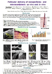
nanoparticles and microcontainers ex vivo and in vivo Sergey Zaitsev 1 Natalia Ksenofontova 1 Yulia I Svenskaya 1 Olga Guslyakova 1 Elina ID: 598902
Download Presentation The PPT/PDF document "Follicular delivery of" is the property of its rightful owner. Permission is granted to download and print the materials on this web site for personal, non-commercial use only, and to display it on your personal computer provided you do not modify the materials and that you retain all copyright notices contained in the materials. By downloading content from our website, you accept the terms of this agreement.


Slide1
Follicular delivery of nanoparticles and microcontainers ex vivo and in vivo
Sergey Zaitsev1, Natalia Ksenofontova1, Yulia I. Svenskaya1, Olga Guslyakova1, Elina A. Genina1,2, Vsevolod Atkin1, Georgy S. Terentyuk1, Alexey N. Bashkatov1,2, Dmitry A. Gorin1, Valery V. Tuchin1,2, and Gleb B. Sukhorukov1,3
1Saratov State University, Russia; 2Tomsk State University, Russia; 3Queen Mary University of London, UK
MotivationThe successful loading of nanoparticles with drugs and their triggered release inside the hair follicle can present an ideal method for localized drug delivery. Depending on the particle size, such a method would permit targeting specific structures in the hair follicles such as stem cells or immune cells or blood vessels found in the vicinity of the hair follicles.The goal of the study is development of the method of follicular delivery of particle and creation of depot in skin.
Methods and MaterialsRat skin ex vivo and in vivoTiO2 particles with the sizes 25 nm, 100 nm, and 5 μm (ex vivo experiments)CaCO3 submicron containers loaded with photodynamic dye Indocyanine Green (in vivo experiments)Ultrasonic (US) low-frequency therapeutical device as enhancer of particle penetration (with power 0.5Wt and treatment time 2 min) and damage of submicron containers (power 2Wt and treatment time 2-4 min)OCT-monitoring of particle penetration in skin was carried out every 2-4 min during US treatment and after 3 daysBiopsy and SEM were used for visualization of submicrocontainers inside follicles
Results of ex vivo experiments
(a)
(b)
Fig. 4
– SEM images of (a) follicle without
submicrocontainers
and (b) follicle wall
Fig 5.
– OCT-image of rat skin with submicrocontainers penetrated into hair follicles after 4 min of US treatment
Fig. 6
– SEM images of (a) follicle wall with the penetrated submicrocontainers after 2-min no-damaging US treatment (just post-treatment)(b) follicle wall with the penetrated submicrocontainers after 4-min damaging US treatment (just post-treatment)
(a)
(b)
(a)
(b)
Fig. 7
- SEM images of (a) follicle with the penetrated
submicrocontainers
after 2-min no-damaging US treatment (after 3 days)(b) CaCO3 submicrocontainers inside the follicle
Fig. 8
-
SEM images of (a) follicle wall with the penetrated submicrocontainers after 4-min damaging US treatment (after 3 days)(b) CaCO3 submicrocontainers inside the follicle
(a)
(b)
Results of in vivo experiments
Fig. 3
- Histogram of temporal dependence of penetration depth of particles with various sizes
Fig. 1
OCT-image of rat skin with 25-nm TiO
2
particles penetrated into hair follicle
Fig. 2 - Histological section of rat skin with 25-nm TiO2 particles penetrated into hair follicle
Follicle filled with nanoparticles
Follicle filled with nanoparticles
Conclusion
The rate of follicular penetration of particles depends on their sizes: 25-nm particles
penetrated faster than 5-μm ones2-min US treatment allowed for deep penetration of submicrocontainers into follicles without damaging4-min US treatment led to recrystallization of CaCO3 containers inside follicles and release of contentsThe work was carried out under the support by Russian Federation Governmental No. 14.Z50.31.0004 designed to support scientific research projects implemented under the supervision of leading scientists at Russian institutions of higher education
Follicular transport
Living
epidermis
Stratum
corneum
Dermis
Follicle
Recrystallization
of particles