
The classic principle of protein folding is that all the information required for a protein to adopt the correct threedimensional conformation is provided by its amino acid sequence Molecular chaperones ID: 236969
Download Presentation The PPT/PDF document "Protein Folding and Processing" is the property of its rightful owner. Permission is granted to download and print the materials on this web site for personal, non-commercial use only, and to display it on your personal computer provided you do not modify the materials and that you retain all copyright notices contained in the materials. By downloading content from our website, you accept the terms of this agreement.

Slide1
Protein Folding and Processing
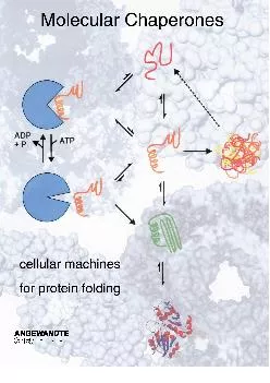
The classic principle of protein folding is that all the information required for a protein to adopt the correct three-dimensional conformation is provided by its amino acid sequence.Molecular chaperones are proteins that facilitate the folding of other proteins.Two specific families of chaperone proteins act in a general pathway of protein folding in both prokaryotic and eukaryotic cells – Heat shock proteins and Chaperonins.Unfolded polypeptide chains are shielded from the cytosol within the chamber of the chaperonin.Slide2
Action
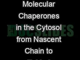
of chaperones during translation and Transportchains that are still being translated on ribosomes, thereby preventing incorrect folding or aggregation of the amino-terminal portion of the polypeptide before synthesis of the chain is finished.Chaperones also stabilize unfolded polypeptide chains during their transport into subcellular organelles.Slide3
The role of N-linked glycosylation in ER protein folding.
3Slide4
The unfolded protein response in yeast Slide5
The export and degradation of misfolded ER proteins Slide6
Protein translocationSlide7
ENDOSITOSisSlide8
Protein folding in the
cellBasics- cell compartments, molecular crowding: cytosol, ER, etc.Folding on the ribosome- co-translational protein foldingMolecular chaperones- concepts, introduction- intramolecular chaperones- chemical chaperones- protein chaperonesSlide9
Folding
in vitro vs. in vivo
folding by dilution
in buffer
protein denatured
in a chaotrope
folded
protein
in vitro
in vivo
folding
folded
proteinSlide10
Problem:
non-native proteins• non-native proteins expose hydrophobic residues that are normally buried within the ‘core’ of the protein • these hydrophobic amino acids have a strong tendency to interact with other hydrophobic (apolar) residues - especially under crowding conditionsintramolecular
misfolding
X
X
X
X
intermolecular
aggregation
X
X
X
X
X
X
incorrect
molecular
interactions
&
loss of activity
exposed
hydrophobic
residues
3-10Slide11
Eukaryotes
ArchaeaBacteria
-
-
Trigger Factor
NAC
NAC
-
Hsp70 system
[Hsp70 system]
Hsp70 system
prefoldin
prefoldin
-
chaperonins (group II)
chaperonins (group II)
chaperonins (Group I)
small Hsps
small Hsps
[small Hsps]
Hsp90
-
[Hsp90]
AAA ATPases
AAA ATPases
AAA ATPases
-
-
SecB
-
-
[PapD/FimC]
Hip, Hop, Bag, clusterin, cofactors A-E, calnexin, calreticulin, etc. etc.
-
-
Overview of chaperone families:
DistributionSlide12
IRE-1
XBP-1The Unfolded Protein Response (UPR) The UPR occurs when proteins are misfolded in the endoplasmic reticulum (ER). Reducing agents, such as DTT, interfere with disulfide bond formation while drugs can interfere with glycosylation
; both agents cause proteins to
misfold
in the ER thus triggering the UPR.
The product of the
ire-1
gene is the sensor of
misfolded
proteins and when activated removes an
intron
from the pre mRNA from the
xbp-1
gene.
Active
xbp-1
protein (from spliced mRNA) activates the genes that code for ER chaperones
,
such as hsp-4.
Hsp4 (grp78)
grp170Slide13Slide14
PROTEIN TURNOVER AND AMINO ACID CATABOLISM
Degradation of proteins1) dietary proteins- amino acids- pepsin in stomach- pancreatic proteases- aminopeptidase Nother peptidases2) endogenous proteins
- protein turnover: synthesis, degradation,
resynthesis
- damaged proteins
- half-lives of proteins: depend on amino-terminal residuesSlide15
Cellular Protein Degradation
Lysosomal Nonspecific Endocytosis Foreign proteins Energy favorable to degrade proteins Non-lysosomal Specificity, requires ATP Conditions of stress
Ubiquitin-
proteosomal
pathway
26S
proteosome
Role in cellular processes/signalingSlide16
Protein turnover; selective degradation/cleavage
Individual cellular proteins turn over (are degraded and re-synthesized) at different rates. E.g., half-lives of selected enzymes of rat liver cells range from 0.2 to 150 hours. N-end rule: On average, a protein's half-life correlates with its N-terminal residue. Proteins with N-terminal Met, Ser, Ala, Thr, Val, or Gly have half lives greater than 20 hours. Proteins with N-terminal Phe, Leu, Asp, Lys, or Arg have half lives of 3 min or less.PEST proteins having domains rich in Pro (P), Glu (E), Ser (S), Thr (T), are more rapidly degraded than other proteins. Slide17
Ubiquitinylation – Proteosome Degradation
E3 determines protein substrateSlide18
8.42 The
ubiquitin-proteasome pathwaySlide19Slide20Slide21
Ubiquitination1) ubiquitin- a 8.5 kd protein (76 residues) formation of an isopeptide bond with ε-amino group of lysine of the proteins - a tag for destruction - polyubiquitin: a strong signal for degradation 2) enzymes for
ubiquitination
- E1 (
ubiquitin
-activating enzyme)
- E2 (
ubiquitin
-conjugating enzyme)
- E3 (
ubiquitin
-protein
ligase
)
- variation: E3 > E2 > E1: more finely tuned substrate discrimination
HPV (human
papilloma
virus) activates a specific E3 enzyme:
tumor suppressor protein p53Slide22
Regulation of ubiquitination
: Some proteins regulate or facilitate ubiquitin conjugation. Regulation by phosphorylation of some target proteins has been observed. E.g., phosphorylation of PEST domains activates ubiquitination of proteins rich in the PEST amino acids. Glycosylation of some PEST proteins with GlcNAc has the opposite effect, prolonging half-life of these proteins. Slide23
19S and 20S
Proteasome Subunits Characteristics20S SubunitBarrelContains 6 proteolytic sites2x Tryptic2x Chymotryptic2x Peptidylglutamyl- peptidase Linearized protein required
19S Subunit
Base and Lid
Contains subunits with known and unknown functions
Tetra-
Ub
(K48) recognition
Deubiquitination
activity
Protein unfolding activity (Chaperone function)Slide24
Ubiquitin AA Sequence
MQIFVKTLTG KTITLEVEPS DTIENVKAKI QDKEGIPPDQ QRLIFAG
K
QL EDGRTLSDYN
IQ
K
ESTLHLV LRLR
GG
48
63
6Slide25Slide26Slide27
Proteasome-1
Proteasome-3Proteasome-4Slide28
Roles of UbiquitinationSlide29
Different Types of Ubiquitin TagsSlide30
Transmembrane
Proteins Regulated by Ub-dependent SortingIn metazoans: Neurotransmission: Ion channels: AMPA glutamate receptors ENaC Glycine receptors ClC-5 Cell-cell contacts: Immune molecules
E-cadherin
downregulated
by viruses:
Occludin
MHC class I
B7-2
Developmental patterning:
ICAM-1
Delta CD4
Notch
RoundaboutSlide31
Poly-Ub Chains
Ub K K
Ub
Ub
Ub
Ub
Ub
Ub
Ub
Ub
K48 Linkage
K63 Linkage
K63
K48
Peters, J.M. 1998
Ubiquitin and the Biology of the Cell
Signal to proteosome
K48, Ub
4
Cell Signaling
K63Slide32
ENaC
function Major ion channel that controls salt and fluid resorption in the kidney Mutations in the PPXY motif cause accumulations of channels at the cell surface and result in Liddle’s syndrome, and inherited form of hypertensionSlide33
ENac
surface Stability Nedd 4 (HECT ligase)-negatively regulates ENaC surface stability Nedd4 WW domains bind PPXY motif of ENaC subunits Nedd4 also interacts with serum and glucocorticoid-regulated kinase (SGK) SGK contains two PPXY motifs that bind to Nedd4 WW domains SGK-dependent Nedd4 P inhibits the Nedd4-ENaC interaction therefore, Nedd4 P increases
ENaC
at the cell surfaceSlide34
ENaC
SubunitsSlide35
Regulation of ENaC Surface StabilitySlide36
Ub
-like ProteinsSUMO-1 (sentrin, smt-3)1996 – covalent modification – RanGAP1RanGAP1 nearly quantitative modifiedCytosolic RanGAP1 to nuclear poreActivate shuttling factorSlide37
Ubiquitin-like Proteins:Slide38
Ubiquitin Superfold and Ubiquitons
Ub – blueSUMO-1 – greenNEDD8 - redUB αβ roll suprfoldSlide39
SUMO
SUMOSUMO-1 & SUMO-2/3Shared characteristicsC-terminal -GG essential for conjugationAffix to lysine residues in targetNOT directly associated with proteasomal degradationSlide40
Competition/Regulation
SUMOReactive Oxygen Species: Oxidizes reactive thiols on SUMO enzymesUba1/Aos1- S – S – Ubc9Thus: SUMO can not attach and proteins not SumoylatedSlide41
Examples of SUMO function
RanGAPIkBc-Junp53 and mdm2Causes nuclear translocationBlocks Ub-conjugation site, prevents degradationInhibits transcriptional activityBlocks mdm2 self-ubiquitination, prevents degradationSUMO-p53 in DNA binding domain apoptotic activity
PROTEIN
SUMO EffectSlide42
Peptide generation in the class I pathwaySlide43
Proteasome specificity
NetChop is the best available cleavage methodwww.cbs.dtu.dk/services/NetChop-3.0