
APPEARANCE AND IDENTIFICATION OF A CAUSATIVE LEAD POINT N MEKKI R KHARRAT S BOURKHIS R BEN NACEUR F BEN AMARA N MNIF CHARLE NICOLLES HOSPITAL TUNIS TUNISIA Introduction ID: 929094
Download Presentation The PPT/PDF document "ADULT BOWEL INTUSSUSCEPTIONS: RADIOLOGY" is the property of its rightful owner. Permission is granted to download and print the materials on this web site for personal, non-commercial use only, and to display it on your personal computer provided you do not modify the materials and that you retain all copyright notices contained in the materials. By downloading content from our website, you accept the terms of this agreement.


Slide1
ADULT BOWEL INTUSSUSCEPTIONS: RADIOLOGYAPPEARANCE AND IDENTIFICATION OF A CAUSATIVE LEAD POINT
N. MEKKI, R. KHARRAT, S. BOURKHIS, R. BEN NACEUR, F. BEN AMARA, N. MNIF.
CHARLE NICOLLE’S HOSPITAL, TUNIS, TUNISIA
Slide2Introduction:Intussusception in adults is an unusual cause of bowel obstruction:
1%
of all bowel obstructions.
5 % of all intussusceptions.
80-90 %: due to an
underlying pathology
.
The
growing use of computed tomography (
CT
) and magnetic resonance imaging (
MRI
) has led to increased detection of intussusceptions
as
mostly unsuspected clinically, presented with non-specific abdominal pain.
Slide3Objectives :
To describe the characteristic radiologic features of
intussusception
according to location.
To illustrate pathologies which cause intussusceptions.
To correlate the different features with the pathologic findings.
Slide4Materials and methods : We made a retrospective study, over 1-year period (2011).
6 cases of adult bowel intussusceptions:
Sex ratio: 4 men/ 2 women.
Age : vary from 21 to 60 years, mean age: 38 years.
Explorations:
5 patients was explored by
abdominal enhanced CT examination General Electric (GE) 16 slices
.
One patient was explored by
MRI GE 1.5 Tesla .
Slide5RESULTS:Clinical
presentation
was
non
specific
for all patients: abdominal pain+++
Radiology
detect:4 ileo ileal intessusception in 3 patients: 2 were in the same one.1 ileo coecal intessusception.1 colo colic intessusception in the transverse colon.1 colo rectal intessuscetion.
ETIOGIES
With
lead
point:
83.4%
Without
lead
point:
16.6%
Neoplastic
Non
neoplstic
1 case :
Post
operative
intessusception
.
Malignant
Benign
1 case :
Crhon’s
disease
Primary
: 2cases:
Secondary
: 1
case:
Metastasis
of
renal
adeno
carcinoma
1 case :
Lipoma
Rectal
adeno
carcinoma
Small
bowel
stromal
tumor
Slide6Case N° 1: A.H. is a 36 year
old
man
who
consult
for
gastro
intestinal bleeding and anemia, contrast enhanced abdominal CT scan was made to identify the cause of hemorrhagia and to search if there is an active bleeding.2
1
RESULTS:
2
Axial slices of CT scan without (
1
) and with contrast enhancement (
2
).
They demonstrate the typical multilayered appearance of a small bowel
intussusception
in the left
hypochondrium
.
Slide7Multiplanar reconstructions: sagittal
and coronal help a lot to see the sausage of the
ileo
ileal
intessusception
.
Sag
Sag Contrast-enhanced CT scan showed invaginated mesenteric fat and vessels . Sag
Sag
RESULTS:
Coro
Slide8At laparotomy, the small bowel intussusception
was confirmed and histology showed the lead point to be a
stromal
tumor.
Coro
Ax
At
the top of the intessusception, we notice a round lesion that enhances heterogeneously after
contrast
injection
( ),
these
one
was
confirmed
by
peroperative
constatation .
Sag
RESULTS:
Slide9Case N°2: R.S. is a 27 Year
old
men
who
was
admitted
in the emergency for abdominal
stab wounds, he was explored by surgery for peritoneal effusion. He was controlled after 10 days by CT without contrast enhancement.
CT
showed
the
target
aspect of
loops
in 2
differnt
sites in relation
with
2
small
bowel
intessusceptions
: the first
exist
under
the
liver
and the second
is
pelvic
.
RESULTS:
Slide10Notice in
this
recontructed
images the
sausage
aspect
Intessusceptions
in this cases were taken as transient
because
the patient
was
asymptomatic
.
Another
control
after
2
weeks
showed
their
persistance,
surgery
did not found a lead point. The final diagnosis
was
post
operative
intessusceptions
.
RESULTS:
Slide11Case N°3: M.A. is a 30 years
old
woman
operated
for
bilateral
kidney adenocarcinoma (radical right nephrectomy and left lumpectomy) consulting for non specific abdominal pain.Enhanced CT examination shows ileo ileal intessusception in the right iliac
fossa
.
RESULTS:
Slide12We
notice the
presence
at
the distal portion of the
intessuceptum
, of an
oval
lesion ( ) that is enhanced by the contrast, it may be the causative lead point. The surgery and the histology find the underlying
pathology
to
be
a
bowel
metastasis
of a
renal
adeno
carcinoma
in the last
ileal loop
.
RESULTS:
Slide13: cortico medullary
hypodense
range in the
upper
pole of the
left
kidney
suggestive of
tumor recurrence.CT shows also other lesions suggestive of metastasis : Intra peritoneal effusion
:
Osteolysis
of the left iliac wing suggestive of to metastases.
RESULTS:
Slide14Case N°4: G.K. is a 21 year
old
men
followed
for
crhon’s
disease
since 6 months, he consult for abdominal pain, he was explored by MRI:CORO SSFSE MRI showed an important inflammatory thickening of the distal ileum heaving a look like tumor with intense heterogeneous enhancement after contrast injectionAX T1 GADORESULTS:
CORO T1
GADO
Small bowel
opacification
showed a masse in the
ileocecal
junction
CORO SSFSE
Slide15SAG T1 GADO Notice the intestinal expansion upstream of the thickening.
Endoscopy
showed an inflammatory
ileo
cecal
valve which is prolapsed in the
caecum
, it showed also ulcerated terminal ileitis. The diagnosis was intessusception on an acute episode of crhon’s disease.RESULTS:SAG T1 GADOCORO T1 GADO The sagittal and coronal sections showed intussusception of the ileocecal valve into the caecum ( ) .
Slide16Case N°5: M.H. is a 60 year
old
men
who
consult
in the emergency for abdominal pain
with
acute
bowel obstruction.RESULTS: CT scan showed the image of bowel-within
-
bowel
in the pelvis
clearly
visible on the axial and sagittal, relevant to a colo rectal
intessusception
( ).
Slide17sag
Ax
Coro
Coro
The
surgery
confirm CT findings and histolo!gy showed
the
lead
point to
be
an
infiltrating
adeno
carcinoma
of the rectum.
RESULTS:
At
the top of the
sausage
,
we
notice an irregular stenosing mass with spontaneous
isoattenuating relative to rectal
wall
which
enhances
heterogeneously
after
contrast
injection .
we
notice
also
a densification of the
mesenteric
fat
surrouding
the rectum
with
lymphadenopathies
.
Slide18Case N°6: M.H. is a 50 years
old
woman
who
was
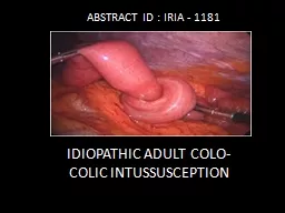
treated for degenerated colic polyp, CT was made to control because of non specific abdominal pain: RESULTS:
Contrast
enhanced
CT scan
showed
a colo
colic
intessusception
( ) in the transverse colon
occuping
the
epigastrium
and the
left
hypochondrium
.
Slide19CT showed in the tip of the
intussusception
an oval
hypodense
mass heaving spontaneously an homogeneous fat density without contrast enhancement, it is characteristic of a
lipoma
.
Surgery
confirmed the large bowel intessusception Histology confirmed the lead point to be a lipoma.RESULTS:
Slide20Definition
:
Intussusception
is
a
progressive
invagination
of a bowel loop with its
mesentery and mesenteric vessels M (intussusceptum) into the lumen of a contiguous portion of bowel (intussuscipiens). It is the results of abnormal peristalsis producing unequal longitudinal forces in the intestinal wall. M
It
may
be
caused
by a mass
pulled forward by normal peristalsis
or by
functional
disturbances
.
Discussion:
Slide21Clinical Presentation:
Symptoms are often chronic: several weeks to months, may be occasionally acute, it
may
be
related to the lead point.
Unlike children, the most common symptoms of
intussusception
in adult are non specific: Abdominal pain, nausea and vomiting +++Less frequently: constipation, fever, weight loss, diarrhea;It is often asymptomatic, especially in chronic invaginations or without leadpoint.Physical examination is often unremarkable, sometimes note palpable mass.Discussion:
Slide22Classification:Intussusceptions are classified according to :
Location:
Small or large bowel
:
more frequent in the small bowel (2/3)than in the colon(1/3).
4 different locations:
Entero enteric
:
confined to the small Bowel. Colo colic:linvolving the large bowel only. Ileo colic: defined as the prolapse of the terminal ileum within the ascending colon.Ileo cecal: the ileo-cecal valve is the leading point of the Intussusception.With
underlying
pathology
: 80-90%:
Neoplastic
: 65%
Benign
Malignant
Non-
neoplastic
: 15-25%
Idiopathic
:10%:
-It tends to be
allmost
transient.
Causes
Discussion:
Slide23Etiologies: in the colon:
the most common underlying
malignant lesions
in the colon: are
primary
malignant
tumors
: adenocarcinoma and lymphoma.Benign lesions : 30% lipoma, leiomyoma, adenomatous polyp, endometriosis and previous anastomosis. Idiopathic intussusception occurs less often than in the small bowel: 10%
Intussusception
in the large bowel is more likely to have a malignant etiology :
50–60%
Discussion:
Slide24Etiologies: in the small bowel
Benign lesions:
65%
benign
neoplasms
:
lipoma
,
leiomyoma, haemangioma, neurofibroma, following abdominal surgery: adhesions,anastomosis,Meckel’s diverticulum, lymphoid hyperplasia and adenitis,Traumatism,coeliac disease, intestinal duplication
Crohn’s
disease
Malignant
lesions
:
15%
:
most often
metastatic
:
Melanoma+++
Idiopathic
intussusception
:
20%
Discussion:
Slide25Radiological findings:
CT constitute the main imaging modality because of its virtually
pathognomonic
appearance:
bowel
-
within
-
bowel: It appears as a complex soft tissue mass, consisting of the outer intussuscipiens and the central intussusceptum. There is often an eccentric area of fat density within the mass representing the intussuscepted mesenteric fat. the mesenteric vessels are often visible within it.Discussion:
Slide26Radiological findings:
When the CT beam is parallel to its longitudinal axis of
the
intussusc
eption
,
it
appears as a sausage-shaped mass.Sometimes as reniform or “pseudokidney” mass: it is due to edema, mural thickening, and vascular compromise.When the beam is perpendicular to the longitudinal axis, it appear as a ‘‘target’’ mass. Discussion:
Slide27Radiological findings:
CT examination allow to:
Detect and confirm the diagnosis of
intessusception
.
Show the exact location: small or large bowel.
Appreciate the viability of
invaginated
loops.
Distinguish between Intussusception without a lead point: no signs of proximal bowel obstruction, target-like or sausage-shaped mass, layering effect.Intussusception with a lead point: signs of bowel obstruction, bowel wall edema with loss of the classic three-layer appearance due to impaired mesenteric circulation and demonstration of the lead mass. CT help reducing the number of unnecessary surgical interventions.Discussion:
Slide28Radiological findings:Intessusception
with
an
underlying
lead
point
Suspected
by the
epidemiological data( age+++, medical history) and the clinical presentation.Ct scan find a mass in addition to the intussusception outlined distal to the tapered lumen of the intussusceptum.Discussion:
Slide29Radiological findings: Intessusception
with
an
underlying
lead
point
The mass’s type is established by the study of its
spontanous density and enhancement: for example:Lipoma: fat density wihout containing blood vessels to be distinguished from mesenteric fat and without enhancement.Other malignant
tumors
(
primary
or
metastatic
):
tissular
density
with
heterogenous
enhancement
.
Neoplastic
lead point VS Non-neoplastic one:significantly longer significantly larger diameter significantly more proximal dilatation of small bowel downstream.Discussion:
Slide30Radiological findings: Transcient intessusception
:
More
frequent
in the
small
bowel
than in the colonIt is most frequently detected incidentally and is presumed to be innocuous.Reported in adults with:Celiac disease Crohn diseaseDiscussion:
Slide31Radiological findings:Magnetic
Resonance
Imaging (MRI)
Recent developments in MRI with ultrafast
multiplanar
techniques now allow for rapid evaluation of bowel obstruction.
The
multiplanar
HASTE (half-
fourier single shot turbo spin echo): SSFSE is particularly useful in the diagnosis of intussusception. The high contrast resolution between the increased signal of the trapped intraluminal fluid and the intermediate to low signal of the bowel wall can clearly demonstrate the pathology.Discussion:
Slide32TreatmentThere is no universal agreement upon the correct treatment of adult
intussusception
,
The surgery decision is based on:
The epidemiological data: age, medical history ..
The
clinical
presentation
: acute abdominal pain, bowel obstruction, digestive hemorrhagia...The imaging findings: if a lead point is found or not,If there is ischemic bowel signs:The type of intervention depends essentially on the intraoperative findings.Discussion:
Slide33Conclusion:Intussusception
in adults is an infrequent cause of
intestinal obstruction.
Preoperative diagnosis is difficult as symptoms can be
intermittent and long standing.
More frequent use of computed tomography in undiagnosed abdominal pain increases the pick up
rates.