
Premier Products for Superior Life Science Research Assay system for measurement of Macrophage Inhibitor Cytokine MIC 1 in biological fluids and extracts where MIC 1 may be present NWLSS TM ID: 942207
Download Pdf The PPT/PDF document "nwkimic1h1rev060514" is the property of its rightful owner. Permission is granted to download and print the materials on this web site for personal, non-commercial use only, and to display it on your personal computer provided you do not modify the materials and that you retain all copyright notices contained in the materials. By downloading content from our website, you accept the terms of this agreement.

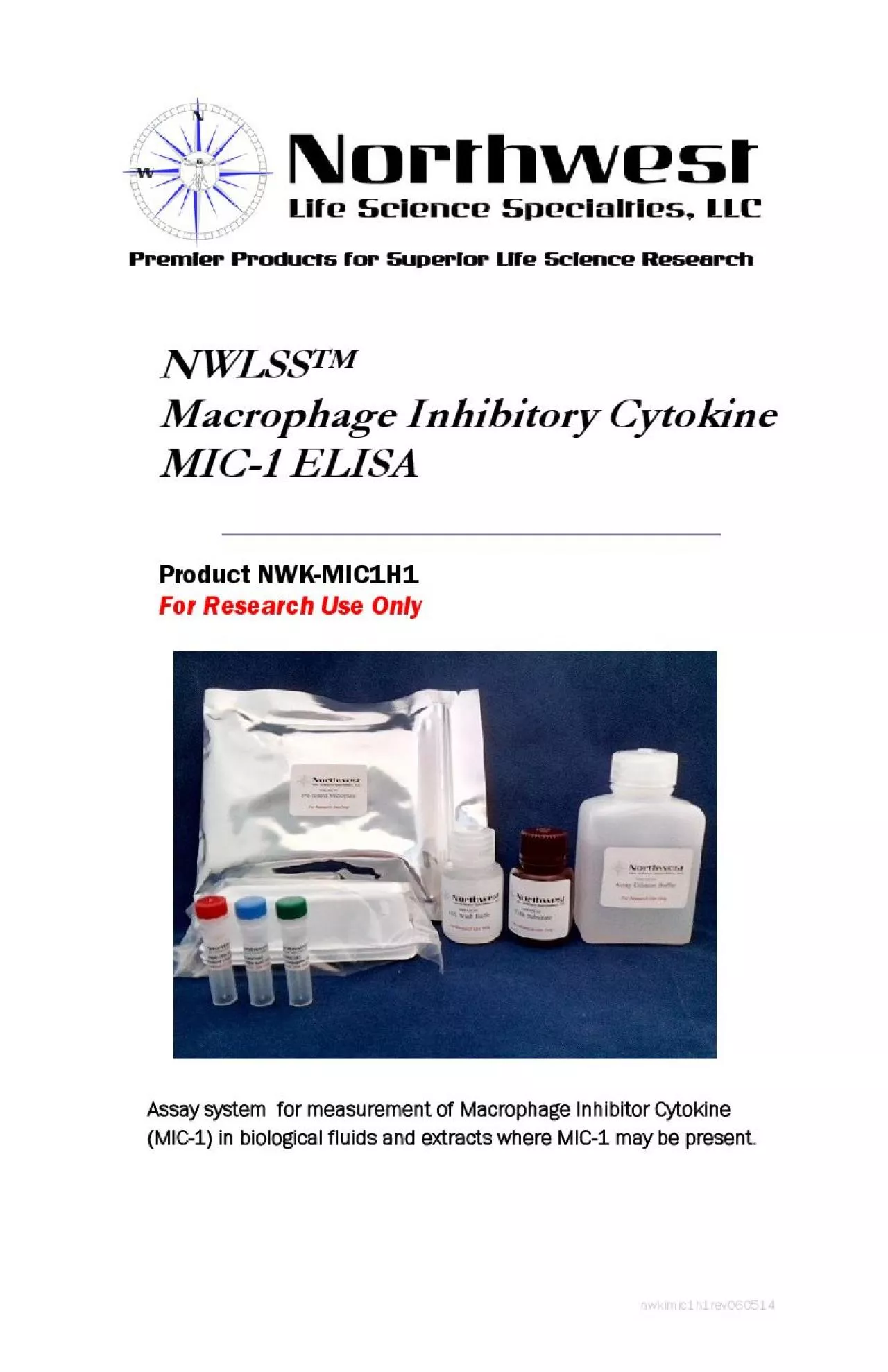
nwkimic1h1rev060514 Premier Products for Superior Life Science Research Assay system for measurement of Macrophage Inhibitor Cytokine (MIC - 1) in biological fluids and extracts where MIC - 1 may be present. NWLSS TM Macrophage Inhibitory Cytokine MIC - 1 ELISA Product NWK - MIC1H1 For Research Use Only nwkmic1h1rev060514 Section Page To Order Call: 1 - 888 - 449 - 3091 Page 2 Table of Contents Introduction 3 Intended Use 3 Test Principle 4 General Specifications 4 Kit Contents 4 Required Materials Not Provided 4 Required Instrumentation 4 Warnings, Limitations, Precautions 4 Storage Instructions 5 Assay Preparation 5 Reagent Preparation 5 Standard Preparation 5 Sample Handling/Preparation 6 Assay Protocol 6 Data Analysis 7 References 8 Statement of Limited Warranty 8 User Notes 9 nwkimic1h1rev060514 Introduction: Macrophage inhibitory cytoki
ne (MIC - 1) is a divergent member of the TGF - β superfamily. The cDNA sequence of MIC - 1 is identical with several other sequences, including growth differentiation factor - 15 (GDF - 15), placental bone morphogenetic protein (PLAB), placental transforming growth factor (PTGF - β ), prostate - derived factor (PDF), and nonsteroidal anti - inflammatory drug - activated protein - 1 (NAG - 1). MIC - 1 mRNA encodes a secreted protein, resulting from cleavage of a propeptide to give rise to the mature form as a 25 - kDa homodimer, which contains seven conserved cysteine residues in the carboxyl terminal. There are at least two known alleles of MIC - 1 that are due to a G C point substitution at position 6 of the mature protein which alters a histidine to an aspartic acid (1). MIC - 1 is distributed in various tissues, being highly expressed in macrophages, choroid plexus, prostate, lung, kidney proximal tubules, placenta and intestinal mucosa. I
t is poorly expressed in the heart although it has been described as a prognostic marker in acute myocardial infarction (2) as well as an independent predictor of chronic heart disease mortality (3). Initially, MIC - 1 was consid- ered to function primarily as a macrophage inhibitor, but recent studies suggest that it is pleiotropic regulating a myriad of cellular processes such as the cell cycle, proliferation, differentiation, and apoptosis. MIC - 1 expres- sion can be induced by stress conditions such as tissue injury, malignancy and inflammation. It has recently been implicated as a cachexia mediator inducing weight loss (4). MIC - 1 is overexpressed by a variety of cancers, which may relate to its antitumorigenic and proapoptotic properties, alt- hough recent studies describe contradictory mechanisms. For example, it has been reported to exhibit both tumorigenic and antitumorigenic activi- ties. MIC - 1 expression is correlated with the tumorigeni
city of melanoma cells where it is highly expressed (5). MIC - 1 may serve as a biomarker for the prediction of gastric cancer progression. Serum concentrations in can- cer patients were 10 - fold higher than those of healthy controls (6). Serum MIC - 1 has been described as a biomarker capable of predicting prostate cancer prognosis (7). Prostate - derived factor (PDF/MIC - 1) may be related to cellular stress through its interaction with p53. The p53 tumor suppres- sor modulates cellular responses in various models of cell stress. Further- more, there appears to be a requirement for functional p53 in PDF induc- tion in these disparate models indicating that PDF may represent a novel target of p53 in response to cell stress (8). Intended Use: This kit is intended for the quantification of macrophage inhibitory cytokine (MIC - 1) in serum, plasma, tissue and cell lysates and other biological fluids where MIC - 1 may be present. To Order
Call: 1 - 888 - 449 - 3091 Page 3 nwkmic1h1rev060514 Test Principle: This assay is a quantitative sandwich enzyme immunoassay. Plates are precoated with a polyclonal antibody specific for native human MIC â 1 (capture antibody). The antibody - bound MIC - 1 in standards and specimens binds to a polyclonal detection antibody. An HRP conjugated anti - detection signal antibody is added followed by substrate. Color development is stopped at the appropriate time and the plates are read. General Specifications: Format: 96 well sandwich ELISA Number of tests: Triplicate = 24 Duplicate= 40 Specificity: Human Macrophage Inhibitory Factor (MIC - 1) Sensitivity: 5 pg/mL Effective Range: 5 pg/mL - 2000 pg/mL Kit Contents Microwells precoated with anti human MIC - 1 antibody: 1 X 96 wells MIC - 1 Standard: Purified MIC - 1 (40 ng/mL) 1 X 60 ï L Primary Antibody (100X Anti human MIC - 1): 1 X
130 ï L 10X Wash Buffer: 1 X 30 mL Assay Dilution Buffer: 1 X 100 mL TMB Substrate: 1 X 25 mL Anti IgG - HRP Conjugate (100X): 1 X 130 ï L Required Materials Not Provided: Adjustable pipettes with a range of 10 µL to 1,000 µL with disposable tips. Glassware for reagent preparation. Deionized water. 3M sulfuric acid. Required Instrumentation: Microtiter plate reader with 450 nm capability. Warnings, Limitations, Precautions: Individual components may be harmful if swallowed, inhaled or absorbed through the skin. Contact should be minimized through the use of gloves and standard good laboratory practices. If contact with skin or eyes occurs, rinse the site immediately with water and consult a physician. To Order Call: 1 - 888 - 449 - 3091 Page 4 nwkimic1h1rev060514 To Order Call: 1 - 888 - 449 - 3091 Storage Instructions: Store the Standard, 100X Primary Antibody and
100X IgG - HRP Enzyme Conjugate at â 20°C. All other components should be stored at 4°C. The kit is stable for 9 months from date of manufacture when stored under these conditions. Assay Preparation 1. Determine the number of wells required to assay standards, samples and controls for the appropriate replicate. 2. Create an assay template showing positioning of standards, controls and samples. Include blank wells also. 3. Next remove the required number of strips and place in the frame sup- plied. Return unused wells to the storage bag, seal and store at 2 - 8ºC. Reagent Preparation: The following instructions are based on the user using the entire kit at one time. Assay Dilution Buffer is supplied ready to use. TMB Substrate is supplied ready to use. 10X Wash Buffer Add the contents of the 10X Wash Buffer to 270 mL deionized H 2 O, mix well and label as Working Wash Buffer. 100X IgG - HRP Enzyme Conjuga
te Briefly centrifuge the vial to remove all liquid from the cap and vial walls. Add 120 ï L conjugate to 12 mL Assay Dilution Buffer. Label as Diluted HRP - Conjugate . 100X Primary Antibody Add 120 ï L Primary Anti â MIC - 1 Antibody to 12 mL Assay Dilution Buffer. Label as Diluted Primary Antibody . Page 5 nwkmic1h1rev060514 To Order Call: 1 - 888 - 449 - 3091 Page 6 Standard Preparation: 1. Label tubes 1 - 8 tubes as: 2000, 1000, 500, 250, 125, 62.5, 31.25 and zero (0) pg/mL. 2. Add 950 ï L Assay Dilution Buffer to tube 1 and 500 ï L Assay Dilution Buffer to each of tubes 2 - 8. 3. Add 50 ï L of 40 ng/mL MIC - 1 Standard to tube 1 and mix well. Note: Unused reconstituted standard can be frozen at - 70 ° C and thawed one time only without significant loss of immunoreactivity. 4. Make a serial dilution by transferring 500 ï L of 2000 pg/mL Standard (tube 1)
into tube 2 mixing thoroughly then 500 ï L of resulting 1000 pg/ mL to tubes 3 and so on to create all Standards down to 31.25 pg/mL. Sample Handling/Preparation Samples should be diluted with Assay Dilution Buffer just prior to assay. Recommended starting dilutions are neat and 1:2 with Assay Dilution Buffer. Urine may require additional dilution. The following ranges of MIC - 1 have been observed with this assay: Serum and Plasma: 85 â 1350 pg/mL Urine: 34 â 32,000 pg/mL Assay Protocol: 1. Add 100 µL of Standard or Sample to each well. Incubate for 1 hour at room temperature. 2. Wash plate with 300µL Working Wash Buffer 3 times allowing plate to stand 2 minutes per wash. Empty plate again by inversion and pat dry upside - down on a lint free towel after final wash. 3. Add 100 µL of Diluted Primary Antibody to each well. Incubate for 1 hour at room temperature. 4. Wash according to Step 2.
5. Add 100 μ L of Working HRP - Conjugate per well. Incubate for 1 hour at room temperature. 6. Wash according to Step 2. 7. Add 150 μ L TMB Substrate per well and allow the color to develop for 20 - 40 minutes at room temperature. 8. Add 50 µL of 3M sulfuric acid to each well to stop the reaction. 9. Record the absorbance at 450 nm using a plate reader. nwkimic1h1rev060514 To Order Call: 1 - 888 - 449 - 3091 Page 7 Data Analysis 1. Average all duplicate well absorbance values. 2. Subtract the average absorbance values for the blank wells (S0) from all other well pairs. 3. Plot a standard curve using the corrected absorbance values of each Standard (y - axis) versus the Standard concentration (x - axis). 4. Determine the concentration of each unknown using the equation of the line. nwkmic1h1rev060514 To Order Call: 1 - 888 - 449 - 3091 Page 8 References: 1. Brown, D.A., et al.; (20
02) Biotechniques 33(1):18 - 126 2. Khan, S.Q., et al.; (2009) Eur Heart J. 30(9):6658 - 64 3. Kempf ,T., et al.; (2009) Circ Cardiovasc Genet. 2(3):286 - 92 4. Ding, Q., et al.; (2009) Endocrinol. 150(4):1688 - 96 5. Boyle, G.M., et al.; (2009) J Invest Dermatol. 129(2):383 - 91 6. Baek, K.E., et al.; (2009) Clin Chim Acta. 401(1 - 2):128 - 33 7. Brown, D.A et al.; (2009) Clin Cancer Res. 15(21):6658 - 64 8. Kelly, J.A., et al.; (2009) Cancer Lett. 277(1):38 - 47 Statement of Limited Warranty: Northwest Life Science Specialties, LLC (NWLSS) makes no guarantee of any kind, expressed or implied, that extends beyond the description of the material in this kit, except that they will meet our specifications at the time of delivery. Customer 's remedy and NWLSSâ sole liability is liïited to, at NWLSSâ option, refund of the purchase price, or the replacement of material not meeting our specifica
tion. By acceptance of our product, customer assumes all liability and will indemnify and hold NWLSS harïless for the consequence of this productâs use or misuse by the cus- tomer , its employees, or others. Refund or replacement is conditioned of customer notifying NWLSS within twenty - one ( 21 ) days of the receipt of product. Failure to give notice within 21 days shall constitute a waiver by the customer of all claims hereunder with respect to said product. nwkimic1h1rev060514 To Order Call: 1 - 888 - 449 - 3091 Page 9 User Notes: nwkmic1h1rev060514 To Order Call: 1 - 888 - 449 - 3091 Page 10 User Notes: nwkimic1h1rev060514 To Order Call: 1 - 888 - 449 - 3091 Page 11 User Notes: nwkmic1h1rev060514 5131 NE 94th Avenue, Suite 201 Vancouver, WA 98662 Phone 360 - 449 - 3091 or Toll Free: 888 - 449 - 3091 Fax 360 - 449 - 3092 E - mail: sales@nwlifescience