
Prepared by I Gede Purnawinadi SKep MKes Organization of the Nervous System Central nervous system CNS Brain and spinal cord Integration and command center Peripheral nervous system PNS ID: 799687
Download The PPT/PDF document "The Central Nervous System" is the property of its rightful owner. Permission is granted to download and print the materials on this web site for personal, non-commercial use only, and to display it on your personal computer provided you do not modify the materials and that you retain all copyright notices contained in the materials. By downloading content from our website, you accept the terms of this agreement.


Slide1
The Central Nervous System
Prepared by I
Gede
Purnawinadi
,
S.Kep
.,
M.Kes
.
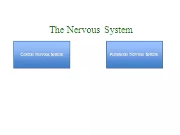
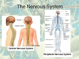
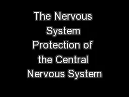
Slide2Organization of the Nervous System
Central nervous system (CNS
)
Brain and spinal cordIntegration and command center Peripheral nervous system (PNS)Paired spinal and cranial nervesCarries messages to and from the spinal cord and brain
Slide3Organization of the Nervous System
The two principal cell types of the nervous system are:
Neurons
– excitable cells that
transmit electrical
signals
Neuroglia
-
supporting cells
Slide4Functional:
Sensory
(afferent) — transmit impulses
toward the CNSMotor (efferent) — carry impulses away from the CNSInterneurons (association neurons) — shuttle signals through CNS pathwaysNeuron Classification
Slide5Neuroglia
Slide6BrainSpinal cord
Central Nervous System:
Slide7Hair, skin, cranium
Meninges
Cerebrospinal fluid
Blood brain barrierCNS Protection
Slide8Meningeal Layers
Meningeal layer of the brain cushion and protect delicate neural tissue
Slide9Cerebrospinal Fluid
Shock absorbing medium
Provides a optimum and
stable environment for
generating
nerve
impulses
Provides a
medium for the exchange of nutrients and wastes between blood and nervous
tissue
Formed by
selective transport
across ependymal cells
Volume
125-150 ml
and is replaced > 3 times/day, flow maintained by
10 mmHg
paressure
gradient
Path: ventricles
subarachnoid space, reabsorbed into blood in
dural
sinuses through arachnoid villi
Slide10Extensive capillaries & sinuses
Tight junctions promoted by astrocyte
Limits permeability for most molecules
except O2, CO2, alcohol, steroids, H2OProtects brain: hormones & circulating chemicalsProtects CNS from chemical fluctuationsPrevents entry of harmful substancesPrevents entry of molecules that could act as neurotransmitters
Brain receives 15% of blood pumped by heart
Brain responsible for about half of body’s glucose consumption
Membrane transporters move glucose from plasma into the brain interstitial fluid
Blood Brain Barrier
Figure 9-6: The blood-brain barrier
Slide11The Central Nervous System (CNS)
The
central nervous system (CNS)
consists of the brain and spinal cord, which occupy the dorsal body cavity. The CNS is the integrating and command center of the nervous system. It interprets sensory input and dictates
motor responses
based on past experience, reflexes, and current conditions.
The Brain
:
t
he average
adult man’s brain
has a mass of about
1600 g
(3.5
lb
); that of a
woman averages
1450 g
(3.2
lb
).
Slide12Brain Development
(a)
Formation of
two major flexures by week 5 of development causes the telencephalon and diencephalon to angle toward the brain stem. Development of the cerebral hemispheres at (b) 13 weeks, (c) 26 weeks, and (d) birth.
Slide13Trillion interneurons fill the brain
Up to 200,000 synapses each
Levels of complexity
Cerebral cortexBasal nucleiThalamusHypothalamusCerebellum
Brain
stem
Brain Organization
Slide14Hypothalamus
Brain stem
Cerebral cortex
Thalamus
(medial)
Basal nuclei
(lateral to thalamus)
Cerebellum
Spinal cord
Midbrain
Pons
Medulla
Brain component
Cerebral cortex
Basal nuclei
Thalamus
Hypothalamus
Cerebellum
Brain stem
(midbrain, pons,
and medulla)
Slide15Major Functions
Brain component
1. Sensory perception
2. Voluntary control of movement
3. Language
4. Personality traits
5. Sophisticated mental events, such as thinking memory,
decision making, creativity, and self-consciousness
1. Inhibition of muscle tone
2. Coordination of slow, sustained movements
3. Suppression of useless patterns of movements
1. Relay station for all synaptic input
2. Crude awareness of sensation
3. Some degree of consciousness
4. Role in motor control
1. Regulation of many homeostatic functions, such as temperature
control, thirst, urine output, and food intake
2. Important link between nervous and endocrine systems
3. Extensive involvement with emotion and basic behavioral patterns
1. Maintenance of balance
2. Enhancement of muscle tone
3. Coordination and planning of skilled voluntary muscle activity
1. Origin of majority of peripheral cranial nerves
2. Cardiovascular, respiratory, and digestive control centers
3. Regulation of muscle reflexes involved with equilibrium and posture
4. Reception and integration of all synaptic input from spinal cord;
arousal and activation of cerebral cortex
5. Role in sleep-wake cycle
Cerebral cortex
Basal nuclei
Thalamus
Hypothalamus
Cerebellum
Brain stem
(midbrain, pons,
and medulla)
Slide16Cerebral Hemispheres
The cerebral hemispheres form the
superior part of the brain
.Together they account for about 83% of total brain mass and are the most conspicuous parts of an intact brain.
Slide17Slide18Cerebral Cortex
Each half of cortex divided into four major lobes
Occipital lobe
- carries out initial processing of visual inputTemporal lobe - initial reception of sound sensation, taste, smellParietal lobe - somatosensory processing
Frontal lobe
responsible for
Voluntary motor activity
Speaking ability
Elaboration of thought
Slide19Primary Motor Cortex
Located in the
precentral
gyrusComposed of pyramidal cells whose axons make up the corticospinal tracts Allows conscious control of precise, skilled, voluntary movementsMotor homunculus – caricature of relative amounts of cortical tissue devoted to each motor function
Slide20Primary Somatosensory Cortex
Located in the
postcentral
gyrus, this area:Receives information from the skin and skeletal musclesExhibits spatial discriminationSomatosensory homunculus – caricature of relative amounts of cortical tissue devoted to each sensory function
Slide21Motor and sensory areas of the cerebral cortex
Slide22Brain Function: Cerebral Lateralization
Each lobe has special functions
Slide23Basal Nuclei
Act by modifying ongoing activity in motor pathways
Primary functions
Regulates muscle tone throughout the bodySelecting and maintaining purposeful motor activity while suppressing useless or unwanted patterns of movementHelping monitor and coordinate slow, sustained contractions, especially those related to posture and support
Controls large automatic movement
Slide24Thalamus
Final relay point for ascending sensory information
Coordinates the activities of the cerebral cortex and basal nuclei
Domain-specific information processing
Slide25Hypothalamus
Receives indirect sensory inputs from all sensory systems
Sends neural outputs to various motor control nuclei
Sends neural outputs to sympathetic and parasympathetic nervous systemsSends both neural and hormonal outputs to pituitary
Slide26Hypothalamus
Controls somatic motor activities at the subconscious level
Controls autonomic function
Coordinates activities of the endocrine and nervous systemsSecretes hormonesProduces emotions and behavioral drivesCoordinates voluntary and autonomic functionsRegulates body temperatureCoordinates circadian cycles of activity4Fs: feeding, fighting, fleeing, and reproductive behavior
Slide27Limbic System
Cingulated
gyrus Coordinates sensory input with emotions Emotional responses to pain Basic, inborn behavioral patterns related to survival and perpetuation of the species
Regulates aggressive
behavior
Hippocampus
- sends
memories
out to the appropriate part of the cerebral hemisphere for long-term storage and retrieving them when necessary, Plays important role in
motivation and learning
Amygdala
- involved in
emotional
responses,
hormonal
secretions, and
memory
,
Slide28Cerebellum
Basic functions:
coordination, balance, motor learning,
etc.Vestibulocerebellum – balance and control of eye movementSpinocerebellum – enhances muscle tone and coordinates skilled voluntary movement – important in synchronization and timingReceives input concerning desired action from motor cortexReceives feedback concerning actual action from proprioceptors, vestibular apparatus, eyes
Compares inputs and sends adjustments or corrective signals to motor tracts
Cerebrocerebellum
– planning and initiation of
voluntary activity by providing input to the cortical motor areas also involved in procedural memories
Slide29Brain Stem: Midbrain, Pons & Medulla
An important link between
spinal cord
and higher brain levels, relays motor and sensory impulses between other “higher” parts of the brain and spinal cordMidbrain – eye movement controlPons/MedullaSignal relay
Involuntary functions
Many cranial nerves
enter
Slide30CervicalThoracic
Lumbar
Sacral
Spinal Cord Regions
Slide31Gray matter
: mostly cell bodies
Dendrites & terminals
Spinal reflex integrating centerWhite matterBundles of myelinated axonsAscending tracts – sensoryDescending tracts – motorDorsal roots
Ventral roots
Spinal Cord Organization
Slide32Spinal Cord Organization
Slide33Spinal Cord: Integrating Center
Neural Reflexes: Overview
Stimulus
Sensory receptor
Sensory
(afferent)
neuron
CNS integration
Efferent
(motor)
neuron
Effector
(target
tissue)
Response
(
movement)
Feedback
to CNS
Slide34Neural Reflex
Slide35Patellar Tendon Reflex: Stretch & Reciprocal Inhibition
Slide36Cross Extensor Reflex/Withdrawal Reflex
Slide37