/
AVIAN  MUSCLE INTRODUCTION AVIAN  MUSCLE INTRODUCTION

AVIAN MUSCLE INTRODUCTION - PowerPoint Presentation

nicole
nicole . @nicole
Follow
65 views
Uploaded On 2023-10-27

AVIAN MUSCLE INTRODUCTION - PPT Presentation

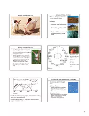
Most birds have approximately 175 different muscles mainly controlling the wings skin and legs The largest muscles in the bird are the pectorals or the breast muscles which control the wings and make up about 15 25 of a ID: 1025478

medial lateral wing muscle lateral medial muscle wing insertion action extends origin dorsal humerus cranial proximal surface vein longus

Share:

Link:

Embed:

Download Presentation from below link

Download Presentation The PPT/PDF document "AVIAN MUSCLE INTRODUCTION" is the property of its rightful owner. Permission is granted to download and print the materials on this web site for personal, non-commercial use only, and to display it on your personal computer provided you do not modify the materials and that you retain all copyright notices contained in the materials. By downloading content from our website, you accept the terms of this agreement.


Presentation Transcript

1. AVIAN MUSCLE

2. INTRODUCTIONMost birds have approximately 175 different muscles, mainly controlling the wings, skin, and legs. The largest muscles in the bird are the pectorals, or the breast muscles, which control the wings and make up about 15 - 25% of a flighted bird’s body weight. They provide the powerful wing stroke essential for flight. The muscle ventral (underneath) to the pectorals is the supracoracoideus. It raises the wing between wingbeats. The supracoracoideus and the pectorals together make up about 25 – 35% of the bird's full body weight.The skin muscles help a bird in its flight by adjusting the feathers, which are attached to the skin muscle and help the bird in its flight maneuvers.There are only a few muscles in the trunk and the tail, but they are very strong and are essential for the bird. The pygostyle controls all the movement in the tail and controls the feathers in the tail. This gives the tail a larger surface area which helps keep the bird in the air.

3. AVIAN MUSCLE

4. Anatomy of avian

5.

6. MUSCLESORIGININSERTIONINNERVATIONACTION

7. UNIQUE MUSCLE CONFIGURATIONSPectoralis majorSupracoracoideusWing mechanics

8. Superficial pectoral (pectoralis)Origin: keel, clavicle, coracoid and sternal ribs Insertion: ventral pectoral crest, proximal humerus Innervation: pectoral nerves Action: downstroke of the wing, depresses leading edge of pars longus of the propatagialis tendon.Detach the pectoralis muscle from its mutliple sites of origin along the keel, beginning distally, and reflect it towards its point of insertion. The supracoracoideus lies deep and medial to the pectoralis -- note airsac that lies between the two muscles.

9. SupracoracoideusOrigin: sternum, clavicle and coracoid .Insertion: by means of a prominent tendon upon the lateral tubercle of the humerus. The tendon passes through the triosseal foramen. Action: elevates the wing

10.

11.

12. Coracobrachialis caudalis (ventralis)Origin: lateral surface of the coracoid.Insertion: medial (ventral) tubercle of the humerus .Action: depresses the wing along with the pectoralis muscle Clinically: frequently torn with shoulder dislocations rendering the bird significantly impaired for flight

13. Propatagialis (pars brevis and pars longus)Origin: Distal end of the coracoid boneInsertion: 2 partsPars longus: inserts on the carpo-metacarpus and forms the leading edge of the wing. The patagium is attached to it.Pars brevis: attaches to apeunerosis arising from dorsal musculature in the proximal 1/3 of the antebrachium. Actions:Tenses the patagium (elastic web)Extends the carpusServes as a check ligament to prevent overextension of the elbow joint.Clinically: frequently contracts following humeral fractures which limits extension of the wing.Origin: Distal end of the coracoid boneInsertion: 2 parts1.Pars longus: inserts on the carpo-metacarpus and forms the leading edge of the wing. The patagium is attached to it.2.Pars brevis: attaches to apeunerosis arising from dorsal musculature in the proximal 1/3 of the antebrachium. Actions:-Tenses the patagium (elastic web)-Extends the carpus-Serves as a check ligament to prevent overextension of the elbow joint.

14. BrachialisOrigin: distal end of the humerusInsertion: proximal portion of the ulnaAction: flexes the elbow jointThere are four muscles of the antebrachium that are important; all are located on the dorsal or lateral aspect

15.

16. Muscle of Pectoral Limbmodifications for flight.comprise 35% of the body weight. shifted craniad under the center of lift have long tendons reaching distally to the portion of the limb controlled by them

17.

18. MUSCULATURE OF PECTORAL LIMBS : HUMERUSBicepsTricepsDeltoid

19. Biceps brachiiOrigin: distal end of coracoid and bicipital crest of humerusInsertion: radius and ulna dorsal aspect Actions: flexes the elbow joint and extends the shoulder

20. Triceps brachiiOrigin: One head from scapula, the other from proximal humerus Insertion: olecranon of the ulna Action: extension of the elbow joint

21. Deltoideus majorOrigin: scapula and clavicle Insertion: dorsal surface of humerus Action: elevates the wing

22. MUSCULATURE OF PECTORAL LIMB : ANTEBRACHIUMExtensor carpi radialisCommon digital extensorExtensor metacarpi ulnarisSupinator

23. Extensor metacarpi radialisOrigin: lateral epicondyle of the humerus.Innervation:radial vein,patagial veinInsertion: dorsal surface of the carpometacarpus Action: extends the metacarpus

24. Extensor digitorum communis (common digital extensor)Origin: lateral epicondyle of the humerus and extend to medial forearm.Innervation:basilic,deep brachial vein.Insertion: dorsal surface of the proximal phalanx of digit III with a short link to the alula Action: extends the carpus, metacarpus, and alula

25. Extensor metacarpi ulnaris (ulnaris lateralis) Origin: lateral epicondyle of the humerus,posterior border and medial side of ulnar and insert at the base of metacarpus.Innervation:ulnar vein,deep brachial vein,basilic vein.Insertion: carpometacarpus Action: flexes the metacarpus (when wing is extended ) only flexor located on lateral (dorsal) surface of the wing.

26. SupinatorOrigin: lateral epicondyle of the humerus (deep) and curved to upper third of radius.Inervation:radial vein,patagial veinbasilic vein,brachial vein.Insertion: cranial surface of the radius Action: elevates the cranial border of the antebrachium and supinates (rotates upward) the cranial edge of the wing

27. PELVIC LIMBPelvic Limb Myology: Movement to center of mass with long tendons going to extemities; tendons are often ossified.

28. MUSCULATURE OF PELVIC LIMB : HIP & FEMURQuadriceps equivalentIliotibialis cranialisIliotibialis lateralisIliofibularisFemerotibialus externus

29. Iliotibialis cranialis (sartorious)Origin: cranial portion of the iliac crest. A strap-shaped muscleInsertion: medial aspect of patella and tibiotarsusAction: flexes the hip joint and extends the stifleClinical: cranial edge serves as a landmark for endoscopic entry point for sexing and general abdominal examination.

30.

31. Iliotibialis lateralisOrigin: most of the length of the iliac crest. . A sheet-like muscle.Insertion: joints the fascia of the femorotibilais and inserts with that muscle upon the patella and tibiotarsusAction: flexes the hip and extends the stifleClinical: large muscle in vultures and fowl, small in psittacines and raptors. Surgical approach to femur lies along its caudal border

32. IliofibularisOrigin: iliac crest caudal to the acetabulumInsertion: proximal end of the fibulaAction: flexes the stifleClinical: Surgical approach to the femur lies between this muscle and iliotibialis lateralis. A large neurovascular bundle lies deep and must be preserved

33.

34. Femorotibialis externusOrigin: adherent to most of the length of the craniolateral femurInsertion: patella and proximal tibiotarsusAction: extends the stifleClinical: typically needs to be periosteally elevated from shaft of femur during surgical approaches.

35. MUSCULATURE OF PELVIC LIMB : TIBIOTARSAL REGION Gastronemius – 3 headsFibularis longusTibialis cranialisExtensor digitorum longus

36. Gastrocnemius complex (3 heads)Origins: 1) lateral head -- lateral femoral condyle2) intermediate head -- area near medial femoral condyle3) medial head -- popliteal area fascia and medial femoral condyleInsertion: All three heads form a common tendon that inserts on the prominent hypotarsal protuberanceAction: strong extender of the hock jointClinical: medial surgical approach to tibiotarsus occurs along cranial edge of medial grastroc.

37.

38. Fibularis longus (peroneus longus)Origin: patella, cranial and lateral cnemial crests and the lateral fibulaInsertion: caudal tarsometarsus and tendon of the flexor perforatus digiti III.Action: extends the intertarsal joint (hock)

39. Tibialis cranialis: lateral and medial headsOrigins: lateral head- lateral condyle of the femur, medial head - lateral and medial cnemial crests and rotular crestInsertion: two heads unite to form a common tendon inserting on the dorsal metatarsal groove.Actions: flexes the hock

40. Extensor digitorum longusOrigin: cranial surface of the tibiotarsus between the medial and lateral cnemial crestsInsertion: the dorsum of the distal-most phalanx of digits II-IVAction: extends the forward digits

41.

42.