

2 TUMORS OF THE BREAST Fibroadenoma Is the most common benign neoplasm of the female breast An increase in estrogen activity is thought to contribute to its development and indeed similar lesions may appear with fibrocystic changes ID: 928461
Download Presentation The PPT/PDF document "BREAST PATHOLOGY LEC. 1" is the property of its rightful owner. Permission is granted to download and print the materials on this web site for personal, non-commercial use only, and to display it on your personal computer provided you do not modify the materials and that you retain all copyright notices contained in the materials. By downloading content from our website, you accept the terms of this agreement.


Slide1
BREAST
PATHOLOGY
LEC. 1
Slide22
Slide3TUMORS OF THE BREAST
Fibroadenoma Is the most common benign neoplasm of the female breast. An increase in estrogen activity is thought to contribute to its development, and indeed similar lesions may appear with fibrocystic changes (fibroadenomatoid changes). The peak incidence is in the third decade of life.
3
Slide4Gross features
The tumor is usually solitary, well-defined, and freely movable. It is variable size & may reach up to 10 cm in diameter. Larger tumors are referred to as giant fibroadenoma. It is firm, with a uniform tan-white color on cut section .
4
Slide5Microscopically
There is a loose fibroblastic stroma containing epithelium-lined duct-like spaces of various forms and sizes. The duct-like spaces are lined with single or multiple layers of benign epithelial cells having an intact basement membrane.
5
Slide6In some lesions the
ductal spaces are open, and fairly regular (pericanalicular fibroadenoma), whereas in others they are compressed by the proliferation stroma and thus appear as slits or irregular, star-shaped structures (intracanalicular fibroadenoma) .
6
Slide7Cytogenetic studies reveal that the
stromal cells are monoclonal and thus represent the neoplastic element of these tumors. It is possible that the neoplastic stromal cells secrete growth factors that induce proliferation of epithelial cells.
7
Slide8The lesion consists of a proliferation of
intralobular stroma surrounding and often pushing and distorting the associated epithelium. The border is sharply delimited from the surrounding tissue.
Fibroadenoma
8
Slide9Phyllodes
Tumors Are much less common than fibroadenomas. They are thought to arise from the periductal stroma. Most grow to large, possibly massive size; the patient typically has a history of a rapidly growing palpable breast mass.
9
Slide10Grossly
, the tumors are lobulated and cystic and on sectioning exhibit leaf-like clefts and slits. The latter feature is responsible for the naming them as phyllodes tumors (Greek for "leaflike"). Microscopically, there is expansion and increased cellularity
of the
stromal
component.
10
Slide11The most ominous change is the appearance of anaplasia and high mitotic activity, usually with invasion of adjacent breast tissue (malignant phyllodes tumor). Most of these tumors remain localized and are cured by excision; malignant lesions may recur, but they also tend to remain localized. Only the most malignant, about 15% of cases, metastasize to distant sites.
11
Slide12The most ominous change is the appearance of anaplasia and high mitotic activity, usually with invasion of adjacent breast tissue (malignant phyllodes tumor). Most of these tumors remain localized and are cured by excision; malignant lesions may recur, but they also tend to remain localized. Only the most malignant, about 15% of cases, metastasize to distant sites.
12
Slide13Microscopic appearance of
phylloides tumor. The tumor has the "intracanalicular" pattern of growth.
Phyllodes
tumor
13
Slide14Intraductal
Papilloma Is a neoplastic papillary growth within a duct. Most lesions are solitary, found within the principal lactiferous ducts or sinuses, thus present clinically with a serous or bloody nipple discharge and/or a small subareolar nodule and rarely, nipple retraction.
Grossly
,
the tumors are usually solitary and less than 1 cm in diameter.
14
Slide15Microscopically
, They consist of delicate, branching papillary growths within a dilated duct (or cyst). Each papillary projection has a connective tissue core covered by double layer of cells; outer cuboidal epithelial cells that overlies myoepithelial cells. Some cases display multiple papillomas (intraductal
papillomatosis
)
.
The latter sometimes become malignant, whereas the solitary
papilloma
is virtually benign.
15
Slide16Papillary carcinoma is distinguished by:
1. The absence of a myoepithelial component and 2. The epithelial cells show either severe cytologic atypia or monotonous ductal morphology.
16
Slide17Intraductal
papilloma. A, Low-power appearance showing complex arborizing
papillary architecture.
High-power view showing dual cell composition, with a well-defined row of
myoepithelial
cells (arrow).
17
Slide18CARCINOMA (BRCA)
Slide19Breast carcinoma in the USA ranks second only to lung cancer as a cause of cancer death in women; in our country it probably ranks first. Despite advances in diagnosis and treatment, almost 25% of women who develop
this neoplasm will die of the disease.19
Slide20This has incite an intense study of the possible causes and origins of this form of cancer and of ways to diagnose it early enough to permit cure. 75% of women with breast cancer are older than age 50; only 5% are younger than the age of 40. 20
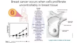
Slide21Epidemiology and Risk Factors
A large number of risk factors have been identified that modify the likelihood of developing BRCA . These risk factors are divided into well-established and less well-established groups.Geographic Variations: the risk for BRCA is significantly higher in North America and northern Europe than in Asia and Africa.
For example, the incidence and mortality rates are five times higher in the United States than in Japan.
21
Slide22These differences seem to be environmental rather than genetic in origin, because migrants from low-incidence to high-incidence areas tend to acquire the rates of their adoptive countries, and vice versa
. Diet, reproductive patterns, and nursing habits are thought to be involved.2. Age: BRCA is uncommon in women younger than age 30. Thereafter, the risk steadily increases throughout life reaching a plateau after menopause.
22
Slide233.
Genetics and Family History: up to 10% of BRCA are related to specific inherited mutations. Women are more likely to carry a BRCA susceptibility gene if they have:- a. BRCA before menopause .b. bilateral cancer. c. Other associated cancers (e.g., ovarian cancer
).
d.
A
significant family history (i.e., multiple relatives affected before menopause) .
23
Slide24About 50% of women with hereditary BRCA have mutations in gene
BRCA1, and an additional 30% have mutations in BRCA2. Both BRCA 1 & 2 seem to be involved in DNA repair and act as tumor suppressor genes. Cancer arises when both alleles are inactive (defective); one due to a germ-line mutation and the second by a subsequent somatic mutation.
It is possible that other mechanisms, such as
methylation
of regulatory regions
, act to inactivate the genes in sporadic (nonhereditary) cancer.
24
Slide253.
Prolonged exposure to exogenous estrogens: short-term use of combined estrogen plus progestin as hormonal replacement therapy in postmenopausal women is associated with an increased risk of breast cancer. However, a large study concluded that birth control pills do not increase the risk of breast cancer.
25
Slide264.
Ionizing radiation: e.g. to the chest increases the risk of breast cancer. Only women irradiated before age 30, during breast development, seem to be affected. For example 20% to 30% of women irradiated for Hodgkin lymphoma in their teens and 20s develop breast cancer, but the risk for women treated later in life is not elevated.26
Slide275.
Other less well-established risk factors, such as obesity, alcohol consumption, and a diet high in fat, have been implicated in the development of breast cancer on the basis of population studies. Obesity is a recognized risk factor in postmenopausal women. Pathogenesis
The exact cause of breast cancer remains unknown. However, three sets of influences seem to be important:
1. Genetic changes.
2. Hormonal influences.
3. Environmental factors
.
27
Slide28Genetic Changes
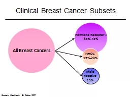
In addition to those producing the well-established familial BRCA, genetic changes have also been implicated in the genesis of sporadic (nonfamilial) breast cancer. Mutations affecting proto-oncogenes and tumor suppressor genes in breast epithelium contribute to the malignant transformation process. Overexpression of the HER2/NEU
proto-oncogene has been found to be amplified in up to 30% of invasive breast cancers.
28
Slide29This gene is a member of the epidermal growth factor receptor family, and its
overexpression is associated with a poor prognosis. Similarly, amplification of RAS and MYC genes has also been reported in some human breast cancers. Mutations of the well-known tumor suppressor genes
RB
and
p53
may also be present
.
Multiple
acquired genetic alterations seem to be involved in the sequential transformation of a normal epithelial cell into a cancerous cell.
29
Slide30Hormonal Influences
Endogenous estrogen excess clearly has a significant role. This is supported by the following observations:-1. Many of the risk factors mentioned (long duration of reproductive life, nulliparity, and late age at birth of first child) imply increased exposure to estrogen peaks during the menstrual cycle. 2. Functioning ovarian tumors that elaborate estrogens are associated with breast cancer in postmenopausal women.
30
Slide313. Estrogens stimulate the production of growth factors by normal breast epithelial cells and by cancer cells
. It seems that estrogen (and progesterone) receptors normally present in breast epithelium, and often in breast cancer cells, may interact with growth promoters (such as transforming growth factor α) produced by human breast cancer cells, to create an autocrine mechanism of tumor development.31
Slide32Environmental
factors: Are suggested by the variable incidence of breast cancer in genetically identical groups and the geographic differences in prevalence. Other important environmental variables include irradiation and exogenous estrogens, described earlier.
32
Slide33Pathological features of BRCA
About 4% of women with breast cancer have bilateral primary tumors. The locations of the tumors within the breast are: Upper outer quadrant 50% Central sector (subareolar
) 20%
Upper inner 10%
Lower outer 10%
Lower inner 10%
33
Slide34Breast cancers are classified into those that have not penetrated the limiting basement membrane (non invasive) and those that have (invasive). The chief forms of carcinoma of the breast are classified as follows:
Non invasive:-
Ductal carcinoma in situ (DCIS).
Lobular carcinoma in situ (LCIS).
Invasive (infiltrating):-
Invasive ductal carcinoma ("not otherwise specified" or NOS).
Invasive lobular carcinoma.
Medullary carcinoma.
Colloid carcinoma (mucinous carcinoma).
Tubular carcinoma.
Others.
34
Slide35Of these,
invasive ductal carcinoma is the most common. Because it usually has an abundant fibrous stroma, it is also referred to as scirrhous carcinoma. There are two types of noninvasive breast carcinoma: ductal
carcinoma in situ (
DCIS) and lobular carcinoma in situ (LCIS)
both usually arise from the terminal duct lobular unit (TDLU).
Both are confined by a basement membrane and do not invade into
stroma
or
lymphovascular
channels.
35