
Cells amp Plasma Cells 1 RBC Erythroid 2 WBC Myeloid Neutrophils Basophils Eosinophils Lymphoid cells Lymphocytes Macrophage system ID: 914444
Download Presentation The PPT/PDF document "Blood Composition of blood" is the property of its rightful owner. Permission is granted to download and print the materials on this web site for personal, non-commercial use only, and to display it on your personal computer provided you do not modify the materials and that you retain all copyright notices contained in the materials. By downloading content from our website, you accept the terms of this agreement.


Slide1
Blood
Composition of blood
Cells & Plasma
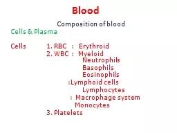
Cells 1. RBC :
Erythroid
2. WBC : Myeloid
Neutrophils
Basophils
Eosinophils
:Lymphoid cells
Lymphocytes
: Macrophage system
Monocytes
3. Platelets
Slide2Hemopoiesis
Committed stem cells
Slide3Hemopoiesis
Pleuripotent
hemopoietic
stem cell differentiate into Committed stem cells maturing in a particular cell eg Colony forming unit (CFU)erythrocyte will mature into an erythrocyte . GM-CFU into granulocytes & Monocyte.
Growth promoters like Interleukin-3 induce growth of all the cells in bone marrow.
Differentiation factors like GM –CSF stimulates the differentiation of
monocytes
and except
basophil
all the granulocytes.
Lymphocytes are differentiate & mature in Thymus (T cell) /
bursal
equivalent(B cell)
eg
liver in mid fetal life & bone marrow in late fetal life & after birth.
Slide4Erythropoiesis
Areas
: yolk sac: primitive embryo
Liver :mid gestation
Spleen & LN also contributeBone marrow : After birth & adultStages of
erythropoiesis
Pleuripotent
& Committed cells ( CFU- E)
Proerythoblast: First identifiable cell of series derived from CFU-E & it give rise basophilic erythroblast (early normoblast) with little Hb. Next generation cell Polychromatophilic erythroblast ( intermidiate normoblast)has Hb saturation of 34% & this gives Orthochromatic erythroblast (Late Normoblast) & subsequently Reticulocyte (Golgi body & mitochondrion turns into reticulum) which disappears within 1-2 days. 1-2 % of circulating RBC are actually reticulocytes.
Early
normoblast
Intermediate
normoblast
Late
Normoblast
Slide5Red Blood Corpuscle (RBC)
RBC : biconcave disk ,7.5
μ
M diameter 2.5
μM thick at periphery , contains 29 pg
Hb
. 5.4 million/
μ
L (male) 4.8 (Female)in number
Characteristics of RBC Variable CalculatnMale Female
Hematocrit
47%
42%
RBC Count
5.4
m/
μ
L
4.8 m/μLHemoglobin16 G%14 G%Mean Corpuscular Volume (MCV)Hct x10 RBC count87 fL 87 fLMean Corpuscular Hemoglobin MCHHb x10RBCcount29 pG29 pGMean corp Hb Conc MCHC Hbx100Hct 34%34%
Slide6Role of erythropoietin, B12 & Folate in
Erythropoiesis
Hypoxia causes increase in erythropoietin production from kidney, erythropoietin in turn enhances RBC production
Formation of Erythropoietin. (EP) 90%
erythropoietin in kidney 10% in liver. In the kidneys the erythropoietin is formed in tubular epithelial cells.
Ep
stimulates
hemopoietic
stem cell to proliferate into
proerythroblast. Vitamin B12 and folic acid are essential for the synthesis of thymidine triphosphate, & DNA ,hence, deficiency cause failure of nuclear maturation & cell division. The erythroblastic cells of bone marrow, fail to proliferate rapidly, produce macrocytes, with weak cell membrane & oval in shape cells.
Slide7Features of Iron, B12 & folate deficiency
anemias
Pathology
Hemoglobin
RBC Count
MCV
MCH
MCHC
Iron deficiencyMale< 13.6Female < 12.0 LessMale <4.3Female 3.5 Less < 75 μ Lreduced < 25 pGreduced
< 27 reduced
B12 & folic acid deficiencyLess
Less > 110
Normal/ reducedNormal
Slide8Bone marrow
Normoblast
Megaloblast
Slide9Classification of Anemia according to Underlying Cause
Blood Loss
Acute: trauma
Chronic: lesions of gastrointestinal tract, gynecologic disturbances
Defect in RBC Increased Destruction (Hemolytic Anemias
)
(A) Intrinsic (
intracorpuscular
) abnormalities
(a)Hereditary membrane abnormalities Membrane skeleton proteins: spherocytosis, elliptocytosis (b) Hereditory Enzyme deficiencies Glycolytic enzymes: pyruvate kinase, hexokinase,Enzymes of hexose monophosphate shunt: glucose-6-phosphate dehydrogenase, glutathione synthetase.
Slide10Classification by underlying Mechanism
Deficiency of dietary factors/ Abnormal
Hb
Synthesis
Iron : Microcytic Hypochromic
B12 :
Macrocytic
or
Megaloblatic
anemiaFolic acid : Macrocytic or Megaloblatic anemia
Slide11Hereditary Spherocytosis
In HS primary abnormality is in supportive skeleton on IC face of RBC wall.
Spectrin
, linked to membrane at two points: through
ankyrin & band 4.2 to membrane protein band 3; & through band 4.1 to protein glycophorin. Horizontal spectrin
-
spectrin
&
spectrin
-intrinsic membrane protein interactions stabilize membrane & are responsible for shape, strength, flexibility of RBC.
Slide12Hereditary Spherocytosis
Most common pathogenic feature of HS is mutation particularly of band3,ankyrin &
spectrin
gene. In all types of HS red cell wall stability is reduced , consequently lose membrane fragments while retaining most of their volume. As a result, ratio of surface area to volume of HS cells decreases until the cells become spherical.
Slide13Disorders of Hb & RBC production
Hemoglobin
Deficient
globin
synthesis :Thalassemia syndrome Abnormal
globin
synthesis: Sickle cell anemia
RBC production
Failure of erythroblast maturation : B12 & Folate deficiencyDefect of Heme synthesis Iron deficiency The most common cause of anemia in India followed by B12 & folate deficiency
Slide14Hemoglobin
Hemoglobin is made up of 4 subunits, each have a
Heme
moiety & polypeptide chain.
HBA has one pair of α & one pair of β
globin
chain(2
α
2 β)HbA2 (2.5%) of Hb has 2α2δHbA1c glycated by glucose in diabetics if > 6.9% indicate poor control of blood sugarFetal Hb (2α2γ) has more affinity for O2 since it bind less avidly to 2,3-DPG and carries more O2 for a given pO2.
Slide15Reactions of Hemoglobin
Each of four iron atoms in hemoglobin can reversibly bind one O
2
molecule. Iron is in ferrous state, so reaction is
oxygenation, not oxidation. Because it contains 4 deoxyhemoglobin (Hb)
units,Hb
molecule represented as Hb
4
,& it actually reacts with four molecules of O
2 to form Hb4O Hb4 + O2↔ Hb4O2, Hb4O2+O2 ↔ Hb4O4 H b4O4+O2 ↔ Hb4O6 Hb4O6 +O2 ↔Hb4
O8 Deoxygenated Hb,
globin is tightly bound in tense state so low affinity for O2. Binding of one O2 loosens the binding & increase affinity for O
2, 500 times when all 4 Hb are bound with O2
Slide16Hemoglobin reactions
Methemoglobin
: Oxidizing agent & drugs convert
Hb
to methHb leading to dusky color of skin. Normally meth hemoglobin formed is converted to Hb by NADH-meth hemoglobin reductase
,. Absence of which in children cause congenital
methemoglobinemia
.
Carboxyhemoglobin
: Hemoglobin has more affinity for Carbon monoxide than for O2 which replaces O2 (CO posioning) withreduced O2 carrying capacity of Hb.
Slide17Sickle cell anemia
In
HbS
, substitution of valine
for glutamic acid at 6th position of β-chain, produces HbS.
Homozygotes
all
HbA
replaced by
HbS. Heterozygote about half is replaced. Deoxygenation, HbS molecules crystallize which distort RBC as elongated crescent or sickle. Sickling initially reversible upon reoxygenation; Later on cell wall damage occurs with each episode of sickling, & finally cells accumulate calcium, lose potassium and water, and become irreversibly sickled.
Slide18Thalassemia
Inherited disorder caused by mutations that decreases synthesis of α- or β-
globin
chains. So deficiency of hemoglobin, red cell abnormalities due to excess of other unaffected globin chain. The α chains are encoded by two α-
globin
genes, which lie in tandem on chromosome 11, while the β chains are encoded by a single β-
globin
gene located on chromosome 16. The mutations that cause
thalassemia are particularly common among Mediterranean, African, and Asian populations.