
The elementary particle that defines light is the photon There are 3 basic dimensions of light Intensity amplitude which is related to the perception of brightness Frequency wavelength perceived as colour ID: 1024651
Download Presentation The PPT/PDF document "What is light? Light is electromagnetic ..." is the property of its rightful owner. Permission is granted to download and print the materials on this web site for personal, non-commercial use only, and to display it on your personal computer provided you do not modify the materials and that you retain all copyright notices contained in the materials. By downloading content from our website, you accept the terms of this agreement.


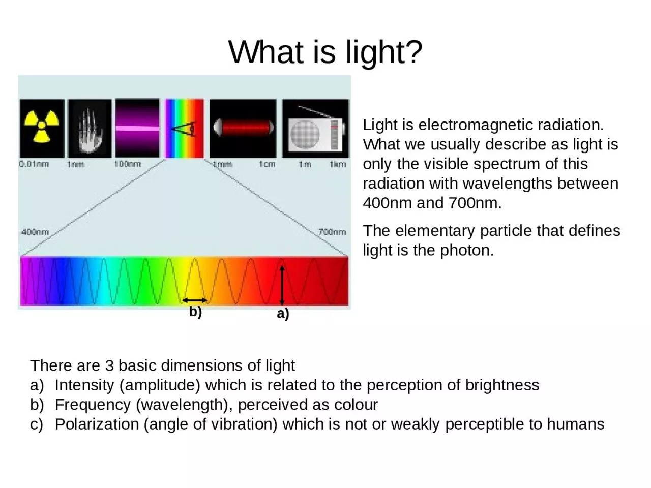
1. What is light?Light is electromagnetic radiation. What we usually describe as light is only the visible spectrum of this radiation with wavelengths between 400nm and 700nm.The elementary particle that defines light is the photon.There are 3 basic dimensions of lightIntensity (amplitude) which is related to the perception of brightnessFrequency (wavelength), perceived as colourPolarization (angle of vibration) which is not or weakly perceptible to humansa)b)
2. What is a microscope?
3. Starting with use of a simple lens in ancient times, to the first compound microscope around 1590, and up to the microscopes you are using in 7th grade life science, the microscope has allowed scientists to make discoveries about the “invisible world.”The microscope has become one of the most recognizable symbols of science.
4. Micro- = “small”; -scope = “to look at”Photographs of cells are taken using a microscope, and these pictures are called micrographs.
5. What is a microscope?Theoretically a microscope is an array of two lenses.Objective lensTube lensEyepiece lensFocal planeImage planeClassic compound microscopeImage plane
6. Your friend - the objective
7. What is magnification?Magnification is a measure of how much larger a microscope (or set of lenses within a microscope) causes an object to appear.Magnification is defined by the magnification by the objective x the magnification by eyepieceBUT maximum magnification does not mean maximum resolution!
8. What is resolution? Resolution describes the minimal distance of two points that can be distinguished. The resolution of a microscope or lens is the smallest distance by which two points can be separated and still be distinguished as separate objects. The smaller this value, the higher the resolving power of the microscope and the better the clarity and detail of the image. Picture taken from http://microscopy.fsu.edu/primer/anatomy/numaperture.html
9. NA = n sin What is the numerical aperture?NA is an estimate of how much light from the sample is collected by the objectiveα1α2Coverslip (n = 1.5)Glass slide (n = 1.5)Oil (n = 1.5)Air (n = 1.0)n = refractive indexα = angle of incident illuminationObjective lens
10. The higher the numerical aperture of a lens, the better the resolution of a specimen will be which can be obtained with that lens.d=0.5 λ/n sin ƟWhere d= resolutionΛ = wavelength of light used
11. Numerical aperture, NOT magnification determines resolution! Increasing NAA lens with a larger NA will be able to visualize finer details and will also collect more light and give a brighter image than a lens with lower NA.
12. Microscopy
13. Light (Optical) MicroscopyVisible light is uses.Glass lens are usedAdvantage: It can often be performed on living cells, so it’s possible to watch cells carrying out their normal behaviors (e.g., migrating or dividing) under the microscope.
14. PrincipleWhen a ray of light passes from one medium to another it bends by phenomena called refraction.Bending of light slows the speed.The bending of light is determined by refractive index of the medium.
15. Types of Light MicroscopesBright field Light MicroscopePhase Contrast Light MicroscopeDark-Field Light MicroscopeFluorescence Light Microscope
16. Contrasting techniquesBrightfieldDarkfieldPhase contrastDICTaken from: http://fig.cox.miami.edu/~cmallery/150/Fallsyll.htm Fibroblast grown in culture
17. BrightfieldPiece of artificially grown skin (www.igb.fhg.de/.../dt/PI_BioTechnica2001.dt.html )Cross section of sunflower root(http://www.zum.de/Faecher/Materialien/beck/12/bs12-5.htm)Principle: Light is transmitted through the sample and absorbed by it.Application: Only useful for specimens that can be contrasted via dyes. Very little contrast in unstained specimens. With a bright background, the human eye requires local intensity fluctuations of at least 10 to 20% to be able to recognize objects.
18. Typical Classroom Microscope
19. Eyepiece
20. EyepieceAlso known as the ocularContains the first lens you look through - usually a magnification of 10xLocated on the top of the body tube
21. Objective Lenses
22. Objective LensesUsed in combination with the eyepiece to provide a range of magnificationMagnification ranges from 40x to 400xLocated on the nose-piece at the bottom of the body tube
23. Nosepiece
24. NosepieceHolds the objective lensesRotates to enable magnificationLocated at the bottom of the body tube
25. Arm
26. ArmSupports the upper parts of the microscopeUsed to carry the microscopeWhen carrying a microscope, always have one hand on the arm and one hand on the base. Use two hands!!
27. Base
28. BaseSupports the whole microscopeUsed to carry the microscopeWhen carrying a microscope, always have one hand on the arm and one hand on the base. Use two hands!!
29. Stage
30. StageSupports the slideThe slide contains the specimen or object that you are viewing with the microscope.
31. Stage Clip
32. Stage ClipHelps to hold the slide in placeUsually one on each side of the hole (stage opening) = 2 stage clipsThe stage opening allows light to pass from the light source to the lenses.
33. Light Source
34. Light SourceProvides light necessary for viewing the specimenUsually either a mirror or illuminatorSends light through the stage opening to the diaphragm
35. Diaphragm
36. DiaphragmWheel or lever located below the stage openingRegulates the amount of light that can enter the lensesMay need to be adjusted based on the thickness of the specimen being studied
37. Coarse Adjustment Knob
38. Coarse Adjustment KnobRaises and lowers the stage or objective lensesUsed only when focusing the low power (4x) objective lens
39. Fine Adjustment Knob
40. Fine Adjustment KnobRaises and lowers the stage or objective lenses a small distance for exact focusingUsed when focusing the medium power (10x) and high power (40x) objective lenses
41. Let’s Review...
42. Phase contrastPrinciple: Incident light [Io] is out of phase with transmitted light [I] as it was slowed down while passing through different parts of the sample and when the phases of the light are synchronized by an interference lens, a new image with greater contrast is seen.II0Phase ringalignednot alignedPhase stopshttps://www.youtube.com/watch?v=fCZw4X7V5Pw
43. Phase contrastApplication: Phase contrast is the most commonly used contrasting technique All tissue culture microscopes and the time-lapse microscopes are set up for phase.wrong phase stopbrightfieldright phase stop
44. ApplicationsDetermine morphologies of living cells such as plant and animal cellsStudying microbial motility and structures of locomotionTo detect certain microbial elements such as the bacterial endospores
45. Fluorescencehttps://www.youtube.com/watch?v=SfzmW7EMdLE
46. ApplicationsUsed in the visualization of bacterial agents such as Mycobacterium tuberculosis.Used to identify specific antibodies produced against bacterial antigens/pathogens in immunofluorescence techniques by labeling the antibodies with fluorochromes.Used in ecological studies to identify and observe microorganisms labeled by the fluorochromesIt can also be used to differentiate between dead and live bacteria by the color they emit when treated with special stains
47. Confocal Microscopehttps://www.youtube.com/watch?v=QFtZFbug1SA
48. DarkfieldPrinciple: The illuminating rays of light are directed through the sample from the side by putting a dark disk into the condenser that hinders the main light beam to enter the objective. Only light that is scattered by structures in the sample enters the objective. Application: People use it a lot to look at Diatoms and other unstained/colourless specimensBrightfieldDarkfieldSymbiotic Diatom colony(www1.tip.nl/~t936927/making.html)
49. ApplicationsIt is used to visualize the internal organs of larger cells such as the eukaryotic cellsIdentification of bacterial cells with distinctive shapes such as Treponema pallidum, a causative agent of syphilis.https://www.youtube.com/watch?v=W9MhkmyfMLQ