
Assistant Professor MBBS Mphil Stimulus amp Modalities A stimulus is a change detectable by the body Stimuli exist in a variety of energy forms or modalities such as heat light sound pressure and chemical changes ID: 774998
Download Presentation The PPT/PDF document " SENSORY NERVOUS SYSTEM Dr. Ayisha Qure..." is the property of its rightful owner. Permission is granted to download and print the materials on this web site for personal, non-commercial use only, and to display it on your personal computer provided you do not modify the materials and that you retain all copyright notices contained in the materials. By downloading content from our website, you accept the terms of this agreement.


Slide1
SENSORY NERVOUS SYSTEM
Dr. Ayisha Qureshi
Assistant Professor
MBBS,
Mphil
Slide2Slide3Stimulus & Modalities
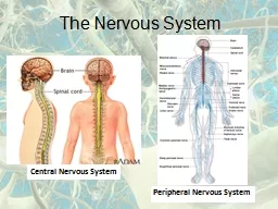
A stimulus is a change detectable by the body.Stimuli exist in a variety of energy forms, or modalities, such as heat, light, sound, pressure, and chemical changes.Sometimes we perceive sensory signals when they reach a level of consciousness, but other times they are processed completely at the subconscious level. All the information regarding all these senses is send to the CNS via AFFERENT NEURONS.
Slide4The conversion of stimulus energy into a graded potential is called Sensory transduction and is done by sensory receptors.
Because the only way
afferent
neurons can transmit information to the CNS about stimuli
is via
action potential
propagation,
these forms of energy must be converted into
electrical signals
.
Slide5Slide6Slide7SENSORY Receptors
Receptors are sensory afferent nerve endings that terminate in periphery as either part of a neuron or in the form of specialized capsulated structures. They act as biological transducers and convert various forms of energy acting on them into action potentials.
Slide8Classification of Receptors
Slide9Slide10Slide11Slide12Slide13Slide14Receptors
are present in the skin, the mucous membranes, fascia and deeper
parts of the body.
They are responsible for 4 different sensations:
Touch-pressure
Cold
Warmth and
Pain
The receptors are:
Encapsulated
receptors:
consist of multilayered capsules of connective tissue which surround a core of cells in which axons end after losing their myelin sheath.
-
Meissner’s
corpuscle:
sensitive to light touch
&
are rapidly adapting. Are present just below the epidermis in the palmer surface of fingers, lips, margins of the eyelids.
-
Pacinian
Corpuscle:
respond to vibration
&
deep pressure
&
is rapidly adapting. Present in deeper tissues and also in pleura, peritoneum, external genitalia and walls of many viscera. Also present in
periostium
, ligaments and joint capsules.
-
Krause’s end
bulbs:
occur in conjunctivae, papillae of lips and tongue.
Expanded
tips on sensory nerve endings:
-
Merkel’s discs:
which detects light, sustained touch and texture,
and
is slowly adapting
. Present in hairless skin e.g. fingertips.
-
Riffini’s
end organs:
in deeper layer of skin and deeper tissues, e.g.
periostium
, ligaments and joint capsules. They respond
to deep, sustained pressure and stretch of the skin, such as during a massage, and are slowly
adapting.
3.
Naked
or free nerve
endings:
are the most widely distributed receptors in the body and can be excited by touch, cold, warmth and pain.
Slide15SENSORY RECEPTORS
Pacinian Corpuscle
Free nerve ending’s
Slide16General properties of receptors
Slide17The following are the properties of the Sensory Receptors:
Receptor Potential.
Specificity of stimulus & the Adequate stimulus.
Effect of strength of stimulus.
Adaptation (also Desensitization).
Muller’s doctrine of specific nerve energies
Law of projection.
Threshold.
Sensory unit
Receptive field.
Slide18The changes in sensory receptor membrane potential is a graded potential called the receptor potential.
1. RECEPTOR POTENTIAL
Slide19SENSORY TRANSDUCTION
Transduction is the conversion of stimulus energy into information that can be processed by the nervous system, which is an action potential. Stimulus↓ Receptor (SENSORY TRANSDUCTION)↓Graded Potential (RECEPTOR POTENTIAL)↓Afferent Neuron ↓Action Potential
Slide20How is a physical or a chemical stimulus converted into achange in membrane potential?
Stimulus
(chemical/ mechanical/ thermal)
↓
Receptor which is either:
Specialized ending of the afferent neuron, OR
A separate receptor cell associated with a peripheral nerve ending.
↓
Membrane permeability altered
(usually by opening of ligand-gated or stimulus sensitive
cation
channels)
↓
A graded potential is generated. It is called RECEPTOR POTENTIAL.
↓
There is summation, and if the stimulus is strong, it leads to a greater permeability change in the receptor which leads to a large Receptor potential.
↓
If the Receptor Potential is large enough
↓
An Action Potential is generated
(by opening of the voltage-gated Na channels in the afferent neuron next to the receptor)
Slide21Slide22THE INITIATION OF THE ACTION POTENTIAL
The initiation site of action potentials in an afferent neuron differs from the site in an efferent neuron or interneuron.In the other two types of neurons (interneuron & the efferent neuron), action potentials are initiated at the axon hillock located at the start of the axon next to the cell body. However, in the afferent neuron, action potentials are initiated at the peripheral end of fiber next to the receptor, a long distance from the cell body.
Slide232. SPECIFICITY OF STIMULUS & ADEQUATE STIMULUS
Slide24If all stimuli are converted to action potentials in sensory neuronsand all action potentials are identical, how can the central nervoussystem tell the difference between heat and pressure, orbetween a pinprick to the toe and one to the hand?
All stimuli once received by the receptor are converted into action potentials and all of them are carried by the afferent neurons. This
means that
the CNS
must distinguish four properties of a
stimulus to be able to specify a stimulus:
(
1) its
nature, or modality
and
(
2) its
location
(3) Intensity
(4) Duration
Slide25Adequate Stimulus
Each
sensory receptor has an
adequate stimulus
, a
particular form
of energy to which it is most responsive. For example,
thermoreceptors
are more sensitive to temperature changes than to pressure, and mechanoreceptors respond preferentially to stimuli that deform the cell
membrane, receptors
in the eye are sensitive to light, receptors in the ear to sound waves, and warmth receptors in the skin to heat energy. Because of this differential sensitivity of receptors, we cannot “see” with our ears or “hear” with our eyes.
Some receptors can respond weakly to stimuli other than their adequate stimulus, but even when activated by a different stimulus, a receptor still gives rise to the sensation usually detected by that receptor type. They respond to most other forms of energy if the intensity is high enough. Photoreceptors of the eye respond most readily to light, for instance, but a blow to the eye may cause us to “see stars”, an example of mechanical energy of sufficient force to stimulate the photoreceptors.
Sensory receptors can be incredibly sensitive to its preferred stimulus.
Slide26Modality/ Nature of the stimulus
The 1:1 association of a receptor with a sensation is called labeled line coding. Stimulation of a cold receptor is always perceived as cold, whether the actual stimulus was cold or an artificial depolarization of the receptor. The blow to the eye that causes us to see a flash of light is another example of labeled line coding. A blow to the eye is seen as “white light” as the photoreceptors were stimulated.
Slide27The table summarizes
how the CNS is informed of the type (what?), location (where?), and intensity (how much?)of a stimulus.
Slide28Location of the stimulus
The location of a stimulus is also coded according to which receptive fields are activated. The sensory region supplied by a single sensory neuron is called a receptive field. For example, touch receptors in the hand project to a specific area of the cerebral cortex. Experimental stimulation of that area of the cortex during brain surgery is interpreted as a touch to the hand, even though there is no contact. Also, lateral inhibition of the less activated regions leads to release of inhibitory NT that inhibits the region around the stimulated area. The contrast leads to a better localization of the stimulated area. (Tactile localization)
Slide29Receptive fields and convergence
Slide303. Effect of Strength & Duration of Stimulus:
Slide31For individual sensory neurons, intensity
discrimination begins
at the receptor. If a stimulus is below threshold, the
primary sensory
neuron does not respond. Once stimulus
intensity exceeds
threshold, the primary sensory neuron begins
to produce
action potentials.
As
stimulus intensity increases, the
receptor potential
amplitude (strength) increases in proportion,
and the
frequency
of action potentials in the primary sensory
neuron increases
, up to a maximum
rate.
Similarly
, the duration of a stimulus is coded by the
duration of
action potentials in the sensory neuron. In general,
a longer
stimulus generates a
longer series
of action potentials
in the
primary sensory
neuron
Slide32Slide33It is the decrease in response of receptors on being continuously stimulated.
4. ADAPTATION
also called Desensitization.
Slide34When a stimulus persists continuously, some receptors adapt, or cease to respond. Thus, the receptor “adapts” to the stimulus by no longer responding to it to the same degree. Receptors fall into one of two classes, depending on how they adapt to continuous stimulation: Tonic receptorsPhasic receptors
Slide35Types of receptors based on their adaptation
TONIC RECEPTORS
Tonic Receptors are slowly adapting receptors that respond rapidly when first activated, then slow down and maintain their response. Pressure sensitive baroreceptors, irritant receptors, and some tactile receptors and proprioceptors fall into this category. In general, the stimuli that activate tonic receptors are parameters that must be monitored continuously by the body. It is important that these receptors do not adapt to a stimulus and continue to generate action potentials to relay this information to the CNS.
PHASIC RECEPTORS
Phasic receptors are
rapidly adapting
receptors that respond when they first receive a stimulus but stop responding if the strength of the stimulus remains constant.
Many of the tactile receptors in the skin belong to this class.
Some phasic
receptors, most
notably the
Pacinian corpuscle,
respond with
a slight depolarization called the
off
response
when
the stimulus is
removed. They are important in situations where it is important to signal a change in stimulus intensity rather than the status quo information.
When
you put something
on, you
soon become accustomed to it, because
of these
receptors’ rapid adaptation. When you
take the
item off , you are aware of its removal
because of
the
“off”
response.
Slide36the nature of perception of a stimulus by the cns is defined by the pathway over which the sensory information is carried. Hence, the origin of the sensation is not important.
5. MULLER’S DOCTRINE OF SPECIFIC NERVE ENERGIES &
Slide37Stimulation of nerve fiber anywhere along its course produces the specific sensation in the area of the body from where it originated.
6. LAW
OF PROJECTION
Slide38All receptors need a minimum strength of stimulus to start showing activity; this strength is called the threshold.
6. THRESHOLD
Slide39The sensory unit is a single primary afferent nerve including all its peripheral branches.
7
. SENSORY UNIT
Slide40THE AREA OF THE BODY WHOSE SENSORY NERVE SUPPLY COMES FROM a single SENSORY UNIT IS CALLED A RECEPTIVE FIELD.
8. RECEPTIVE FIELD.
Slide41Summary
1. Each receptor is most sensitive to a particular type
of stimulus
.
2. A stimulus above threshold initiates action potentials in
a sensory
neuron that projects to the CNS.
3. Stimulus intensity and duration are coded in the
pattern of
action potentials reaching the CNS.
4. Stimulus location and modality are coded according
to which
receptors are
activated.
5. Each sensory pathway projects to a
specific
region of
the cerebral
cortex dedicated to a particular receptive
field
.
The brain can then tell the origin of each incoming signal.
Slide42SENSORY CLASSIFICATION OF THE NERVE FIBERS
Type A fibers are the typical large and medium-sized myelinated fibers of spinal nerves. Type C fibers are the small unmyelinated nerve fibers that conduct impulses at low velocities. The C fibers constitute more than one half of the sensory fibers in most peripheral nerves, as well as all the postganglionic autonomic fibers. Note that a few large myelinated fibers can transmit impulses at velocities as great as 120 m/sec, a distance in 1 second that is longer than a football field.Conversely, the smallest fibers transmit impulses as slowly as 0.5 m/sec, requiring about 2 seconds to go from the big toe to the spinal cord.
Slide43THE SENSE OF TOUCH: TACTILE SENSE
Slide44Sense of touch is also called the Tactile sense. Sense of pressure is not separate from the sense of touch as it is only sustained touch.
Receptors:
Free nerve endings.
Pacinian corpuscles.
Meissner’s
corpuscles.
Ruffini’s
end organs
Merkel’s discs.
Hair end organs.
Location:
All cutaneous receptors (skin)
Dermal tissue
Within the mouth (tip of tongue esp.)
Tendons
Periostium
Nerve fibers carrying the tactile sensations:
A-beta nerve fibers
C nerve fibers
Slide45Tactile Localization
This is the capacity to localize the area where a touch stimulus is applied. The lips and the fingers have the best developed tactile localization and also possess a low touch threshold.
Slide46TWO-POINT DISCRIMINATION
This is the capacity to distinguish two tactile stimuli when an area of skin is stimulated by two stimuli simultaneously at a certain distance from each other.
A high tactile discrimination is said to present when one can distinguish between the two points.
Slide47Slide48THE DORSAL COLUMN MEDIAL LEMNISCUS SYSTEM
Slide49`
DCML is a crossed system. It originates from
mechano
-receptors located in the body wall and projects to the contralateral cerebral hemisphere via a 3-neuron projection system.
Also called:
- Dorsal white column system
- The
Lemniscal
system
It is
constituted
of 2 tracts called:
- Fasciculus
Gracilis
- Fasciculus
Cuneatus
The
dorsal column-medial
lemniscal
system is composed of large,
myelinated
nerve fibers that transmit signals to the brain at velocities of 30 to 110
m/sec.
The
dorsal column-medial
lemniscal
system, as its name implies,
mainly
in the
dorsal columns
of the cord.
FUNCTIONS:
It carries the following sensations:
Fine tactile sensations
Tactile localization
Tactile discrimination
Sensation of vibration
Conscious kinesthetic sense (sensation or awareness of various muscular activities in different parts of the body).
Stereognosis
(It is the ability to recognize the known objects by touch with closed eyes).
Slide50Touch receptor/proprioceptor- Fasciculus Gracilis fibers from sacral, lumbar & lower thoracic ganglion- Fasciculus Cuneatus contains fibers from Upper thoracic and cervical ganglion ↓First order neuron ↓Posterior root ganglion (Cell bodies) ↓Spinal cord (post. Column. Same side)↓Medulla Oblongata (the cuneate & gracile nuclei)↓Second order neurons ↓Internal Arcuate Fibers arise tocross over to form the SENSORY DECUSSATION ↓Ascends as the MEDIAL LEMNISCUS through the pons & midbrain on the contralateral side ↓THALAMUS (Ventral Posterolateral nucleus of thalamus)↓Third order neurons ↓Cerebral cortex(Primary Somatosensory Cortex)
Slide51Slide52Lesion of the DCML at the level of T8 on the left side causes what kind of impairment?
Slide53Lesion of the DCML at the level of C3 on the left side produces what kind of impairment?
Slide54Lesion of the right medial
lemniscus
produces what impairment?
Slide55Lesion of the right somatosensory cortex or the right internal capsule produces what impairment?
Slide56Effects produced by lesions of the posterior white columns leads to:
The lesion produces the following effects on the same side:
Loss of tactile localization and two-point discrimination.
Loss of vibratory sense.
Astereognosis
(loss of appreciation of difference in weight and inability to identify objects placed in hand by feeling them)
Position and movement sense is lost leading to impairment in the performance of voluntary motor functions.