
The Synthesis of DFO8 for Positron Emission Tomography Aaron Chung Mahmoud Abdalrahman Dr Planalp ac2017wildcatsunhedu Department of Chemistry University of New Hampshire Durham New Hampshire 03824 ID: 768675
Download Presentation The PPT/PDF document "The Synthesis of DFO-8 for" is the property of its rightful owner. Permission is granted to download and print the materials on this web site for personal, non-commercial use only, and to display it on your personal computer provided you do not modify the materials and that you retain all copyright notices contained in the materials. By downloading content from our website, you accept the terms of this agreement.


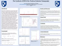
The Synthesis of DFO-8 for Positron Emission TomographyAaron Chung, Mahmoud Abdalrahman, Dr. Planalpac2017@wildcats.unh.edu, Department of Chemistry, University of New HampshireDurham, New Hampshire, 038245/4/17 Introduction Positron Emission Tomography (PET) is a powerful clinical imaging technique that is used to detect various pathogens, including cancers. PET scans are an important technique in clinics because they allow for a physician to diagnose a patient’s illness, prescribe treatment, and monitor treatment efficacy. Traditional PET isotopes include 18F, 15O, 13N and 11C, but due to their lengthy radiosyntheses and short half-lives only early time points were available for imaging, leaving investigations of biological processes, which occur over duration of hours or days, difficult to explore.1 As a result, an ideal radioisotope used in PET scans involves matching the physical half-life to the pharmokinetic half-life of the immunoglobulin.2 The desire for an ideal isotope in antibody-based imaging has led to the emerging research on 89Zr based imaging. 89Zr has favorable physical characteristics for antibody-based imaging, with a half-life of 78.4 hrs and a relatively low positron energy of 395.5 keV. The current ligand employed to 89Zr is Desferrioxamine (DFO), which binds six times through oxygen atoms, leaving two binding sites on the metal exposed to solvent (H2O). Not filling all of the coordination sites on the zirconium metal creates instability in the complex and will result in demetallation.3 To combat this, a better ligand can be designed and synthesized; a ligand that will fill all eight binding sites on Zr+4, creating a stronger chelate effect. This will be done by attaching aminomalonic acid, which will be synthesized by the hydrolysis of dimethyl aminomalonate, to the existing DFO ligand by HATU Coupling reaction. This will result in an octadentate ligand, yielding a greater chelate effect and an ideal binding agent. If successful, this ligand will minimize the release of free zirconium ions targeting the bones of organisms, providing a safer way of utilizing PET scans. ExperimentalA multistep synthesis was performed starting with dimethyl aminomalonate (DMA) to yield DFO-8. The hydrolysis of DMA yielded aminomalonic acid (AMA). Scheme 1. Hydrolysis of Dimethyl AminomalonateAminomalonic acid was attached to Desferrioxamine by HATU Coupling reaction to yield DFO-8.Scheme 2. HATU Coupling with DFO and AMA Results and Discussion Aminomalonic acid was recovered with an 82% yield. 1 H NMR reveals a 3:1 ratio of starting material to product. 13C NMR illustrates three peaks, suggesting that only one side of the molecule was protonated. IR spectrum suggests aminomalonic acid was successfully prepared as the carbonyl of the carboxylic acid is shifted more upfield than the carbonyl of the ester. DFO-8 was recovered with a 61% yield. MALDI TOF mass spectroscopy shows a major peak of a molecular weight (MW) of 660, matching the predicted MW of the desired product. References1. Thaddeus J. Wadas, Edward H. Wong, Gary R. Weisman, Carolyn J. Anderson; Coordinating Radiometals of Copper, Gallium, Indium, Yttrium and Zirconium for PET and SPECT Imaging of Diseases. Chemical Reviews. 2010, 110, (5).2. Positron Emission Tomography (PET) Scan | Cleveland Clinic. Cleveland Clinic. 2014.3. Melissa A. Deri, Brian M. Zeglis, Lynn C. Francesconi, Jason S. Lewis; PET Imaging with 89Zr: From Radiochemistry to the Clinic. Nuclear Medicine and Biology. 2013. (3-14).4. Francois Guerard, Yong-Sok Lee, Martin W. Brechbiel; Rational Design, Synthesis, and Evaluation of Tetrahydroxamic Acid Chelators for Stable Complexation of Zirconium(IV). Chemistry a European Journal. 2014, 20, (5584-5591). AcknowledgementsI would like to thank the UNH Chemistry Department, Mahmoud Abdalrahman, Luke Fulton, Zane Relethford and Dr. Planalp. ConclusionThe success of the multi-step synthesis is inconclusive as MALDI TOF mass spectroscopy, alone, is insufficient in determining the identity of the compound. Additional characterization techniques, such as ligand binding assay (LBA) need to be performed in order to identify the ligand. Due to time constraints the hydrolysis of the starting material, Dimethyl Aminomalonate (DMA), was not performed in a sufficient way to yield a significant turnover of desired product. Future WorkOnce the success of the synthesis of DFO-8 can be confirmed, binding affinity will be measured through ligand binding assay (LBA). Further studies will be made in preparation of the Zr+4 complexation with DFO-8. If proven successful, preparations may be made to distribute the chelator to clinics for trials. Figure 1. 13C NMR of Aminomalonic Acid Figure 3. 1H NMR of Aminomalonic Acid Figure 5 . MALDI TOF Mass Spectroscopy of DFO-8 Figure 2 . IR of Aminomalonic Acid vs Dimethyl Aminomalonate Figure 4 . 1H NMR of Dimethyl Aminomalonate