
DISORDERS Urinary obstruction can occur in persons of any age and can involve any level of the urinary tract from the urethra to the renal pelvis The two most damaging effects of urinary obstruction are ID: 1014308
Download Presentation The PPT/PDF document "Dr.NADIA HAMEED OBSTRUCTIVE" is the property of its rightful owner. Permission is granted to download and print the materials on this web site for personal, non-commercial use only, and to display it on your personal computer provided you do not modify the materials and that you retain all copyright notices contained in the materials. By downloading content from our website, you accept the terms of this agreement.


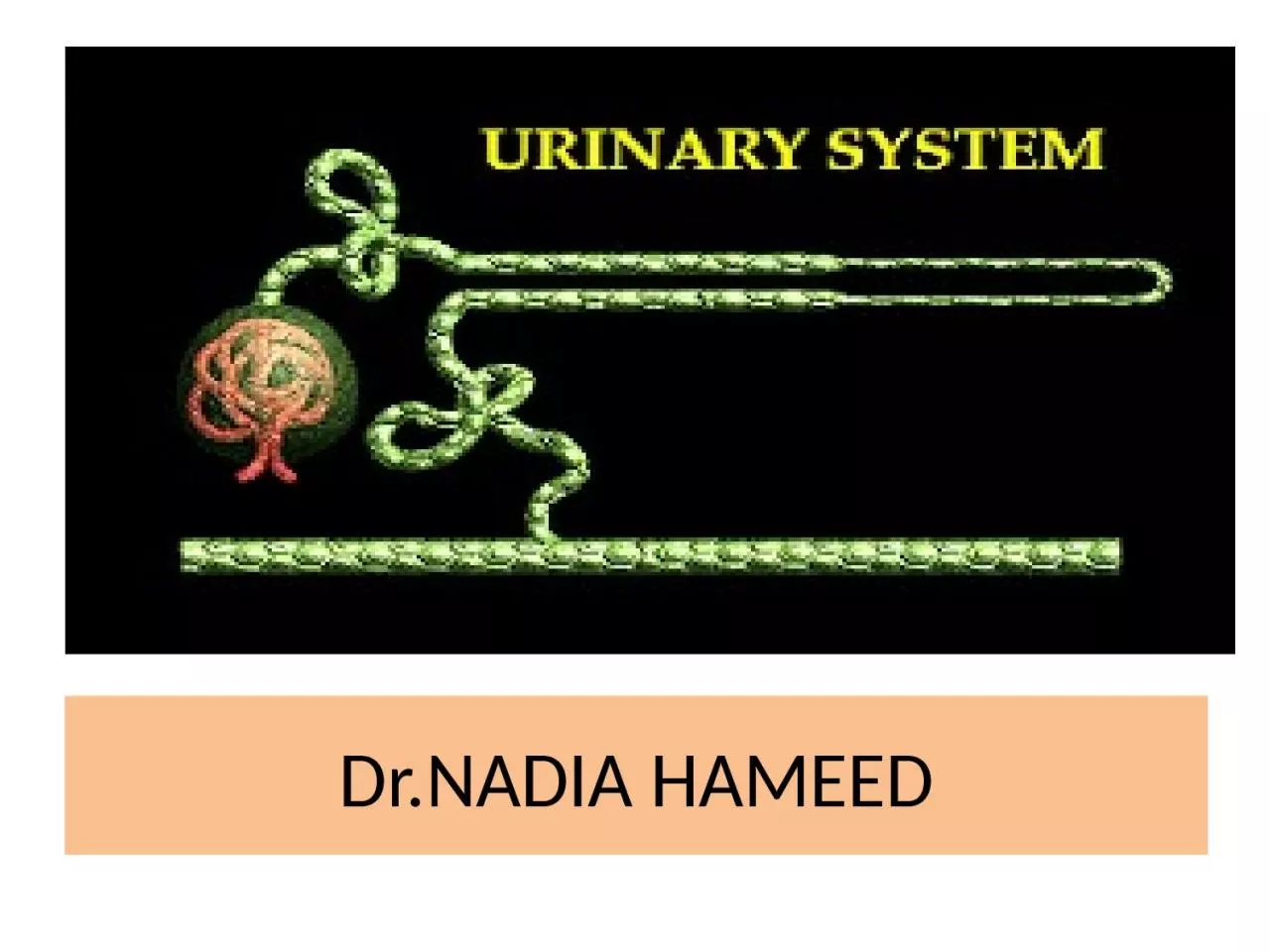
1. Dr.NADIA HAMEED
2. OBSTRUCTIVE DISORDERSUrinary obstruction can occur in persons of any age and can involve any level of the urinary tract from the urethra to the renal pelvis. The two most damaging effects of urinary obstruction are (1) stasis of urine, which predisposes to infection and stone formation, and (2) development of backpressure, which interferes with renal blood flow, destroys kidney tissue, tubules are distended with urine and predisposes to hydronephrosis: (Hydronephrosis refers to dilation of the renal pelvis and calices, with atrophy of renal tissue, that is caused by obstruction to the outflow of urine. The obstruction may be sudden or insidious in onset and may occur at any level of the urinary tract).
3. HYDRONEPHROSIS
4.
5. Renal pelvisRenal stonesPapillary necrosisUreterRenal stonesPregnancyTumors that compress the ureterUreteral strictureCongenital disorders of the ureterovesical junction and ureteropelvic junction stricturesBladder and urethraBladder cancerNeurogenic bladderBladder stonesProstatic hyperplasia or cancerUrethral stricturesCongenital urethral defectsCauses of Urinary Tract Obstruction
6. Renal stones (Calculi) Kidney stones are crystalline structures that form from components of the urine. The most common cause of upper urinary tract obstruction is urinary stones. Although stones can form in any part of the urinary tract, most develop in the kidneys. Men are more frequently affected than women, with a ratio of 4/1. Kidney stones require a nidus, or nucleus, to form and a urinary environment that supports continued precipitation of stone components to grow.Predisposing factors for stone formation: Increases blood and urinary levels of stone components ca++ stones associated with hypercalcemia Anatomic changes in urinary tract structures.Metabolic and endocrine influences Dietary factors.Renal obstructive disorders.Urinary tract infections.
7.
8. Types of Stones: There are four basic types of kidney stones: Calcium stones (i.e., ca-oxalate or ca-phosphate)Magnesium ammonium phosphate stones Uric acid stonesCystine stones.
9.
10.
11. Calcium Stones. Most kidney stones (70% to 80%) are calcium stones—calcium oxalate, calcium phosphate, or a combination of the two materials. Calcium stones usually are associated with increased concentrations of calcium in the blood and urine. Excessive bone resorption caused by immobility, bone disease, hyperparathyroidism, and renal tubular acidosis all are contributing conditions. Magnesium Ammonium Phosphate Stones. Magnesium ammonium phosphate stones, also called struvite stones, form only in alkaline urine and in the presence of bacteria that possess an enzyme called urease, which splits the urea in the urine into ammonia. The ammonia that is formed takes up a hydrogen ion to become an ammonium ion, increasing the pH of the urine so that it becomes more alkaline. Because phosphate levels are increased in alkaline urine and because magnesium always is present in the urine, struvite stones formed. These stones enlarge as the bacterial count grows, and they can increase in size until they fill an entire renal pelvis. Because of their shape, they often are called staghorn stones. Struvite stones in usually are too large to ben passed and require lithotripsy or surgical removal.
12. Uric Acid Stones. Uric acid stones develop in conditions of gout and high concentrations of uric acid in the urine. Unlike radiopaque calcium stones, uric acid stones are not visible on x-ray films. Uric acid stones form most readily in urine with a pH of 5.1 to 5.9. Thus, these stones can be treated by raising the urinary pH to 6 to 6.5 with potassium alkali salts.Cystine Stones. Cystine stones are rare. They are seen in cystinuria, which results from a genetic defect in renal transport of cystine. These stones resemble struvite stones except that infection is unlikely to be present.
13.
14. Theories of urinary stones formation:The saturation theory states that the risk of stone formation is increased when the urine is supersaturated with stone components (e.g., calcium salts, uric acid, magnesium ammonium phosphate, cystine).The matrix theory proposes that organic materials, such as mucopolysaccharides derived from the epithelial cells that line the tubules, act as a nidus for stone formation. This theory is based on the observation that organic matrix materials can be found in all layers of kidney stones.The inhibitor theory suggests that persons who have a deficiency of proteins that inhibit stone formation in their urine are at increased risk for stone formation. Kidney cells produce at least three proteins that are thought to slow the rate of calcium oxalate crystallization: nephrocalcin, Tamm-Horsfall mucoprotein, and uropontin. Nephrocalcin inhibits nucleation, aggregation, and growth of calcium oxalate stones. Tamm-Horsfall mucoprotein is impair crystal aggregation. Uropontin inhibits the growth of calcium oxalate crystals.
15. Pathogenesis of renal obstructive disorders in calcium stone formation: Urinary obstruction and stagnation of urine predisposes to infection. If calculi formed first, it serves as foreign bodies and contribute to the infection. Once established, the infection is difficult to treat. If infection occur first it often caused by urea-splitting organisms (e.g., Proteus, staphylococci) or other organisms. Calcium salts precipitate more readily in stagnant infected urine and calcium stone formed.
16. URINARY TRACT INFECTIONSUrinary tract infections (UTIs) are the second most common type of bacterial infections seen by health care providers (respiratory tract infections are first). Etiologic bacteriaMost UTIs are caused by Escherichia coli. Other common pathogens include gram negative enterbacteriacae, Proteus, Klebsiella pneumoniae, and gram positive Staphylococcus saprophyticus and Enterococcus species. Normal flora of skin can cause infection in immune compromised patients or after instrumentation. Bacteria can enter the kidneys either through the bloodstream or as an ascending infection from the lower urinary tract. Most infections are of the ascending type. Among the factors that contribute to bacterial virulence is the type of fimbriae (pili) that the bacteria possess.
17. Risk (predisposing factors):Urinary obstruction: renal stones, neurogenic bladder a disorder that impair bladder emptying, men with diseases of the prostate benign prostatic hyperplasia BPH) .Urine reflux: Reflux occurs when urine from the urethra moves into the bladder (i.e., urethrovesical reflux) or from the bladder into the ureters (i.e., vesicoureteral reflux).Sexual activity: women more affected than men because of short urethra. Urinary catheterization is the most common predisposing factors for nosocomial UTIs.
18. Host Defenses against UTI:washout phenomenon, in which bacteria are removed from the bladder and urethra during voiding protective mucin layer that protects mucosa against bacterial invasion peristaltic movements of ureter facilitate the movement of urine secretory immunoglobulin A (IgA)Phagocytic blood cells further assist in the removal of bacteria from the urinary tract.
19. Signs and symptoms of urinary tract infections:Urgency: A strong, persistent urge desire to urinateDysuria: A burning sensation when urinatingFrequency: Passing frequent, small amounts of urineHematuria: urine that appears red a sign of presence of blood in the urinePelvic pain— suprapubic in lower UTI and loin pain radiate to the back (costovertebral) in upper UTI
20. Part of urinary tract affectedSigns and symptomsKidneys (acute pyelonephritis)Upper back and side (flank) painHigh feverShaking and chillsNauseaVomitingBladder (cystitis)Pelvic pressureLower abdomen discomfortFrequent, painful urinationBlood in urineUrethra (urethritis)Burning with urinationDischarge
21. Classification UTI:• Lower UTI Infections of the lower urinary tract (urethra, bladder). The most prominent symptoms are dysuria, frequency, urgency and urinary incontinence. Cystitis: This type of UTI is usually caused by Escherichia coli (E. coli), a type of bacteria commonly found in the gastrointestinal (GI) tract. Other common pathogens include gram negative enterobacteriacae eg Proteus, Klebsiella pneumoniae, or grame positive Staphylococcus saprophyticus and Enterococcus species.Honey moon cystitis: cystitis due to sexual activity of newly married couples. Urethritis: sexually transmitted infections, such as herpes, gonorrhea, chlamydia and mycoplasma, are the most common cause of urethritis. G-ve enterobacteriace also cause of urethritis
22. Upper UTIPyelonephritisRefers to an inflammation of the kidneys and renal pelvis. There are two forms of pyelonephritis: acute and chronic. Acute pyelonephritis represents a patchy interstitial suppurative inflammatory process. Infection may occur through bloodstream or ascend from the bladder. Bacteria such as E. coli often cause the infection. However, any serious infection in the bloodstream can also spread to the kidneys and cause acute pyelonephritis. The onset of acute pyelonephritis typically is abrupt, with chills, fever, headache, back pain, tenderness over the costovertebral angle, and general malaise. It usually is accompanied by symptoms of bladder irritation, such as dysuria, frequency, and urgency.Complications:Chronic pyelonephritisabscess formation tubular necrosis.
23. Chronic pyelonephritis represents a progressive process usually associated with unresolved obstructive renal diseases. There is scarring and deformation of the renal calices and pelvis due to chronic pyelonephritis
24. Glomerulus structuresE=Endothelial cellsP=podocytes (epithelia)C=CapillaryM=MesengymeBM=Bacement MembraneBC=Bowman CapsuleBM
25.
26.
27. DISORDERS OF GLOMERULAR FUNCTIONGlomerular disorders affect the glomerular capillary-membrane structures that filter materials from the blood. It is the leading cause of chronic renal failure in the United States, accounting for one half of persons with end-stage renal disease. Causes and pathogenesis:I// Primary (idiopathic)II// Secondary: due to immune mediated injury. Two types of immune mechanisms have been implicated in the development of glomerular disease: injury resulting from antibodies reacting with fixed glomerular antigens (type II HSR)injury resulting from circulating antigen-antibody complexes that become trapped in the glomerular membrane (type III HSR)
28. Antigens responsible for development of the immune response may be: Endogenous origin, such as DNA in SLEExogenous origin, such as streptococcal membrane antigens in poststreptococcal glomerulonephritis. The source of the antigen is unknown.The pathological changes that occur with glomerular disease includeProliferative changes: refers to an increase in the cellular components of the glomerulus regardless of origin (mesengial cell proliferation)Sclerotic changes: increase in the non-cellular components of the glomerulus primarily collagen and fibrous tissue Membranous changes: an increase in the thickness of the glomerular capillary wall caused by immune complex deposition.
29. Patern of glomerular changes (area that involved)Glomerular changes can be diffuse, involving all glomeruli and all parts of the glomeruli; focal, in which only some glomeruli are affected and others are essentially normal; segmental, involving only a certain segment of each glomeruli; or mesangial, affecting only the mesangial cell.
30. Glomerular diseases include broadly two conditions: nephritis and nephrotic syndroms. Nephritic syndromes are caused by diseases that produce proliferative inflammatory responses that decrease the permeability of the glomerular capillary membrane.The nephrotic syndrome is caused by derangment disorders that increase the permeability of the glomerular capillary membrane, causing massive loss of protein in the urine.
31.
32. Nephritic Syndromes (glomerulonephritis)Glomerulonephritis is characterized by hematuria oliguria and diminished glomerular filtration rate (GFR), azotemia (presence of nitrogenous wastes in the blood), hypertension. It is caused by diseases that provoke a proliferative inflammatory response of the endothelial, mesangial, or epithelial cells of the glomeruli. The inflammatory process damages the capillary wall, permitting red blood cells to escape into the urine and producing hemodynamic changes that decrease the GFR. The nephritic syndromes include: acute proliferative glomerulonephritis and rapidly progressive glomerulonephritis.
33. A//Acute Proliferative GlomerulonephritisThe glomerular changes are proliferative and the pattern is diffuse (diffuse proliferative glomerulonephritis), which follows infections caused by strains of group A β-hemolytic streptococci. Diffuse proliferative glomerulonephritis also may occur after infections by other organisms, including staphylococci and a number of viral agents, such as mumps, measles, and chickenpox. With this type of nephritis, the proliferative inflammatory response is caused by an immune reaction that occurs when circulating immune complexes become entrapped in the glomerular membrane. The disease primarily in children but adults of any age also can be affected.
34. A//Acute Proliferative Glomerulonephritiscola-colored urine may be the first sign of the disorder. Sodium and water retention gives rise to edema, particularly of the face and hands, and hypertension.Important laboratory findings include an elevated streptococcal exoenzyme (antistreptolysin O) titer, a decline in C3 complement. The immediate prognosis is favorable, and approximately 95% of children recover spontaneously
35. B// Rapidly Progressive GlomerulonephritisRapidly progressive glomerulonephritis is a clinical syndrome characterized by signs of severe glomerular injury that does not have a specific cause. As its name indicates, this type of glomerulonephritis is rapidly progressive, often within a matter of months. The glomerular changes are proliferative and the pattern of involvement is focal or segmental (focal or segmental proliferation glomerulonephritis).Rapidly proliferative glomerulonephritis may be caused by a number of immunologic disorders, some are systemic eg (SLE, vasculitis) and others are restricted to the kidney eg Goodpasture’s syndrome (It is a rare disease and is associated with a triad of pulmonary hemorrhage, iron-deficiency anemia, and glomerulonephritis. It is caused by antibodies to the glomerular basement membrane (GBM), accounts for approximately 5% of cases of rapidly progressive glomerulonephritis).
36. Nephrotic SyndromeThe nephrotic syndrome is characterized by:massive proteinuria(>3.5 g/day) hypoalbuminemia (<3 g/dL)generalized edemahyperlipidemia (cholesterol >300 mg/dL) and lipiduria (e.g., free fat, oval bodies, fatty casts).The initiating event in the development of nephrosis (=nephrotic)is a derangement in the glomerular membrane that causes increased permeability to plasma proteins. Generalized edema, which is a hallmark of nephrosis, results from salt and water retention and a loss of serum albumin below that needed to maintain the colloid osmotic pressure of the vascular compartment.
37. Nephrotic Syndrome
38. Nephrotic SyndromeThe hyperlipidemia that occurs in persons with nephrosis is characterized by elevated levels of triglycerides and low-density lipoproteins (LDL). Levels of high-density lipoproteins (HDL) usually are normal. These abnormalities are thought to be related, in part, to increased synthesis of lipoproteins in the liver secondary to a compensatory increase in albumin production. Thrombotic complications can occur in persons with nephrotic syndrome due to renal loss of coagulation and anticoagulation factors. Renal vein thrombosis, deep vein thrombosis and pulmonary emboli are the major thrombotic complications of nephrotic syndrome.
39. Diabetic nephropathy (Glomerulosclerosis)Diabetic nephropathy, or kidney disease, is a major complication of diabetes mellitus. It affects approximately 30% of persons with type 1 diabetes and accounts for 20% of deaths in diabetic patients younger than 40 years of age. The glomerulus is the most commonly affected structure in diabetic nephropathy. Diabetic patients clinically pass through the following stages: non-nephrotic proteinuria, nephrotic syndrome, and renal failure.
40. Diabetic nephropathy (Glomerulosclerosis)Pathologically diabetic glomerulosclerosis changes include:Widespread thickening of the glomerular capillary basement membrane occurs in almost all persons with diabetes and can occur without evidence of proteinuria (arteriolosclerosis).This is followed by a diffuse increase in mesangial matrix, with mild proliferation of mesangial cells (diffuse glomerulosclerosis) . Then nodular deposition of hyaline in the mesangial portion of the glomerulus leads to (nodular glomerulosclerosis).The sclerotic process progresses in both the diffuse and nodular forms of glomerulosclerosis, there is complete obliteration of the glomerulus, with impairment of renal function.
41. Diabetic nephropathy (Glomerulosclerosis)arteriolosclerosisdiffuse glomerulosclerosisnodular glomerulosclerosismild proliferation of mesangial cells
42. Diabetic nephropathy (Glomerulosclerosis)Pathophysiology of diabetic glomerulosclerosis changes Defective synthesis of the glomerular basement membrane and mesangial matrix elevations in blood glucose produce an increase in GFR and glomerular intracapillary pressure that leads to an enlargement of glomerular capillary pores by a mechanism that is at least partly mediated by angiotensin II.This enlargement impairs the size-selective function of the membrane so that the protein content of the glomerular filtrate increases, which in turn requires increased endocytosis of protein by the tubular endothelial cells, a process that ultimately leads to nephron destruction and progressive deterioration of renal function.
43. Diabetic nephropathy (Glomerulosclerosis)The clinical manifestations of diabetic glomerulosclerosis are closely linked to stage of changes. microalbuminuria, defined as urinary albumin excretion greater than 30 mg/24 hours and no more than 300 mg/ 24 hs. Microalbuminuria is an important predictor of future diabetic nephropathies.EdemaHypertensionRenal failure In many cases, these early changes in glomerular function can be reversed by careful control of blood glucose levels. Inhibition of angiotensinII by angiotensin-converting enzyme inhibitors (e.g., captopril) has been shown to have a beneficial effect, possibly by reversing increased glomerular pressure.
44. Hypertensive Glomerular DiseaseRenal failure and azotemia occur in 1% to 5% of persons with long-standing hypertension. Hypertension is associated with sclerotic changes in glomerular structures. As the glomerular vascular structures thicken and perfusion diminishes, the blood supply to the nephron decreases, causing the kidneys to lose some of their ability to concentrate the urine. Blood urea nitrogen levels also may become elevated, Proteinuria may occur as a result of changes in glomerular structure.
45. Drug-Related NephropathiesMechanisms of drug related nephropathies:Some drugs and toxic substances damage the kidneys by causing a decrease in blood flow eg NSAIDs inhibit prostaglandin synthesis p(articularly PGI2 and PGE2) Direct toxic damage to tubulointerstitial structures (aspirin) Damage by producing acute drug related hypersensitivity reactions ends with tubulointerstitial nephritis. sulfonamide drugs, furosemide and the thiazide diuretics methicillin and other synthetic antibiotics included
46. 4. can cause kidney damage by obstructing urinary flow and crystal formation (e.g. methoprim + vit c)5. multifactorial mechanisms that involve direct vasoconstriction, altered systemic hemodynamics, and myoglobulin-induced renal failure (cocaine intoxication) .
47. ACUTE RENAL FAILURE ARFAcute renal failure is caused by conditions that produce an acute shutdown in renal function. It represents a rapid decline in renal function sufficient to increase blood levels of nitrogenous wastes and impair fluid and electrolyte balance. It is a common threat to seriously ill persons in intensive care units, with a mortality rate ranging from 42% to 88%.
48. ACUTE RENAL FAILURE ARFThe most common indicator of acute renal failure is azotemia, an accumulation of nitrogenous wastes (urea nitrogen, uric acid, and creatinine) in the blood.Because of the high morbidity and mortality rates associated with acute renal failure, identification of persons at risk is important to clinical decision making. Acute renal failure often is reversible, making early identification and correctionof the underlying cause (e.g., improving renal perfusion, discontinuing nephrotoxic drugs) important.
49.
50. Causes of acute renal failureprerenal failure It can result from decreased blood flow to the kidney;I// HypovolemiaHemorrhageDehydrationExcessive loss of gastrointestinal tract fluidsExcessive loss of fluid due to burn injuryII// Decreased vascular fillingAnaphylactic shockSeptic shock
51. prerenal failureIII// Heart failure and cardiogenic shockIV// Decreased renal perfusion due to vasoactive mediators, drugs, diagnostic agents (contrast media): endotoxins, radiocontrast agents as those used for cardiac catheterization, cyclosporine (an immunosuppressant drug that is used to prevent transplant rejection), amphotericin B (an antifungal agent), epinephrine, and high doses of dopamine. NSAIDs) can reduce renal blood flow through inhibition of prostaglandin synthesis.Normally, the kidneys receive 22% of the cardiac output. As renal blood flow falls, the GFR decreases, the amount of sodium and other substances that are filtered by the glomeruli is reduced, and the blood flow needed for the energy-dependent mechanisms that reabsorb these substances is reduced. Because of their high metabolic rate, the tubular epithelial cells are most vulnerable to ischemic injury. Improperly treated, prolonged renal hypoperfusion can lead to ischemic tubular necrosis with significant morbidity and mortality.
52. intrinsic or intrarenal failure disorders that disrupt the structures in the kidney( Acute tubular necrosis)Prolonged renal ischemia (prerenal ischemia ends with intrarenal damage)Exposure to nephrotoxic drugs, heavy metals, and organic solventsIntratubular obstruction resulting from hemoglobinuria, myoglobinuria, myeloma light chains, or uric acid castsAcute renal disease (acute glomerulonephritis, pyelonephritis) and acute renal shutdown in pathient with nephritis for first presentation.
53. postrenal failurePostrenal failure results from obstruction of urine outflow from the kidneys. The obstruction can occur in the: ureter (i.e., calculi and strictures), bladder (i.e., tumors or neurogenic bladder)urethra (i.e., prostatic hyperplasia). Prostatic hyperplasia is the most common underlying problem.
54. Chronic Renal Failure CRFChronic renal failure results from the destructive effects of many forms of renal disease. Regardless of the cause. the consequences of nephron destruction in ESRD are alterations in the filtration, reabsorption, and endocrine functions of the kidneys. The progression of chronic renal failure usually occurs in four stages: diminished renal reserve, renal insufficiency, renal failure, and End Stage Renal Diseases ESRD.
55. Diminished renal reserve occurs when the GFR drops to approximately 50% of normal. At this point, the serum BUN and creatinine levels still are normal, and no symptoms of impaired renal function are evident.Renal insufficiency represents a reduction in the GFR to approximately 20% to 50% of normal. During this stage, azotemia, anemia, and hypertension appear.Renal failure, a reduction to less than 20% to 25% of normal. Renal failure develops when the GFR is less than 20% of normal. At this point, the kidneys cannot regulate volume and solute composition, and edema, metabolic acidosis, and hyperkalemia develop. These alterations affect other body systems to cause neurologic, gastrointestinal, and cardiovascular manifestations.
56. ESRD, a decrease in GFR to less than 5% of normal. Histologic findings of an end-stage kidney include a reduction in renal capillaries and scarring in the glomeruli. Atrophy and fibrosis are evident in the tubules. The treatment of ESRD can be divided into two types:conservative management of renal insufficiency (measures to prevent or retard deterioration in remaining renal function) renal replacement therapy with dialysis or transplantation.
57.