
Presenter Rohan Yewale DNB MGE Registrar SIMS Hospital Chennai Guide Dr BS Ramakrishna Case capsule 43 year old gentleman seen by our department in August 2018 Presented with complaints of ID: 999524
Download Presentation The PPT/PDF document "Story of a man with an unusual gastric m..." is the property of its rightful owner. Permission is granted to download and print the materials on this web site for personal, non-commercial use only, and to display it on your personal computer provided you do not modify the materials and that you retain all copyright notices contained in the materials. By downloading content from our website, you accept the terms of this agreement.


1. Story of a man with an unusual gastric massPresenter: Rohan Yewale, DNB MGE Registrar, SIMS Hospital, ChennaiGuide : Dr. B.S. Ramakrishna
2. Case capsule43 year old gentleman seen by our department in August 2018Presented with complaints of – “chronic indigestion”Poor appetiteEarly satiety x 1 year Post prandial, vague, upper abdominal discomfortVomiting few hours after meals – non bilious, undigested food particles Acute onset pain in left lower abdomen, radiating to back with multiple episodes of non bilious vomiting x 3 daysNo h/o weight loss, hematemesis, melena, hematochezia, jaundice, heartburn, dysphagia, bloating, altered bowel habits, hematuria, feverH/o NSAID use on and off for abdominal pain
3. Past historyDiabetic, hypertensive since 3 years on regular medicationH/o multiple hospitalizations under various departments since the last 1 yearDenied any form of addictionsNo significant family history
4. Timeline
5. ExaminationWell builtOverweight, BMI 30 kg/m2Vitals normalPer abdomen - soft, mild left lumbar tenderness +Other systems normal
6. InvestigationsLab investigations – e/o acute kidney injury with elevated serum creatinine (1.94), urea (55) and raised Total WBC count (14,500)Urine routine analysis normal, no growth on cultureOther baseline investigations essentially normalCT abdomen – bilateral bulky kidneys with left perinephric inflammatory changes and a microlith in left kidney, no obstructive changesFocal, heterogeneously enhancing wall thickening with ulceration and calcification in the posterior wall of antrum of stomach along the lesser curvature – incidental findingImp: Gastric ulcer and antral submucosal lesion ? Fibro-inflammatory lesion Left Pyelonephritis
7.
8. Treated symptomatically with iv fluids, antibioticsAKI (? pre-renal azotemia / ? NSAID induced tubulo-interstitial nephritis / ? Acute pyelonephritis) settledHe was then evaluated for the incidentally detected gastric antral submucosal lesion
9. Upper GI endoscopy - Antral submucosal lesion along the lesser curvature with central excavated ulcer Endoscopic biopsy - HPE Mild chronic gastritis with focal active inflammation and ulcerationEndoscopic Ultrasound Submucosal mass lesion in antrum arising from the muscular layer (layer 2), FNAB – insufficienct tissueProvisional diagnosis Gastric antral submucosal mass lesion Etiology …….. ?????
10.
11. Surgical gastroenterology opinion soughtIn view of ? GIST like appearance of the lesion without any evidence of other intra abdominal lesions on CT, laparotomy converted to open “partial gastrectomy/mass resection” was doneOperative findings – 7 x 5 cm mass in the greater curvature extending over posterior wall up to lesser curvatureMass adherent posteriorly to the mesocolonShowed symptomatic improvement and was discharged
12. Histopathology / IHCFibroinflammatory lesion with diffuse lymphoplasmacytic infiltrateOn the basis of histology, a diagnosis of IgG4 related disease was suggestedIHC - positive for IgG4 (35-40%)Serum IgG 4 levels markedly elevated (4.36g/l; n-0.03-2.01)
13.
14. Course after discharge4 months later - October 2018Readmitted with c/o –Epigastric pain, continuous, unrelated to meals, dull aching type, non radiating x 7 daysA/w occasional episodes of non bilious vomitingWeight loss of 12kg x 3 monthsNo h/o back pain, GI bleeding, jaundice, altered bowel habitsPer abdomen soft, non tender, no guarding or rigidityBaseline labs, amylase, lipase normal
15. Upper GI endoscopy – submucosal lesion esophagus (previous HPE - leiomyoma), post - operative stomach with a linear gastric ulcer at the site of resection and a deep excavating ulcer in duodenumStarted on iv pantoprazole infusion with syrup sucralfateThought to be a recurrence of IgG4 – RD, hence started onInj hydrocortisone 100mg q8h for 3 days and later changed to a tapering dose of T. PrednisoloneT. Azathioprine 50mg Symptoms settled over the next few weeks
16. 2 months later - December 2018Readmitted with similar complaints (on Prednisolone 30mg and Azathioprine 50mg)Upper GI endoscopy – Excavating clean based ulcer in antrum parallel to resection site and a clean based duodenal ulcerHPE – Marked reduction in plasma cell infiltration in gastric mucosa, H. Pylori +Serum IgG 4 levels reduced (2.71 g/l)Oral steroids, Azathioprine continuedPain management
17. Readmitted after 1 month - Feb 2019Persistent epigastric, left upper quadrant pain, occasional vomitingUpper GI endoscopy – partially healed residual gastric ulcer, healing duodenal ulcerHPE - Very few plasma cellsIHC – IgG4 10 cells/HPF (1+)Diagnostic dilemma Was IgG 4 – RD in remission ?If not, should Rituximab be considered ?Were we missing out on some other condition ?
18. CT abdomen with oral contrast – retention of contrast material in stomach – severe gastroparesis, gastric outlet obstructionGastric scintigraphy – Delayed gastric emptyingDiagnosis – Post operative gastroparesisUnderwent gastric antral resection with truncal vagotomy and gastrojejunostomyLast follow up in April 2019 – asymptomatic, regained appetite and weight
19.
20.
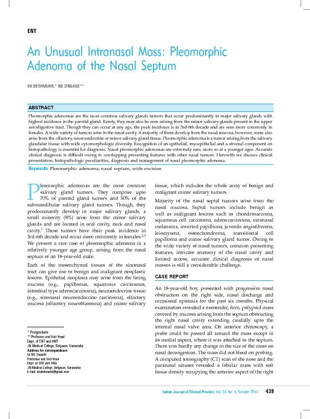
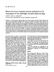
21. IgG-4 RD presenting as isolated gastric lesions – A literature reviewSkorus. U et al; Pol Przegl Chir; Jun 2018
22.
23. Our experience so far…Case NoAge/SexSymptomsGastric locationLesion type in endoscopic examinationPre-operative diagnosisLesion sizeHPE/IHCSerum IgG4 levelTreatmentFollow up1.43/MDyspepsia vomiting,Poor appetite,Recurrent admissions for multi system illnessesAntrumSubmucosal lesion with a central excavated ulcerGIST /Malignancy7 x 5 cmDense lymphoplasmacytic infiltrate,IHC – Positive for IgG 4 cells (35-40%)4.36g/lelevatedSurgical resectionf/bTapering steroids and IMStormy course,Gastric ulcer,Post operative gastroparesisNo recurrence at 11 months2.58/MEpigastric fullness, heartburnDistal body along lesser curveSubmucosal lesionGIST4 x 4.5 cmLymphoplasmacytic infiltrate, storeiform fibrosisLap wedge resectionNo recurrence at 6 months
24. Take home messageOver the last 6 months, we have come across 2 cases of gastric submucosal lesions which on surgical resection turned to be IgG 4 - RD on HPE and IHCThus, although IgG4-RD itself is a rare entity, mainly presenting as autoimmune pancreatitis, it can also present as gastrointestinal submucosal mass lesionsWhen a patient has a history of multisystem involvement of doubtful etiology dispersed in time, IgG4 – RD should be considered as a differential diagnosisSurgery may not be the final cure since it is a multisystem disease with relapses and remissions Monitoring disease remission remains to be a challenge
25. What is IgG4 - RDChronic,Relapsing, Multi-organ, Fibro-inflammatory syndrome Of presumed autoimmune etiologyCharacterized by increased serum levels of IgG4 and tissue infiltration by IgG4+ cells
26. What makes it so unique ?Pathognomonic antigens probably differ among different manifestationsDifferent antigens or autoantibodies produce Similar immune reactions in different organs
27. EpidemiologyEstimated prevalence of fewer than 1 per 100,000 in the general populationlargely a disease of men, with a 3:1 male predominanceTypically diagnosed late in life (mean age older than 60 years)Commonly affected organs Men – pancreatitisWomen – salivary glands
28. Pathogenesis2 parallel immunologic processesTissue-destructive process mediated by activated lymphocytes, macrophages and fibroblastsAnti-inflammatory response thatinvolves IgG4 and regulatory T (T reg) cellsImmunological triggers Potential autoantigens – carbonic anhydrase, trypsinogens, annexins A11)Chronic exposure – microbes, chemicalsSolvents, metal dusts
29.
30. Organ involvement – multisystem Gastro-intestinalPancreas – autoimmune pancreatitits type I Bile duct – sclerosing cholangitisRetroperitoneumLiverGall bladderSmall, large bowelStomachExtra - GastrointestinalLacrimal glandsSalivary glandsKidneysLungsOrbitThyroidSinuses>50% patients have more than 2 organs involved - Pooled analysis of 5 large studies in 758 patients
31.
32. Modified HISORt criteria for diagnosis of IgG4-RDHISORtDomainFeaturesLevel 1Level 2Not suggestiveHHistology andimmuno-stainingDense lymphoplasmacyticinfiltrate, Storiform fibrosis,Obliterative venulitis,Abundant IgG4+plasma cells,IgG4+/IgG+ plasmacells >40%Any 4 of 5Any 2 of 5Involved organ shows any of:Scant IgG4+ plasma cellsIgG4+/IgG+ plasma cells <40%Malignant cellsFeatures of infection (neutrophils/ abscess/organisms)
33. HISORtDomainFeaturesLevel 1Level 2Not suggestiveIImagingTypical imaging featuresTypicalpancreasinvolvement Bilateral involvement of 2 sets of salivary/lacrimal glandsRenal lesionsBilateral involvement of a single salivary set of salivary / lacrimal glandRetroperitoneal fibrosisLung lesionsInvolved organ shows features suggestive of malignancy withworkup for malignancy not done or inconclusiveLong-bone abnormalities suggestiveof Erdheim-Chester diseaseSSerologySerum IgG4,autoantibodies,complementSerum IgG4 levels >3 X ULN>=1 of:Serum IgG4 levels 1–2 X ULNLow complementCryoglobulinemiaAutoantibodies to Ro, La,double-stranded DNA,ribonucleoprotein, Smith,neutrophil cytoplasm (ANCA)Other disease-specific antibodies
34. HISORtDomainFeaturesLevel 1Level 2Not suggestiveOOrganTypical organsinvolved: pancreas,bile duct, salivaryglands, lacrimal glands,retroperitoneum,kidneys, lungs>=2 organsinvolved1 organ involvedNone of typical organs involvedRtResponse to steroid therapyReduction/resolutionof inflammatory component after 4 wk of treatment with at least20 mg/d prednisoneRapid responseof inflammatorycomponentPartial responseNo response to therapy
35. DIAGNOSTIC COMBINATIONSClassic clinical profileAll 4:Imaging: Level 1 feature in >= 1 organOrgan involvement: level 1 featureAnother domain: >1 level 1 or 2 featuresRemaining domains: no relevant not suggestive featuresDiagnostic histologyBoth:Histology/immunostaining: >=4 featuresAnother domain: >= level 1 or 2 featuresResponse to steroidsAll 3:Response to steroids: level 1Other domains: level 1 or 2 in >= 3 domainsNo: not suggestive criteriaOrgan specific diagnostic criteriaMeets international consensus diagnostic criteria for IgG4-related pancreatitis
36. Meets diagnostic criteria for IgG4 RDH/o or potential for steroid resistance / Multiorgan involvement / Proximal biliary diseasePrednisone 40 mg/day × 4 weeks, then taper by 5 mg/weekReassess at 6-8 weeksRapid, complete/near complete resolutionIncomplete response, requirement >20mg/d and/or steroid intoleranceTaper prednisone without maintenance therapyRemission achievedContinue IM × 12-18 monthsReassess every 3–6 months for relapseStart IM:Taper prednisone with ~8 weeks overlapUnable to wean off prednisoneConsider increasing IM,if possibleSwitch IMIM intoleranceRelapse (IM resistant AIP)RituximabNoAlternative diagnosisYesTREATMENTALGORITHM2 doses1000mg/dose 2 weeks apart
37. Thank You