
Bilaterally symmetrical body with tagmosis and cephalization Body 3 tagmata or divisions head thorax and abdomen Cephalization fusion of anterior segments Cephalothorax covered by ID: 1034087
Download Presentation The PPT/PDF document "PHYLUM ARTHROPODA Joint footed animals" is the property of its rightful owner. Permission is granted to download and print the materials on this web site for personal, non-commercial use only, and to display it on your personal computer provided you do not modify the materials and that you retain all copyright notices contained in the materials. By downloading content from our website, you accept the terms of this agreement.


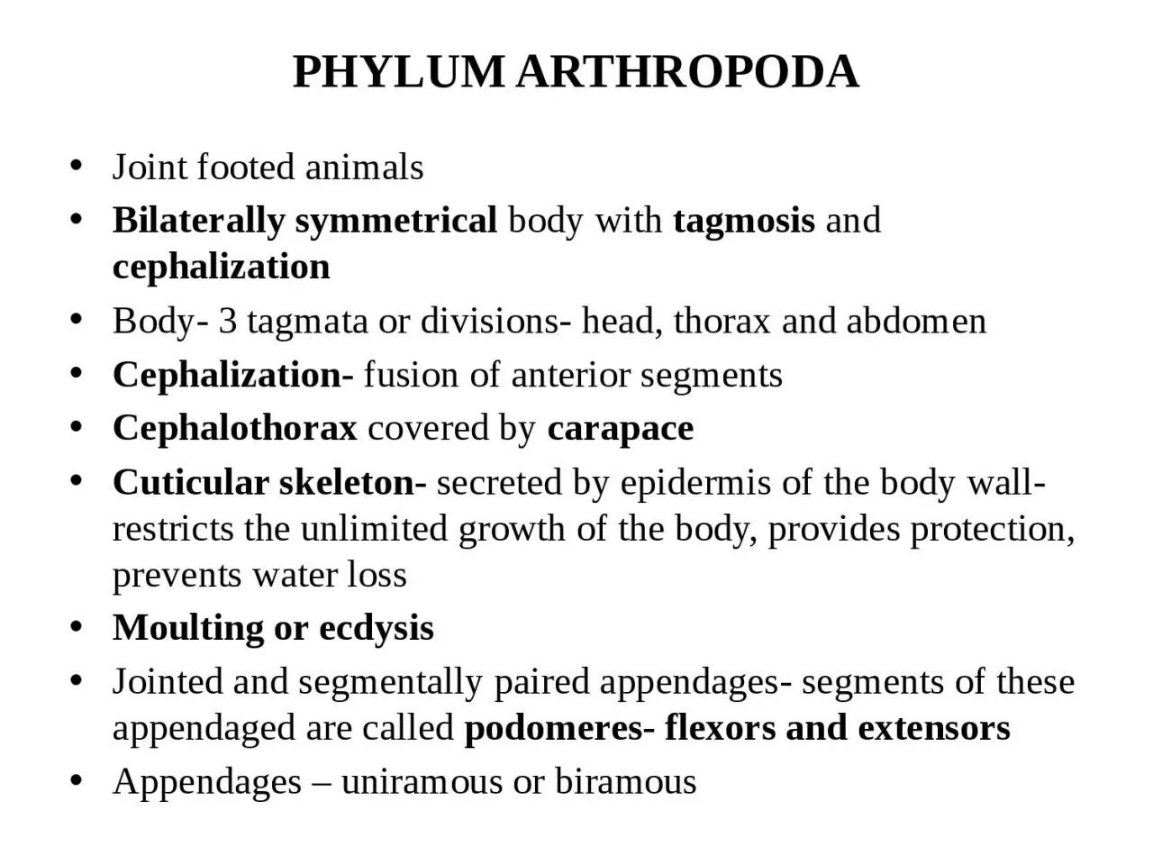
1. PHYLUM ARTHROPODAJoint footed animalsBilaterally symmetrical body with tagmosis and cephalizationBody- 3 tagmata or divisions- head, thorax and abdomenCephalization- fusion of anterior segmentsCephalothorax covered by carapaceCuticular skeleton- secreted by epidermis of the body wall- restricts the unlimited growth of the body, provides protection, prevents water lossMoulting or ecdysisJointed and segmentally paired appendages- segments of these appendaged are called podomeres- flexors and extensorsAppendages – uniramous or biramous
2. Striped musclesPerivisceral haemocoel and reduced coelomOpen circulatory system with dorsal heart – blood flows through open channels and spaces and not through closed blood vesselsRespiratory organs include branchiae, tracheae, book gills and book lungsExcretory organs - Malpighian tubules and green glandsAnnelidan type of nervous systemAbsence of locomotor cilia
3. PENAEUS INDICUS(class malacostraca)Cephalothorax (fusion of 13 segments-5 cephalic and 8 thoracic) and abdomen(6 segments) with a telsonCuticular skeleton, secreted by underlying epidermis(formed of chitin and protein)- has infolding called apodemesSegments called scleritesAbdominal region – terga (dorsal sclerite) and sterna (ventral sclerite)Pleuron extension from tergumEpimeron – the part between pleuron and abdominal appendage13 tergal plates fuse to form carapaceSternal plates form ventral shieldThelycum on femaleRostrum – compound eyesBranchiostegite - gill cover
4.
5.
6.
7. Appendages 19 pair- 5 cephalic , 8 thoracic and 6 abdominalAttach to sternum of exoskeletonSegmented (podomeres ) and biramous Base called protopodite- 2 segmented- coxa and basisRami- exopodite(unsegmented) and endopodite (segmented in some)Cephalic appendagesAntinnule and antennae- tactile structuresMandible – masticatory structures1st maxillae and 2nd maxillae- feeding jaws
8.
9. Thoracic appendages8 pairsMaxillipeds or foot jaws- first 3Pareopods or walking legs- rest 5All are biramous with 2 segmented protopodite5 segmented endopodite- ischium, merus, carpus, propodus and dactylusMaxillipeds- exopodite well developed and unsegmentedTo the coxa of maxillipeds and chelipeds is a respiratory process called epipoditePareopods- 1st 3 are chelate legs or chelipeds(formed by propodus and dactylus)Last 2 non chelate pareopods- no epipodite
10.
11.
12.
13. Abdominal appendagesFor swimming- called pleopods or swimmeretsBiramous- 2 segmented protopodite, unsegmented exopodite and endopoditeLast pair – uropods- serve as tail finsIn uropod coxa and basis fuse to form single segment – sympodIn male prawns- 1st pair of pleopods- endopodite has small hooks- with this right and left appendages interlock and form petasma (used for transferring sperms to the thelycum of female)
14. Digestive systemAlimentary canal and digestive gland, called hepatopancreas or liverAlimentary canal 3 divisions- stomodaeum, mesodaeum and proctodaeumStomodaeum (fore gut) consists of mouth, buccal cavity, oesophagus and stomachMesodaeum (mid gut)- intestineProctadaeum (hind gut) – rectum and anusIntima – internal cuticular lining of stomodaeum and proctodaeumMouth Situated in between 3rd and 4th cephalic segmentsGuarded infront by chitinous plate, called labrum or upper lip, on sides- mandibles and behind by labium or lower lipLabrum is bilobed- 2 lobes called paragnathae
15. Buccal cavity (compressed chamber) and oesophagusStomach Spacious chamberDivisions- cardiac (for storage and grinding of food) and pyloric stomach(for sorting and straining of food)On the floor of cardiac stomach- there are cuticular folds with setae and spiculesAlso has many tooth like denticles- serve as internal masticatory apparatus- called gastric millPyloric stomach also has cuticular folds- called lappets or valvulaeIt also has a filtering apparatus, formed of chitinous plates and comb like bristlesThen comes intestine, leads to rectum, opens out by anus(guarded by muscular sphincter)
16. Digestive glandOnly digestive gland is the bilobed hepatopancreasSurrounds the stomachDevelop from mid gut as hepatic caecaFormed of branching tubules held together by connective tissue called tunica propriaFrom each lobe is hetatopancreatic ductServes the function of liver, pancreas and intestinal glands of vertebratesSecretes proteolytic, amylolytic and lipolytic enzymesStores glycogen, fats and calcium and absorbs digested food
17. Feeding and digestionOmnivores – planktonic organismsChelipeds(food collection) – 2nd maxialle(push food into mouth) – mandibles(food cut into pieces - mastication) – maxillae and maxillipeds(masticated food is pushed to oesophagus) – reach stomach through peristaltic movements of oesophagusIn stomach- food mixed with digestive enzymes and get pulverized, strained and sortedIn cardiac stomach- internal mastication and churning – further contraction and expansion of stomach leads to crushing and grinding of food by gastric mill – churning(mixing with hepatopancreatic secretions)Digestion mostly in cardiac stomach – sometimes in hepatopancreas wherein they are ingested and digested by some phagocytic cellsAbsorption mostly in hepatopancreas- some in intestine
18. After gastric digestion, the pyloric filter separates the digested food and diverts it into hepatopancreas via hepatopancreatic ductsThe absorptive cells of hepato pancreas absorb itExcess nutrients are stored in the hepatopancreas as glycogen and fatsUndigested residue or faeces is 1st forced from stomach to intestine and finally passed to outside by peristaltic movements of gut wall
19. Respiratory systemInclude gills or branchiae –principalEpipodites or mastigobranchiae and the vascular lining of branchiostegites- accessory respiratory structuresGills - vascular and feathery or plumose outgrowths from the thoracic appendages18 gills on each side, lodged in the branchial chamber in between the branchiostegite and the thoracic wallEach gills is a crescentic structure, with a central axis or stem, called gill axisOn this there are numerous thin, flat and bilaterally arranged plates, called gill lamellae, which in turn divided into gill filaments- this gill of tree like fashion- called dendrobranchs or dendrobranchiaeGill axis and gill lamellae – is vascularised and covered by chitin layer
20. Each gill is attached to the body by a root through which blood vessels and nerves pass. Based on this 3 types of gillsPodobranchs- foot gills- attach to the coxa of the thoracic appendagesArthrobranchs- joint gills- remain attached to the arthrodial membrane, seen at the junction between the thoracic wall and appendagePleurobranchs- side gills or wall gills- remain attached to the inner wall of the branchial chamberIn penaeus1 pair of podobranchs- attached to 2nd maxillipeds 11 pairs of arthrobranchs, attached to the 2nd and 3rd pair of maxillipeds and 1st 4 pairs of walking legs6 pairs of pleurobranchs, attached to the segments to which the 3rd pair of maxillipeds and 1st 5 pairs of walking legs are connected
21. Mechanism of respirationVentilation of gills- scaphognathites (exopodite of 2nd maxillae) or balers vibrate- constant current of water over the gills , epipodites and branchiostegal plates- gaseous exchange takes placeSetose hairs at the entrance of gill chambers prevent entry of foreign particles into the gill chambers
22. Blood vascular systemOpen or lacunar typeBlood flows through open channels and spaces, sinuses and lacunaeConsists blood, heart, arteries and sinuses and lacunaeCapillaries and veins are absentBlood or haemolymph, is a light blue fluid - consists of plasma(with cu containing pigment haemocynanin) and colourless and amoeboid leucocytes. Hb and erythrocytes absentHeart triangular organ- in cephalo thoracic region- in the pericardial cavity below dorsal shield. Has 5 pairs of valvular lateral openings, called ostiaFrom heart arise many arteries, its branches open to blood channels and blood spaces, which collectively constitute the haemocoel
23. Heart arteries blood sinuses lacunae ventral sinuses afferent branchial channels gills efferent branchial channels pericardial sinus again to heart…so on
24. Excretory systemInclude a pair of antennary glands(coxal glands, green glands, or renal glands)4 parts- end sac, labyrinth, glandular tube and bladderEnd sac- closed mesodermal sac with a coelomic cavity. Lined by excretory cellsConnects with labyrinth through a small openingLabyrinth or glandular plexus- made of coiled renal tubulesGlandular tube – coiled excretory tube that leads from labyrinth and opens to bladderBladder – chamber- lined by excretory cells- renal duct – opens to outside by an renal pore
25. Green gland functionExcretion and osmoregulationExcretion and elimination of nitrogenous wastes and the excess of water and mineral ions and also in the regulation of the ionic composition and volume of bloodAmmonia + urea + uric acid – in prawnElimination occurs by diffusion across gills+From blood by ultrafiltration- reach end sac = called primary urine or ultrafiltrate – undergoes reabsorption in labrynth – the fluid left out in labrynth is called final urine, which contains water, ammonium compounds, uric acid, urea etc Urine collected in bladder and pass out through renal opening
26.
27. Nervous systemComposed of Circum oesophageal nerve ring , fused and ganglionated double ventral nerve cord and a large number of nervesCircum oesphageal nerve ring consists of Supra oesophageal ganglionic massAbove oesophagusUnspecialized brain – formed by fusion of several gangliaFrom it arise, a pair of antennular nerves to antennules, statocysts etc, a pair of antennary nerves to antennae and a pair of optic nerves to compound eyes
28. Sub oesophageal ganglionic massBelow oesophagusFormed by fusion of a few gangliaFrom it arise, 5 pairs of nerves – to mandibles, 1st maxillae, 2nd maxillae, 1st maxillipeds and 2nd maxillipedsCircum oesophageal connectives Around oesophagusTransverse nerve – called oesophageal commissure, commisural nerve or visceral loop Just below oesophagus
29. Double ventral nerve cordFusedHas 6 thoracic ganglia and 6 abdominal gangliaFrom each ganglion – 3 pairs of nerves – 2 pairs to muscles and 1 pair to appendagesBetween 11th and 12th body segments, 2 nerve cords remain separate, leaving a passage for sternal artery
30.
31.
32. Sense organsStatocystsFor orientation and equilibriumA pair of them in the pre coxal segment of antennulesConsists of a chitinous sac, attached to pre coxa through a short stalk.Chitinous sac encloses a central mass of sand grains (act as statoliths), surrounded by ring of sensory setaeA sensory setae has 2 parts – a swollen base (attached to statocyst wall through an arthrodial membrane) and a filamentar shaft (has many sensory bristles). Any change in the swimming position of the animal, or any movement in the surrounding water, will cause the displacement of sand grains. They press against and stimulate sensory setae. From setae impulses are transmitted to brain. Brain responds and enables the animal to properly orient itself
33.
34.
35. 2. Compound eyesA pair of stalked and movable compound eyesSeen at the base of rostrumCannot see distant objects as it has no perfect focusing mechanism or power of accomodationFormed of a large number of (2000 – 2500)light perceiving visual units or simple eyes called ommatidiaOmmatidiumAre long, rod shaped and closely packed structuresHas its own focusing system, light transmitting system, photoreceptor cells and a field of vision (overlap in case of adjacent ommatidia)Adjacent ones are separated by a layer of black pigmentSpecialized for refracting light rays and also for eliciting sensory impulsesIt has a transparent cuticular covering, which serves as a cornea or as biconvex lens
36. The cuticular cornea is rigid in shape- so it has a fixed focal length and is incapable of accomodation in relation to the changing distance of the objectsJust below cornea- a pair of epidermal cells, known as reticular cells or corneagen cells, which secretes the cuticular corneaInner to this is a group of 4 elongated and inwardly tapering crystalline cells, called cone cells or vitrellaeThese cells secrete and surround a central transparent and refractive body called cyrstalline coneThe vitrellae, inturn maybe surrounded by a pigmented sheath (that separates adjacent ommatidia), known as iris sheath or distal pigment sheath – formed of pigment cells- called chromatophores.
37. Beneath vitrellae – is a group of 7 or 8 elongated cells, called retinular cells or retinulae – they contain light sensitive visual pigment rhodopsin –Rhodopsin when sensitized by light undergoes chemical change which elicits an action potential transmitted to brain as a nerve impulseRetinular cells surround a central refractile rod – rhabdome, formed by interdigitation of microvilli of retinular cellsRetinular cells are surrounded by a pigmented sheath, called retinular sheath or proximal pigment sheathFrom each retinular cell is an axon at its terminal portion7-8 axons from each ommatidium join optic ganglionAxons from all ommatidia together form an optic nerve – which joins the brain
38. Dioptric apparatus or focusing apparatus – the portions of ommatidium that comprises the cuticular lens, lenticular layer and vitrellaeRemaining part –i.e, rhabdome, retinular cells and the reticular sheath forms receptor apparatus or receptor regionEach ommatidum rests on a basal membrane or basal lamina
39. Working of compound eye2 mechanismsMosaic visionIn bright lightPigment cells fully expand and spread all over the ommatidium – isolating adjacent ommatidia from each other- thus each becomes an individual visual unitThey will be stimulated only by the parallel or perpendicular rays falling on the cornea- thus each can form an image of only a portion of the objectAll the images obtained are integrated in the brainComplete image is formed by the apposition of fusion of of several partial images – called apposition image or mosaic imageSuch a vision called mosaic vision or apposition vision
40. Superimposed visionIn dim light or nightPigment cells contract very much – so adjacent ommatidia overlap each other – forming a visual complexImages formed by adjacent ommatidia overlap and vision becomes blurred – called superimposition image or super imposed imageObject cannot be seen clearly – but can detect moving objects
41.
42. ReproductionSexes are separateAlso some degree of sexual dimorphismMale has petasma and female has thelycumMale genital openings are seen on the coxal segment of the last pair of walking legs, but female openings are seen on coxa of the 3rd pair of walking legsMale organsInclude a pair of testes – one on each side of thoraxTestes - formed of numerous long and coiled seminiferous tubules2 Testes are connected anteriorly, while separate posteriorlyHas testicular diverticulaA vas deferens or sperm duct- which leads to a seminal vesicle or ejaculatory bulb- then comes ejaculatory duct that opens out to coxa of last walking leg
43. Secretions of seminal vesicle glue sperms together and form sperm clusters- spermatophores- stored in seminal vesicleFemale organsA pair of ovaries, one on each side of thoracic and abdominal regionsUnited posteriorly while separate anteriorlyOvarian diverticulaOviduct- opens to coxa of 3rd walking leg
44.
45.