
Ultrasound Bladder ScannerUser and Service Manual wwwvitaconcomVerRel NoRelease dateMod ByMod DateRev byrelevantstakeholdersRev dateAuth byAuth dateRemarkRevision details20320Dec19 MW MW MW Initia ID: 865470
Download Pdf The PPT/PDF document "VitaScan PD" is the property of its rightful owner. Permission is granted to download and print the materials on this web site for personal, non-commercial use only, and to display it on your personal computer provided you do not modify the materials and that you retain all copyright notices contained in the materials. By downloading content from our website, you accept the terms of this agreement.

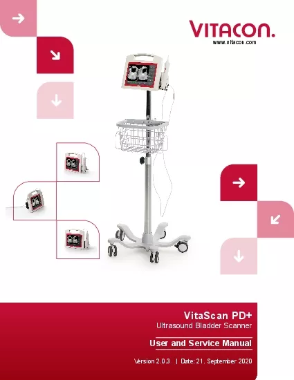
1 VitaScan PD+ Ultrasound Bladder Scanner
VitaScan PD+ Ultrasound Bladder Scanner User and Service Manual www.vitacon.com Ver/Rel. No Release date Mod. By Mod. Date Rev. by relevant stakeholders Rev. date Auth. by Auth. date Remark/Revision details 2.0.3 20-Dec-19 MW MW MW Initial release 20-Dec-19 20-Dec-19 20-Dec-19 2.0.3 7-Jan-20 MW MW MW Added required software version page 4. 7-Jan-20 7-Jan-20 7-Jan-20 2.0.3 21-Sep-20 MW MW MW Updated accuracy assessment procedure and recommendation (page 15) 21-Sep-20 21-Sep-20 21-Sep-20 The Information contained in this user and service manual is proprietary to Vitacon. It is only used for convenience of our customers. It may be changed in whole or in part without written notice. Any service work performed by persons who are not authorized by Vitacon may void your warranty. www.vitacon.com Vitacon provides this publication as is without warranty of any kind, either expressed or implied, including but not limited to the implied warranties of merchantability or �tness for any particular purpose. Further, Vitacon reserves the right to revise this publication and to make changes from time to time to the content hereof, without obligation to Vitacon or its local representatives to
2 notify any person of such revisions or
notify any person of such revisions or changes. Some jurisdictions do not allow disclaimers of expressed or implied warranties in certain transactions; therefore, this statement may not apply to you. In Europe: O�ce Address: Vitacon AS Vegamot 8B 7049 Trondheim, Norway E-mail: sales@vitacon.com http://www.vitacon.com In North America: Vitacon US 600 Twelve Oaks Center Drive Suite 103, Wayzata MN 55391, USA E-mail: info@vitacon.us http://www.vitacon.us Manufacturer: UAB Vitacon LT Lazdyneliu g. 12-43 04126 Vilnius Lithuania Copyright 2020. All rights reserved. PN: 6100-500 Vitacon warrants that the VitaScan PD+ will substantially conform to published provided that it is used for the purpose for which it was designed. Vitacon will, for a period of sixty (60) months from the date of purchase, replace or repair any defective device, if the fault is due to a manufacturing defect. In no event will Vitacon or its local representatives be liable for direct, indirect, special, incidental, or consequential damages arising out of the use of or inability to use the VitaScan PD+, even if advised of the possibility of such damages. Vitacon or its local representatives are not
3 responsible or claims by third parties
responsible or claims by third parties due to use of, or inability to use the VitaScan PD+. Neither Vitacon nor its local representatives will accept, nor be bound by any other form of guarantee concerning the VitaScan PD+ other than this guarantee. Some jurisdictions do not allow disclaimers of expressed or implied warranties in certain transactions; therefore, this statement may not apply to you. Disclaimer Contacting Vitacon Limited Warranty www.vitacon.com Tables of Contents 1 Introduction 2 Important Information 2 Indications for Use 3 Product Features 3 Unpacking and Inspection 3 Content of the packaging ....................................................................................................... 4 Storage ................................................................................................................................. 4 Technical Speci�cations 4 AC/DC Adaptor 5 Battery 5 Product Upgrades and Updates 5 System Familiarization 6 About the System Software ................................................................................................ 10 Electrical Safety 10 Equipment Safety 11 Safety and Performance Summary 1
4 1 Labeling Symbols 12 Applicable Stan
1 Labeling Symbols 12 Applicable Standards/Approvals .......................................................................................... 13 Applying the Ultrasound Gel 14 Measuring Bladder Volume 14 Regular Inspections and Maintenance 15 Care, Cleaning and Disinfecting 16 Powering up the System ..................................................................................................... 16 Scan Operation .................................................................................................................... 18 Adjust result 20 Setup 21 Patient infomation 22 Service ................................................................................................................................ 23 Manufacturer 26 1 www.vitacon.com Table of Contents PAGE VitaScan PD+ - Ultrasound Bladder Scanner Real-time bladder scanning is a safe and easy, non-invasive method to measure bladder volume. Bladder scanning a patient’s body and di�erentiates the urinary bladder from the surrounding tissues. The VitaScan PD+ is a B-mode ultrasound instrument, portable and battery operated, intended for the non- invasive measurement of urinary bladder volume. A me
5 chanical sector scanning transducer pro
chanical sector scanning transducer provides cross-sectional images of the bladder from up to twenty- four scan planes. Based on these images the VitaScan PD+ automatically calculates the estimated bladder volume in milliliters and displays it on a screen. VitaScan PD+ is applicable in many clinical areas to determine bladder volume, time for bladder emptying and detection of post-void residual volume (PVR). A real-time image of the bladder during pre-scan makes it easier to detect the bladder before scanning. Notice To All Operators The VitaScan PD+ should be used only by individuals who have been trained and authorized by a physician or the institution providing patient care. All operators should read this manual prior to using the VitaScan PD+. Failure to comply with these instructions may compromise the performance of the instrument and the safety of the patient. Biological Safety To date, exposure to pulsed diagnostic ultrasound has not been shown to However, ultrasound should be used only by medical professionals when clinically indicated, using the lowest exposure times possible commensurate with clinical utility. The ultrasound output power of the VitaScan PD+ is not use
6 r-adjustable and is limited to the mini
r-adjustable and is limited to the minimum level necessary acoustic output levels can be found in the section titled, “Technical Speci�cations” in this manual. It is recommended that users read the Health Canada Guidelines for the Safe Use of Diagnostic Ultrasound before using this, or any other diagnostic ultrasonic device. (http://www.hc-sc.gc.ca/ ewh-semt/alt_formats/hecs-sesc/pdf/ pubs/radiation/01hecs-secs255/01hecs- secs255-eng.pdf, note this link may change over time). Statement of Intended Use The VitaScan PD+ projects ultrasound energy through the lower abdomen of the patient to obtain an image of the bladder. This image is used to determine bladder volume noninvasively. Contraindications The VitaScan PD+ is not intended for fetal use or pregnant patients. Warning: Exposure to low power diagnostic ultrasound has not been shown to produce adverse e�ects. However, medical professionals should use ultrasound only when clinically indicated. Warning: There is the hazard of possible explosion if the VitaScan PD+ First time users: We advise new operators to use the VitaScan PD+ on patients with moderately full bladders, rather than initially attempting
7 to locate nearly empty bladders. Intr
to locate nearly empty bladders. Introduction Important Information ! ! 2 PAGE www.vitacon.com Caution: The VitaScan PD+ should not be used on a patient with open skin or wounds in the suprapubic region. • The manual measurement function should be used on patients with ultrasound signals that can lead to an inaccurate volume measurement. • User care with suprapubic/pelvic surgery patients, Scar tissue, incisions, • peritoneum. Indications for Use This manual is directed toward the reader who is familiar with Ultrasound techniques. Sonography training and clinical procedures are not included here. This manual is not intended as training material for the principles of ultrasound, anatomy, scanning techniques, or applications. You should be familiar with all of these before attempting to read this manual or using the device. Product Features • Real-time 3D Ultrasound Bladder Scanner. • Automatic Urinary Bladder volume calculation in large digits • Simple, intuitive software with Touch- Screen support • Touch Screen • USB port to save on external memory device • Integrated carry handle • Battery operated Unpacking and Inspection There are
8 no special unpacking instructions, but
no special unpacking instructions, but be careful not to damage the instrument when unpacking it. When unpacking the VitaScan PD+ to check for damage during shipment: • Inspect the shipping carton for damage. If the shipping carton is damaged, carefully continue unpacking the instrument and note any dents and scratches on the VitaScan PD+. Save the damaged shipping carton and packing material for the carrier’s inspection and contact the respective carrier. If there is any damage to the scanner equipment, contact Vitacon. • If there is no shipping damage, continue removing the VitaScan PD+ from the shipping case. Save the box and packing materials; they will be needed when returning the VitaScan PD+ to Vitacon for recalibration or future service. • Verify that all items listed on the packing list have been received and are in good condition. ! Note: PD+. Save these for future shipment of the unit for service or calibration. 3 PAGE www.vitacon.com The content of the packaging • VitaScan PD+ Console with thermal printer • VitaScan v.2 ultrasound probe and probe holder. • Power cord Storage If the system is to be stored, pack it in the original container, and ke
9 ep it in an environment , and vibration
ep it in an environment , and vibration and shock. Storage Requirements Storage temperature from -30°C to 50°C Relative humidity of 20% to 90% @ 30°C, non-condensing Atmospheric pressure from 700 hPa to 1060 hPa As with most electronic equipment, the unit should be operated in a dry area within normal temperature limits (+10°C to +45°C, 10% - 80% humidity). Dispose of electronic waste VitaScan PD+ complies with the WEEE Directive (2002/96/EC) marking requirements. domestic household waste. Product category: With reference to the equipment types in To return unwanted products, contact Vitacon at the address mentioned at the front of this manual or your local VitaScan distributor. Technical Speci�cations • Display type LCD Touchscreen Display - 10.1 Inch • Input method Touchscreen • Volume range 0 – 1000 ml • Accuracy ± 7.5 % on volumes greater than 100 ml ¹ • ....................................................................... ± 7.5 mL on volumes less than 100 ml ¹ • Frequency ....................................................................................................... 3.40 MHz • Acoustic Output ................
10 ...................................... M
...................................... MI max: 0.38, Power: 0.25 mW/cm² • Thermal Index – TI • Scanning method Sector, 180 degrees • Rotation positions 12 or 24 rotating positions • Sweep angle 130 degrees • Max detection depth 100, 160 or 230 mm • Max probe temperature 35°C @ 22°C ambient temperature • IP rating IPX7 • Input Voltage 100 ~ 240VAC, 50 ~ 60Hz • Dimension (D x W x H) 9 x 27 x 23.5 cm - 3.5 x 10.6 x 9.3 in • Weight 2.2 kg - 4.8 lbs • Operating conditions +10°C to + 45°C, 10% - 80% relative humidity • Storage temperature .............................................................................. -30°C to +50°C phantom and software version 2.0.3 and above. 4 PAGE www.vitacon.com AC/DC Adaptor Input 100-240VAC Frequency50 - 60Hz Input current0.7- 0.35A Output Maximum Power25W Table 0-0 Battery Speci�cations Product Upgrades and Updates Vitacon may o�er software upgrades and new features that may improve system new features on system performance, will accompany the upgrades. Number Item Speci�cations 1 Rated Capacity (minimum) 13200mAH with 0.2C Charging & 0
11 .2C Discharging 2 Nominal Capacity 132
.2C Discharging 2 Nominal Capacity 13200mAH with 0.2C Charging & 0.2C Discharging 3 Normal Voltage 11.1V 4 Open circuit voltage when shipped 11.40V - 11.85V 5 Charge ending voltage 12.60V 6 Discharge ending voltage 9.0V 7 Battery run time 5 - 6 hours 8 Charging time 4 – 5 hours 5 PAGE www.vitacon.com Battery 1 Touch Display 2 Ultrasound probe 3 ON/OFF button System Familiarization Introduction The VitaScan PD+ is an Ultrasound Urinary Bladder Scanner System. The VitaScan PD+ consists of an inbuilt LCD Display with Touch Screen input and utilizes a VitaScan v.2 Ultrasound Probe. Front View The front view of the VitaScan PD+ is as shown in the �gure below: 6 PAGE www.vitacon.com 1 1 2 2 3 3 7 PAGE www.vitacon.com 1 VESA mount - 75 x 75 mm 2 Probe holder mount 3 Thermal printer 4 DC Power Inlet – 15V Back View The back view of the VitaScan PD+ is as shown in the �gure below: 1 1 2 2 3 3 4 4 8 PAGE www.vitacon.com 1 Power ON/OFF switch 2 Thermal printer Left Side View The left side view of the VitaScan PD+ is as shown in the �gure below: 1 1 2 2 9 PAGE www.vitacon.com 1 USB port for external USB memory stick 2 USB port for VitaScan v.2 pr
12 obe 3 Probe holder Right Side View T
obe 3 Probe holder Right Side View The right side view of the VitaScan PD+ is as shown in the �gure below: 1 1 2 2 3 3 About the System Software The VitaScan PD+ system contains software that controls its operation. Vitacon will provide software updates on micro SD card. Typically, the new software provides new capabilities. Electrical Safety This system meets EN60601-1, Class I and Type BF isolated patient-applied parts safety requirements. This system complies with the applicable medical equipment requirements published in the European Norm Harmonized Standards, Underwriters Laboratories (UL) and the Canadian Standards Association (CSA). For maximum safety observe the following warnings and cautions: 10 PAGE www.vitacon.com Warning: To avoid the risk of electrical shock or injury, do not open the system enclosure. All internal replacements must be made by a To avoid the risk of injury, do not operate the or anesthetics. To avoid the risk of electrical shock, use only properly grounded equipment. Shock hazards exist if the power supply is not properly grounded. Grounding reliability can only be achieved when equipment is connected to a receptacle marked “Hospital The
13 grounding wire must not be removed or
grounding wire must not be removed or defeated. Connect equipotential ground terminal whenever integrity of the external protective earth conductor arrangement is in doubt. To avoid the risk of electrical shock, before using the VitaScan PD+, inspect the housing, cable, and probe. Do not use the VitaScan PD+ if these are damaged. To avoid the risk of electrical shock, always disconnect the power inlet before cleaning the system. To avoid the risk of electrical shock, do not use any transducer that has been accidentally immersed in any liquid, or has been immersed in any liquid for cleaning or any other purpose. To avoid the risk of electrical shock, do not touch USB port or Ethernet port and the patient at the same time. Caution: Although your system has been manufactured in compliance with existing EMC/EMI requirements (EN60601-1-2), use of the system in the cause degradation of the ultrasound image. If this occurs often, Vitacon suggests a review of the system environment. Identify and remove the possible sources of the emissions or move your system. by portable or mobile RF communication devices. Turn OFF any portable or mobile RF device before operating your system. Elect
14 rostatic discharge (ESD), or static sho, or static
sho")
rostatic discharge (ESD), or static shock, is a naturally occurring phenomenon. ESD is common in conditions of low humidity, which can be caused by heating or air conditioning. Static shock is a discharge of electrical energy from a charged body to a lesser or non-charged body. The degree of discharge to a transducer or an ultrasound system. The following precautions can help reduce ESD: anti-static spray on carpets, anti-static spray on linoleum, and anti-static mats. Do not use the system if an error message appears on the display: note the error code; turn o� the system; call Vitacon or your local representative. ! ! 11 PAGE www.vitacon.com Equipment Safety To protect your ultrasound system, scanner and accessories follow these precautions. Caution : • To avoid the risk of excessive heating or damage to the system, use the system in a well-ventilated environment. • If the operating environmental temperature exceeds 25°C, limit scans to 5 minutes and allow a 10 minute cooling period between scans. • Excessive bending or twisting of cables can cause a failure or intermittent operation. • Do not submerge the VitaScan PD+ in any solution, follow the cleani
15 ng instructions. • To avoid damagi
ng instructions. • To avoid damaging the power supply, verify the power supply input is within the correct voltage range. • Do not short the USB or Ethernet terminals. • Always charge the battery before using the system, to avoid the risk of the • Incorrect cleaning or disinfecting of any part of the system can cause permanent damage. • Do not use solvents such as thinner or benzene, or abrasive cleaners on any part of the system. • Do not spill liquid on the system. • Do not use the system if it exhibits erratic or inconsistent behavior. Turn Customer Service. • • Immediately discontinue use of the battery if, while using, charging or storing the battery, the battery emits an unusual smell, feels hot, changes color or shape, or appears abnormal in any other way. Contact a customer service representative if any of these problems are observed. • Do not use the VitaScan PD+ if probe head or cable is damaged. • Do not use the VitaScan PD+ if there is evidence of leakage of internal liquids. Wash hands immediately in warm, soapy water. Consult the MSDS on Polypropylene Glycol for additional information/precautions. • To avoid the ris
16 k of electrical shock, do not use any V
k of electrical shock, do not use any VitaScan PD+ that has been immersed in liquid. Safety and Performance Summary The VitaScan PD+ computes the volume of the urinary bladder based upon twenty-four cross-sectional ultrasound images (or less). For maximum accuracy, be sure to hold the Scan head motionless while scanning. The most accurate measurements are obtained when the patient rests quietly in the supine position. Accuracy is compromised if the user does not obtain an optimal, repeatable image. Errors in usage tend to result in the under- estimation of bladder volume, except in cases where the Scan head is moved during scanning. In this case, the measurement may overestimate the patient’s bladder volume. The patient being scanned should not have a catheter in his/her bladder. This could create micro bubbles in the bladder, which Do not use the VitaScan PD+ on patients with open skin or wounds in the suprapubic region. Use care when scanning suprapubic and pelvic surgery patients. Scar tissue, surgical ! 12 PAGE www.vitacon.com Warning : There is a possible hazard of explosion if the VitaScan PD+ is used in the presence of Labeling Symbols ! ! CE mark – Noti�ed body N
17 o. 2274 Warning, consult accompanying
o. 2274 Warning, consult accompanying documents Read the documentation Ultrasound radiation Drip proof Alternating current input Test Agency Certi�cation Mark – North America BF type (Body Floating) WEEE - Waste Electrical and Electronic Equipment Applicable Standards/Approvals 93/42/EECCouncil Directive concerning medical devices EN ISO 13485:2012+ AC:2012 Requirements for regulatory purposes Medical devices – Application of risk management to medical devices EN ISO 14155:2011 — Good clinical practice EN 60601-1:2006+ AC:2010 + A1:2013 General requirements for basic safety and essential performance EN 60601-1-1:2001Medical electrical equipment. Part 1-1: General requirements for safety. Collateral standard: Safety requirements for medical electrical systems (IEC 60601-1- 1:2000) EN 60601-1-2:2015Medical electrical equipment Part 1: General requirements for safety 2.Collateral Standard: Electromagnetic compatibility - Requirements and tests EN 60601-1-4: 1996+ A1:1999 requirements for safety- Collateral Standard: Programmable electrical medical systems EN 60601-1-6:2010Medical electrical equipment-Part 1-6: General requirements for safety- Collateral Standard: Usability
18 EN 60601-2-37:2008+A11:2011 requirements
EN 60601-2-37:2008+A11:2011 requirements for the safety of ultrasonic medical diagnostic and monitoring equipment) EN 62304:2006+АС:2008 Medical device software – Software life-cycle processes Medical Devices. Application of usability engineering to medical devices EN 60529:1991+A1:2000 Degrees of protection provided by enclosures (IP code) 13 PAGE www.vitacon.com Applying the Ultrasound Gel Palpate the patient’s symphysis pubis (pubic bone) and apply the Gel immediately superior to the patient’s symphysis pubis, as shown in images below. Or apply the Gel around the dome of the Probe. Smooth the gel out and remove any air bubbles, which may block ultrasound transmission. Measuring Bladder Volume Palpate the patient’s symphysis pubis and place the Scanhead midline on the patient’s abdomen, approximately 4 cm (1.5 inches) superior to the symphysis pubis, as shown in images below. • Aim the Scanhead so the ultrasound is projected toward the expected location of the bladder. For most patients, this means aiming the tip of the Scanhead toward the patient’s coccyx. • Press and release the scan button, located on the Scanhead. • Locate the bladder.
19 • Press and release the scan butto
• Press and release the scan button and hold the Scanhead steady throughout the scan. Note: While scanning, avoid making any changes in the position, angle or pressure of the Scanhead. 14 PAGE www.vitacon.com Regular Inspections and Maintenance VitaScan PD+ is a Medical Electric Equipment and therefore needs special precautions regarding EMC. VitaScan PD+ needs to be installed and put into service according to the EMC information provided in the accompanying documents. Weekly Inspections Once a week, you should inspect the Scanhead and cable for physical faults or cracks. performance of the instrument. Any apparent faults or cracks must be referred to your authorized VitaScan Service Center or your local VitaScan distributor. Accuracy assessment : Caution: In the event of changes in the performance of the instrument, discontinue use and contact your authorized VitaScan Service Center or your local VitaScan distributor. Whenever accuracy assessment is desired or in question, the accuracy of the VitaScan should be tested using one of the following procedures: 1. Perform an o�-line veri�cation test using a Veri�cation Test Tool; or 2. Perform an online integrity an
20 d calibration service using a Calibratio
d calibration service using a Calibration Test Tool; or 3. Take a measurement on a Vitacon Phantom. 4. Compare patient measurement Take a Pre-void measurement of bladder volume. Void or catheterize into a measuring beaker. Take a post-void measurement of bladder volume to check for post-void residual phantom. Vitacon recommends preforming an accuracy assessment once a year. No annual inspection and maintenance is required on your VitaScan PD+. If accuracy assessment fails, you should: • Contact your local dealer to learn about options for sending in the device for manufacturer calibration and service. ! 15 PAGE www.vitacon.com Care, Cleaning and Disinfecting Clean the VitaScan PD+ with a soft cloth soaked in a mild liquid detergent solution. Rinse with clean water and carefully dry with a clean soft cloth. Dampen a soft cloth with 70% ethanol. Wipe the probe with the dampened soft cloth and let the ethanol evaporate. If the VitaScan PD+ needs to be disinfected, we recommend CIDEX® OPA Solution, PDI Sani-Cloth AF3 Germicidal Disposable Wipes, or other comparable disposable wipe designed for use on non-porous plastic surfaces. You may also use any glutaraldehyde based hospital
21 disinfection solution or Clorox Healthca
disinfection solution or Clorox Healthcare bleach products. Dampen a soft cloth and wipe the instrument thoroughly. To remove all traces of disinfection solution, wipe the VitaScan PD+ with a clean soft cloth dampened with sterile water or cleaning solution. Carefully dry the VitaScan PD+ with a clean soft cloth before use. Use appropriate hand protection according to the labeling on the disinfectant to avoid skin reactions. Warning: • Do not subject any part of the VitaScan PD+ to steam sterilization or ethylene oxide sterilization. • Do not immerse the instrument in any cleaning or disinfecting solution. Powering up the System Running on Mains Power. Push the Mains Switch ON - located at the left-hand side. Press the ON/OFF button on the front. Once the VitaScan PD+ is powered ON, the system boots up and the Home Screen is displayed. Running on Battery: Push the Main Switch OFF – located at the left-hand side. ! 16 PAGE www.vitacon.com Figure 0-1 Home Screen • Tap on the screen, to navigate through the screen. • Tap Woman or Man option • For Woman tap Hysterectomy if needed • Tap Scan Depth option • Tap Start to start Prescan and locate the bladder.
22 • Tap Restart to start a new sca
• Tap Restart to start a new scan • Tap Setup to enter setup menu 17 PAGE www.vitacon.com Scan Operation To make a measurement, press the Start button in the Home Screen. The Prescan displayed as shown below. Figure 0-2 PreScan • Tap Scan to start scanning of the whole bladder. • Tap Restart to stop pre-scan. • Tap Icon to colorize the detected area. 18 PAGE www.vitacon.com Figure 0-3 Result • Tap Start to do a new scan • Tap Clear/Restart to clear screen • Tap Print to print result • Tap Save to save result to USB memory stick. (Only enabled when USB-memory stick is connected) • Tap Adjust to adjust result. (Adjustment might be needed if bladder edge is not detected properly) 19 PAGE www.vitacon.com Adjust result • Tap to Adjust transversal plane. (Move blue marker points to bladder edge) • Tap to Adjust sagittal plane. (Move blue marker points to bladder edge) • Tap Done to end adjustment 20 PAGE www.vitacon.com Setup Figure 0-5 Setup • Tap H:M:S to toggle between Hour – Minutes – Second. Change setting by – or + • Tap Y-M-D to tog
23 gle between Year – Month – Day
gle between Year – Month – Day. Change setting by – or + • Tap “ 6 12 • Easy-Mode (toggle between Yes or No ) to set only one slices in a scan. • Tap Live-Update to set continuous reading in pre-scan • Phantom (toggle between Yes or No ) to measure on Vitacon bladder phantom 21 PAGE www.vitacon.com Patient: ID, Name and Facility 22 PAGE www.vitacon.com Figure 0-6 Setup • • • • To enable factory defaultsettings: type “DEFAULT” in PatentID �led. • To disable facotry default settings: type “DEFAULT” in PatentID �led again. The Information contained in this service manual is proprietary to Vitacon. It is supplied solely for the convenience of our customers. It may be changed in whole or in part without the written notice. This manual is not intended as support or any unauthorized servicing, disassembly, VitaScan PD+ by unauthorized third parties. Introduction This section contains the IVBM VUFE design description. IVBM VUFE is the electronic module in the VitaScan PD+ bladder volume scanner. VitaScan PD+ is a 3-dimensional ultrasonic scanning device used to measure the bladder volume and quantity of
24 urine remaining in the bladder; safely
urine remaining in the bladder; safely and comfortably through a non-invasive method. The VitaScan v.2 probe consists of mechanics and electronics. The VitaScan PD+ contains the mechanical components needed to execute the 3 dimensional scanning of the bladder, and the electronics to obtain the ultrasound image needed to calculate the volume of urine in the bladder both pre and post void. The VitaScan v.2 probe is attached to the computer via a USB connection, which also provides the necessary power to the probe. This document only covers electronics, IVBM VUFE. Overview The drawing below shows an overview of the complete VitaScan PD+ bladder scanner system. All modules are physically located inside the VitaScan PD+. Service Block diagram for VitaScan v.2 Probe Vitascan v.2 Probe AnalogFrontendTransducerSweepMRotatio FPGSignalProcessingPush Button MicrControlle Motor Driver USBPSU M 23 PAGE www.vitacon.com Functional Description The IVBM VUFE is a single element ultrasound scanner with the electronic module and scanning mechanics, designed to �t in a hand held scanner, having the following main functionality: • USB communication interface for computer • Ultrasoun
25 d Pulse generator • Ultrasound rece
d Pulse generator • Ultrasound receiver and signal processing • Motor control and positioning of the ultrasound transducer element The module is realized in analog and digital hardware, FPGA con�guration, and microcontroller �rmware. The following are the main parts of the design: • Microcontroller • FPGA • PSU • USB • Motor driver • Analogue Receiver and transmitter Microcontroller The microcontroller is the master in the IVBM VUFE-module, controlling all functionality and safety. It is also a communication interface between the probe and the computer. The microcontroller is a slave with respect to the computer, and will only communicate in response to a command from the computer. Microcontroller states of operation: Power up: At normal power up, a self-test and initiation procedure will start. The FPGA con�guration will be downloaded from the computer before ultrasound acquisition may start. The transducer element positions will be initialized. At power up with the push-button pressed, the module will enter connection. This is a maintenance function, and should only be used by trained service personnel. Idle: In
26 Idle state, the module is waiting for a
Idle state, the module is waiting for a command from the computer or the pushbutton. The current consumption is reduced to an absolute minimum. Pre-Scan: In Pre-Scan mode, the module will move the transducer sweep-motor continuously between the end positions, and ultrasound data for one vector will be transmitted to the The rotation-motor is parked in the initial position. This will give the computer the possibility to present a “live” image of the bladder, making it possible to move the VitaScan v.2 probe to the correct position before making a Full-Scan with volume calculation. Full-Scan: In Full-Scan mode, the module will move the transducer sweep-motor from one end position to the other, transmitting ultrasound data for one vector to the computer at each de�ned position. The rotation motor will then move one step, and a new sweep will be performed. When the rotation motor has moved a total of 180 degrees, and the computer has a set. FPGA: The FPGA (Field Programmable Gate Array) is responsible for all signal- processing in the IVBM VUFE module. FPGA with “Bitstream” data and computer. The microcontroller initiates an Trigger signal. The FPGA will generate
27 an ultrasound pulse according to the an
an ultrasound pulse according to the and send data for the vector to the microcontroller after signal processing. PAGE www.vitacon.com P SU: The PSU (power supply unit) in the IVBM-VUFE module get its input voltage from the USB-connection. The computer will provide all power needed by the IVBM-VUFE module, including motors and transducer that are driven from this module. The power consumption must be lower than the maximum rating of power to be provided by the computers USB port. 1.2V: FPGA 3.3V: Microcontroller, 5.0V: Analog receiver, Motor driver logic Motor: HW-programmable voltage for the motors to be used USB: The USB controller is an integral part of the microcontroller. The IVBM-VUFE USB will be the SLAVE, and the computer will be MASTER. Implementation of the USB �rmware follows the USB 2.0 standard. Motor driver: The motor driver is designed for running Stepper-motors, and consists of three parts: • A HW part that will transform logical control signals into currents driving the motors. • A SW part in the microcontroller that will keep track of position of each motor, and generate logical control signals to move the motors to correct positions dep
28 endent on operating mode. • A HW HA
endent on operating mode. • A HW HAL-sensor and magnet to be used as a reference to calibrate sweep- motor position. • The motor driver is designed to support Full-step, Half-step and Micro-step modes, but for maximum motor-torque the Full-Step mode is used. The motor supply voltage is made HW- motors. Analog Receiver and Transmitter: The transmitter is a bipolar “square- wave” pulse transmitter. The voltage level is settable to tree levels by software/con�guration data control. The waveform is controlled by a sequence generator in the FPGA. The input signal from the transducer is attenuated by a t/r-switch during the pulse transmit period. The t/r-switch is controlled by a sequencer in the FPGA. The receiving signal is ampli�ed by a TGC-ampli�er chain. The gain control signal is set by an analog ramp signal, controlled from the FPGA. The ampli�ed analog receive L-C �lter before it is fed to the A/D- converter. The A/D converter is clocked from the FPGA. Resolution is 12 bits, and sampling speed is 12.5MHz. The parallel output data is fed to the FPGA for further digital processing. Safety Mechanisms: The following sa
29 fety mechanisms are implemented in the
fety mechanisms are implemented in the IVBM VUFE module: TX-Voltage and Input voltage monitoring Software Watchdog: The microcontroller is supervising the safety, and a watchdog is supervising the microcontroller program execution. In case of a SW/HW error that causes abnormal program execution, a HW- reset condition will occur and the High Voltage will be disabled. The HW has a measurement circuit allowing the microcontroller to measure the High Voltage and the Input voltage. The microcontroller will disable the TX Voltage if the voltage is out of the pre- an error will be sent to the computer. If the Input voltage reaches the minimum value (4.2V), an error message will be sent to the computer. If the input voltage is too low to drive the IVBM VUFE, a HW-reset condition will occur and the High Voltage will be disabled. 25 PAGE www.vitacon.com Vitacon will upon request make available other technical personnel to repair the equipment. Repair should be performed only by Vitacon authorized service organization. 2274 Copyright 2020 by Vitacon. All rights re served . www.vitacon.com Manufacturer: UAB Vitacon LT 04126 Vilnius Lithuania VitaScan PD+ User and Service Manual VitaScan PD+