
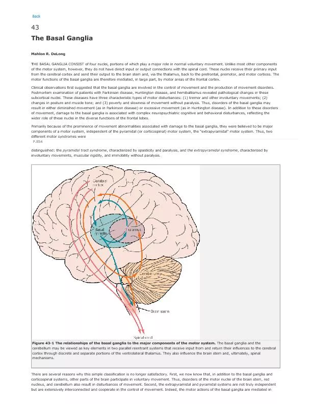
SR JNMC ALIGARH BASAL NUCLEI The basal nuclei or ganglia are large subcortical masses of grey matter located inside the white matter in the basal part of the cerebral hemisphere ID: 928397
Download Presentation The PPT/PDF document "Basal nuclei By Dr. Roberton Gautam" is the property of its rightful owner. Permission is granted to download and print the materials on this web site for personal, non-commercial use only, and to display it on your personal computer provided you do not modify the materials and that you retain all copyright notices contained in the materials. By downloading content from our website, you accept the terms of this agreement.


Slide1
Basal nuclei
By Dr. Roberton Gautam
SR, JNMC ALIGARH
Slide2BASAL NUCLEI
The
basal nuclei (or ganglia) are large
subcortical
masses
of grey
matter located inside the white matter in the basal
part of
the cerebral hemisphere
.
Anatomically, the term
basal ganglia include:
(a) corpus striatum,
(b)
claustrum
, and
(c)
amygdaloid
body
.
Functionally, basal ganglia also include
substantia
nigra
,
red nucleus
, and
subthalamus
.
Slide3Horizontal section of brain
Slide4Slide5AXIAL SECTION OF BRAIN
Slide6CORONAL SECTION OF BRAIN
Slide7CORPUS STRIATUM
The
corpus striatum is situated lateral to the thalamus.
D
ivided
into
the
caudate
nucleus and
the
lentiform
nucleus by a band
of
nerve
fibres
, the
internal capsule.
The
lentiform
nucleus
consists of two
parts
a darker lateral
part
putamen
and
a
medial paler part
globus
pallidus
.
Phylogenetically
, corpus striatum forms two
distinct functional units
the
paleostriatum
and the
neostriatum
.
The
globus
pallidus
is relatively ancient and
termed
paleostriatum
/
pallidum
.
The
caudate nucleus and
putamen
being
recent in development, together form the
neostriatum
/striatum
.
Slide8The anterior aspect of a coronal section through the leftcerebral hemisphere.
Slide9CAUDATE NUCLEUS
Caudate
nucleus is a large comma-shaped mass of
grey matter
, which surrounds the thalamus and is
itself surrounded
by the lateral ventricle.
The
whole length of
its convexity
projects into the cavity of lateral ventricle
.
Its rounded anterior part in front of
interventricular
foramen
is called its
head
.
The
head
gradually
and
imperceptibly
tapers caudally into the
body
then
into
a tail
,
which merges at its anterior extremity with an
almond shaped
mass
of grey matter called
amygdaloid
body.
Slide10The striatum within the left cerebral hemisphere
Slide11LENTIFORM NUCLEUS
A
large lens-shaped (biconvex) mass
of grey
matter beneath the
insula
forming the lateral
boundary of
the internal capsule.
In
horizontal section of cerebrum,
it appears
wedge shaped with broad convex base
directed laterally
.
D
ivided
into two parts by a vertical plate of
white matter
(external
medullary
lamina):
an
outer darker part,
the
putamen
and
an
inner lighter part the
globus
pallidus
.
The larger lateral part, the
putamen
consists of
densely
packed
small cells, and is structurally similar to the
caudate nucleus
.
The
globus
pallidus
is smaller medial part and consists
of
large
(motor) cells.
The
globus
pallidus
is
further
subdivided by an
internal
medullary
lamina of white
matter
into
outer and inner segments.
Slide12CONNECTIONS OF CORPUS STRIATUM
The
striatum (caudate nucleus and
putamen
) is the
receptive part
while
globus
pallidus
is the
efferent part
(
outflow centre
) of the corpus striatum.
The striatum
receives
fibres
mainly from the
cerebral cortex
, thalamus, and
substantia
nigra
.
The
globus
pallidus
sends
fibres
to the thalamus,
subthalamus
,
substantia
nigra
, reticular
formation, and red nucleus.
Different pathways involve different
neurotransmitters, which
include dopamine, acetylcholine, glutamate,
and
γ-
aminobutyric
acid (GABA).
Slide13Slide14CLAUSTRUM
Claustrum
is a thin saucer-shaped mass of grey matter
situated between the
putamen
and
insula
.
Its significance is not known.
Slide15AMYGDALOID BODY (OR AMYGDALA)
Amygdaloid
body is an almond-shaped mass of grey
matter in
the temporal lobe, lying
anterosuperior
to the tip
of inferior
horn of lateral ventricle.
It
is situated deep to
uncus
.
The
fibres
arising from
amygdaloid
body form
stria
terminalis
.
The
stria
terminalis
is the main efferent tract of the
amygdaloid
body
.
Developmentally it is related to the basal nuclei
but functionally
it is included in the limbic system, and
therefore, shares
its
functions.
Slide16SUBSTANTIA NIGRA AND RED NUCLEUS
SUBTHALAMUS (SUBTHALAMIC NUCLEUS)
This small nucleus in the ventral part of the diencephalon,
which looks like a biconvex lens in coronal section. It
is located
caudal to the lateral half of the thalamus
and
inferomedial
to
globus
pallidus
.
It
is separated from
thalamus by
a smaller nucleus called
zona
inserta
.
Slide17Caudate nucleus, Amygdaloid Body and
Stria
Terminalis
Slide18FUNCTIONS OF THE BASAL NUCLEI
Concerned
with planning and programming
of voluntary movements.
Determine
how rapidly a movement is to be
performed and
how large the movement must
be.
Decrease
muscle tone and inhibit unwanted
muscular activity.
Regulate
the muscle tone and thus help in
smoothening the
voluntary motor activities of the
body.
Control
automatic associated movements, like
swinging of
arms during
walking.
Control
group of movements responsible for
emotional expression.
Control
reflex muscular activity.
Slide19Clinical
Lesions of basal ganglia: The lesions of basal
ganglia
result
in various forms of unwanted involuntary
movements and
disturbance in the muscle tone.
These
disorders
include Parkinsonism
chorea,
athetosis
, and
ballismus
.
The Parkinsonism
being the commonest
disorder.
•
Parkinsonism (also called Parkinson’s
disease/paralysis
agitans
): This disease usually occurs after 50 years of
age due
to deficiency of the neurotransmitter dopamine in
the corpus
striatum following a lesion in
substantia
nigra
and/ or
its
nigrostriate
fibres
.
Characteristic
features of Parkinsonism:
–
Resting tremors.
–
Lead-pipe or cogwheel type of muscular rigidity.
–
Pill-rolling movements of hands.
–
Mask-like face or loss of facial expression.
–
Stiff, shuffling gait.
–
Stooped posture.
–
General slowing-down of movements and absence
of associated
movements, such as arm swinging
during walking
.
Slide20THANK YOU