
External Features Body is elongated fusiform or spindleshaped and laterally compressed Head merge into trunk without a neck is dorsoventrally flattened and produced in front into a pointed ID: 997744
Download Presentation The PPT/PDF document "Scoliodon Morphology, Exoskeleton, Dige..." is the property of its rightful owner. Permission is granted to download and print the materials on this web site for personal, non-commercial use only, and to display it on your personal computer provided you do not modify the materials and that you retain all copyright notices contained in the materials. By downloading content from our website, you accept the terms of this agreement.


1. Scoliodon Morphology, Exoskeleton, Digestive, Circulatory and Urinogenital system
2. External Features Body is elongated, fusiform or spindle-shaped and laterally compressed. Head merge into trunk without a neck, is dorsoventrally flattened and produced in front into a pointed rostrum or snout.Trunk is almost oval in transverse section and gradually tapers behind. . Body surface is rough due to backwardly projecting spines of placoid scales embedded in the skin. Colour of body is dark grey
3. Fins. AppendagesTwo sets of fins occur : unpaired or median and paired or lateral Median fins. Median fins comprise two dorsals, one caudal and one ventral. (b) Lateral fins. Two pairs of triangular finsare attached ventro-laterally to the trunk region,the larger pectoral fins anteriorly and the muchsmaller pelvic fins posteriorly. In the male dogfish, the medial part of each pelvic fin is produced into a dorsally grooved, stiff and rod-like organ called as clasper or myxipterygium which is used as copulatory organ.
4. Body apertures. The following important apertures are present on the body surface.(a) Mouth. It is a transverse somewhatcrcscentic opening lying ventrally on head near its anterior end. It is bounded by upper and lower jaws, each bearing I or 2 rows of sharply pointed and backwardly directed teeth adapted for holding and tearing but not for chewing.(b) Nares. the nares or nostrils, are present ventrally and anterior to mouthc) External gill slits. a series 5 external gill slits or branchial clefts. They lead internally into pharyngeal cavity via gill pouches and are respiratory in function.(d) Cloacai apertures. At the root of tailbetween two pelvic fins is an elongated medial groove or cloacai aperture. It leads into a small chamber, the cloaca, which is the common exit for digestive and uninogenital systems. (e) Ampullary pores. On the head and snout open several groups of minute ampullary pores ofthe receptors called ampullae of Lorenzini.
5.
6. ExoskeletonThe entire surface of the body is covered by oblique rows of placoid scales or odontoids .A typical placoid scale has a basal plate made up of calcified tissue which remains embedded in the skin and a backwardly directed spine projecting out of skin.The basal plate is held in the dermis by Sharpey’s and other fibres. A perforation is present at the base of the spine which communicates the pulp cavity of the basal plate with the pulp cavity in the spine.
7. The spine is composed of dentine coated externally with enamel..The scales covering the body extend inside the jaws, where they act as teeth.
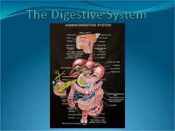
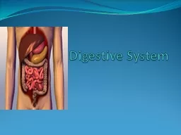
8. Digestive System
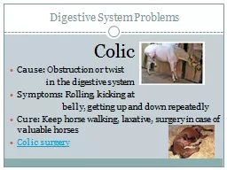
9. The digestive system consists of the alimentary canal and the digestive glands. The alimentary canal starts with the mouth and terminates in the anus.
10. The major digestive gland is the liver which is a massive yellowish gland and consists of two lobes. A thin-walled V-shaped gall-bladder is present in the anterior part of the right lobe of liver. The pancreas is a pale compact irregular body. The pancreatic juice is poured into the intestine by pancreatic duct situated opposite to the aperture of the bile duct.The functional significance of rectal gland is not properly known. It discharges a fluid into the lumen of the intestine but its actual role is not known.The spleen is located dorsal to the distal end of the body of the stomach. The spleen is functionally associated with the circulatory system, but remains connected with the alimentary canal.
11. Urinogenital System of Scoliodon
12. The excretory organ consists of a pair of elongated kidneys. The functional adult kidneys are called opisthonephros. The anterior portion of the kidney is non-functional and the posterior portion becomes greatly developed.The kidneys are composed of coiled glandular uriniferous tubules or nephrons. Each tubule consists of a double-walled cup (or Bowman’s capsule) enclosing glomerulus and a much coiled renal tubule. The collecting tubules of the anterior nonrenal portion of the kidney open to the Wolffian duct and the posterior tubules open into the ureter which in turn opens into the urinogenital sinus. The Wolffian duct in males, becomes the vas deferens which is connected with the vasa efferentia from the testis.
13. Male reproductive system
14. The testes are paired elongated organs. The sperm cells escape by vasa efferentia into the vas deferens which becomes extremely coiled in the anterior portion of the kidney.Posteriorly the vas deferens becomes very much dilated to form the seminal vesicle. The seminal vesicles open into the urinogenital sinus which in turn opens into the cloaca. The wall of the urinogenital sinus has become evaginated to form a sperm sac. A pair of sacs designated as siphons are present. The siphons are located under the skin on the ventral aspect of the body.
15. Female reproductive system
16. In females there is no connection between the kidneys and the genital organs. The ovaries are two in number and are kept in position by peritoneal folds called mesovarium.The shape, size and colour of the ovaries vary greatly according to the age of the individual. A pair of epigonial organs is present between the ovary and the rectal gland. The oviducts are very long tubes and remain united both posteriorly and anteriorly. Posteriorly, two oviducts unite to form the vagina which opens into the cloaca.
17. Anteriorly the oviducts converge and open into the coelomic cavity by a longitudinal slit-like opening designated as oviducal funnel. At the anterior portion of each oviduct there is a dilated shell gland which has but little signifiance in Scoliodon. As Scoliodon is ovo-viviparous the posterior portion of the oviduct becomes dilated to form the uterus for the development of the young.
18. Circulatory System of Scoliodon:The circulatory system consists of:(a) The circulatory fluid, called blood,(b) The heart,(c) The arteries and(d) The veins.
19. Blood:The blood consists of a colourless plasma and corpuscles are suspended in the plasma. Two kinds of corpuscles are encountered; the RBC (or erythrocytes) and the WBC (or leucocytes). The erythrocytes are oval bodies containing a nucleus. The haemoglobin is present in the erythrocytes. The leucocytes are amoeboid in structure.
20. Heart:The heart is a bent muscular tube and consists of:sinus venosus auricle,ventricleconus arteriosus. Receiving parts of the heart:The sinus venosus is a thin-walled tubular chamber. Two great veins, the ductus Cuveiri, open into the sinus venosus, one on each lateral side. The sinus venosus opens into the auricle by sinuauricular aperture which is guarded by a pair of valves. The auricle is a large, triangular and thin-walled chamber situated dorsal to the ventricle but in front of the sinus venosus.The auricle communicates with the ventricle through a slit-like auriculoventricular aperture guarded by two lipped valves. The receiving chambers, (sinus venosus and auricle) receive the venous blood from all parts of the body..
21. Forwarding parts of the heart:The ventricle has a very thick muscular wall, the inner surface gives many muscular strands, thus giving it a spongy texture. It is an oval chamber and constitutes the most prominent part of the heart. The conus arteriosus is a stout median muscular tube arising from the ventricle.The conus arteriosus is continued forward as the ventral aorta
22. 1. Vental aorta and afferent branchial arteries112. Efferent branchial and epibranchial arteries123. Hypobranchial blood plexus1111344. Dorsal aorta and its branches5. Arteries of head5
23. 1. Anterior cardinal systemInferior JSInternal JSBuccopharyngeal region, gill pochesDorsal region of head and gill 11112. Posterior cardinal or renal portal system113.Hepatic portal system4.Lateral abdominal systemPelvic finsPectoral fins5. Cutaneous system123
24. Flight muscles in birds
25. Flight musclesFore arms modified into wingsThree categories of flight muscles1. Pectoralis major Pectoralis minor2. Accessory3. Tensor
26. Pectoralis major: Largest and most powerful flight muscles One side attached to keel and on other side to the head of humerus Dark red in colour due to rich blood supply It is depressor muscles causing downstroke of the wings. When it contracts, the wing is pulled downwords and forwards so that body lifted upward and propels itself through the air
27. Pectoralis minor: Elevation of wings is performed by this muscles also known as supracoracoideus or subclavius It lies on the sternum, in the angle between keel and Body , beneath the pectoralis major. It tapers anteriorally into a long strong thin tendon which passes upword through Foramen triosseum (a slit or gap) Causing upstoke of the wings When it contract foramen triosseum acts like pulley for its tendon. pulling the humerus backwards and upward, thus raising the wing during flight.
28.
29. Accessory muscles Several small accessory muscles also help in elevating or depressing the wingscoraco-brachialis longus and the coraco-brachialis brevis: They help to rotate the wing in the glenoid cavity.Biceps and triceps: perform adjustment during flightExtensor carpi radialis and extensor carpi ulnaris: helping in stretching and folding of the wingsBrachioradialis muscles: Tensor muscles: keep the body stretched condition during flight