
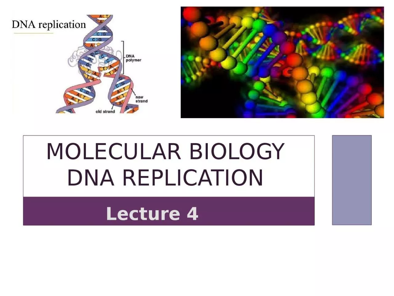
Lecture 4 DNA replication Before each cell division new copies must be made of each of the many molecules that form the cell including the duplication of all DNA molecules DNA replication ID: 916505
Download Presentation The PPT/PDF document "Molecular biology DNA Replication" is the property of its rightful owner. Permission is granted to download and print the materials on this web site for personal, non-commercial use only, and to display it on your personal computer provided you do not modify the materials and that you retain all copyright notices contained in the materials. By downloading content from our website, you accept the terms of this agreement.


Slide1
Molecular biology DNA Replication
Lecture 4
Slide2DNA
replication
Before
each cell
division,
new copies
must be made of each of the many molecules that form
the cell
, including the duplication of all DNA molecules.
DNA replication
is the name given to this
duplication process
,
which enables
an organism’s genetic information — its genes — to
be passed
to the two daughter cells created when a cell
divides.
DNA
replication is the process of producing two identical copies from one original DNA molecule. This biological process occurs in all living organisms . It is the basis for
biological inheritance
. DNA is composed
of
two strands and each strand of the original DNA molecule serves as template for the production of the complementary strand,
a
process referred to as
semiconservative
replication.
Slide3After Watson and Crick proposed the double helix model of DNA, three models for DNA replication were proposed:
conservative, semiconservative and dispersive.
Conservative ModelAfter DNA replication, the
parental DNA remains together, and the newly formed daughter strands are together.
Semiconservative Model
The semi-conservative method suggests that each of the two parental DNA strands act as a template for new DNA to be synthesized; after replication, each double-stranded DNA includes one parental or “old” strand and one “new” strand.
Dispersive
Model both new copies of DNA have double-stranded segments of parental DNA and newly synthesized DNA interspersed.
Slide4Meselson
–Stahl experiment
:
An experiment by Matthew
Meselson
and Franklin Stahl in 1958, which supported the hypothesis of DNA replication was
semiconservative.
It has been called "the most beautiful experiment in biology. Each of the parent strands will serve as a template to synthesis the complementary strand. The following steps were performed to prove this theory:Experiment
1-They cultured
the bacterium E .coli in culture medium containing a heavy Nitrogen ( N15) which is an isotope of nitrogen ( N14 ). (Since only heavy nitrogen was the only source of Nitrogen for the bacteria they had to use it during new DNA synthesis). Therefore their DNA would slowly be replaced with DNA made of heavy nitrogen only. They maintained the culture in a heavy nitrogen medium for several generations, they also isolated the DNA of the heavy Nitrogen ( N15 ) grown bacteria and run centrifuged on a salt density gradient. They got a single band of DNA that had very high density.
How is semiconservative replication model discovered ?
Slide52-
Then, they sub-cultured those N
15 containing
E
. coli
on light nitrogen ( N14 ) containing normal culture media. These bacteria can now use only
N14 as nitrogen source. Then they isolated the DNA of E. coli after one round of replication and performed a centrifuged on a salt density gradient. This time they got a single band of DNA in the salt centrifuge tube. This band was higher, intermediate in density between the heavy N15 DNA and the light N14 DNA.The conservative model would have predicted two distinct bands in this generation (a band for the heavy original molecule and a band for the light, newly made molecule). This result fit with the dispersive and semi-conservative models, but not with the conservative models.
Slide63-The
second round of replication produced two bands of different molecular weights. One DNA band was at the intermediate position between
N
15
and
N
14, and the other band was higher (appeared to be labeled only with the light N14
)
. These results could only be explained if DNA replicates in a semi-conservative manner. In contrast, in dispersive replication, all the molecules should have bits of old and new DNA, making it impossible to get a "purely light" molecule. Therefore, the other two modes were ruled out
Slide7Slide8DNA replication process
the process of DNA replication followed by proofreading or error-checking mechanisms to ensure correct reading of the genetic code,. like all biological polymerization processes (Transcription and Translation, will be discussed
later),
the process involve 3 stages :
1- Initiation 2-Elongation and 3- Termination
1- Initiation
The replication of both prokaryotic and eukaryotic DNAs starts at a unique sequence called the origin of replication, which serves as a specific binding site for proteins that initiate the replication process.
Slide9In
E.coli
, which has a single origin of replication on
its one chromosome (as do most prokaryotes), it is approximately 245 base pairs long and is rich in AT sequence (
rich
in adenine and thymine bases), because A-T base pairs have two hydrogen bonds (rather than the three bond in a C-G pair) and easy to break in this
site.
The origin of replication (oriC) is recognized by certain proteins that bind to this
site called
Initiator proteins. These proteins (DnaA in prokaryotes, origin recognition complex in yeast ) binds specifically to the AT-rich replicator sequence oriC to form a specific DnaA-oriC complex. An enzyme called helicase unwinds the DNA by breaking the hydrogen bonds between the nitrogenous base pairs. ATP hydrolysis is required for this process.
Slide10As the DNA opens up, Y-shaped structures called
replication forks are formed. Two replication forks are formed at the origin of replication and these get
extended bi-directionally
as replication proceeds. (two replication forks begin at a single replication origin in bacteria and proceed in opposite directions around the chromosome forming
θ theta
shape
,
which look like a bubble , moving away from the origin till reaching the opposite direction in one point called Ter Terminus(
Teri).
Slide11Single
-strand binding proteins (SSBPs)
bind to the single strands of DNA near the replication fork to prevent the
ssDNA
strands
from winding back into a double
helix,
thus maintaining the strand separation
Slide12http://biology-
pictures.blogspot.com/2011/10/bidirectional-replication.html
Slide13The
mechanism of eukaryotic DNA replication is similar to that of prokaryotic DNA replication but it is more complex. There are multiple origins of replication on the eukaryotic
chromosome
so
multiple replication bubbles will
form.
In
yeast, which is a eukaryote,
special sequences known as Autonomously Replicating Sequences (ARS) are found on the chromosomes. These are equivalent to the origin of replication in E. coli.
Slide14One of the key
players in DNA replication is the enzyme DNA polymerase
, also known as DNA pol
(
there are many types of DNA polymerases in prokaryotes and eukaryotes will be discussed later
).
DNA
polymerase is able to add nucleotides only in the 5' to 3' direction (a new DNA strand can be only extended in this direction). It also requires a free 3'-OH group to which it can add nucleotides by forming a phosphodister bond between the 3'-OH end and the 5' phosphate of the next nucleotide. This essentially means that it cannot add nucleotides if a free 3'-OH group is not available.
The
problem is solved with the help of an RNA sequence that provides the free 3′-OH end. RNA primase, synthesizes an RNA primer that is about five to ten nucleotides long and complementary to the DNA template .
Slide15Because this sequence primes the DNA synthesis
, it
is appropriately called the
primer
.
DNA
polymerase can now extend this RNA primer, adding nucleotides one
by one that are complementary to the template strand. ( example: A in the template strand is complement
to
T in new growing strand strand , and G in the template strand is complement to C in new growing strand strand)… The primer is RNA rather than DNA because DNA polymerases cannot start chains de novo
Slide162- Elongation
stepDNA
double helix is anti-parallel; that is, one strand is in the
5' to 3
' direction and the other is oriented in the
3' to 5'
direction. both strands of parental DNA serve as templates for the synthesis of new
DNA. A new DNA strand is always synthesized in a 5’ to 3’ direction. Thus, the replication of both the strands goes in two different ways .
One
strand, which is complementary to the 3' to 5' parental DNA strand, is synthesized continuously in 5----3 direction towards the replication fork because the DNA polymerase III can add nucleotides in this direction. This continuously synthesized strand is known as the leading strand. in prokaryote, DNA polymerase III begins the synthesis of the leading strand by using the RNA primer formed by primase ( from 5’-3’direction or the same direction as the replication fork movement) and add the nucleotides according to complement base-paring to the template( A=T,T=A; G≅ C; C≅G)
Slide17Slide18However
, all known DNA polymerases synthesize DNA in the 5′ → 3′ direction but not in the
3′ → 5′ direction.
How then does one of the daughter ( lagging ) DNA strands appear to grow in the 3′ → 5′ direction?
The answer is
The
other strand, complementary to the 5' to 3' parental DNA, is extended away from the replication fork discontinuously;
in small fragments known as Okazaki fragments, each requiring a primer to start the synthesis (this strand needs a new primer for each of the short Okazaki fragments) . Okazaki fragments are then synthesized via extension of these RNA primers by DNA polymerase. An important consequence of such RNA priming is that newly synthesized Okazaki fragments contain an RNA-DNA joint, the discovery of which provided critical evidence for the role of RNA primers in DNA replication. Okazaki fragments are named after the Japanese scientist Reiji Okazaki (1968) who first discovered them. The strand with the Okazaki fragments is known as the lagging strand.
Slide19Slide20Most
current evidence indicates that DNA polymerases epsilon
ε
and
Delta
δ
, respectively, perform the bulk of leading and lagging strand replication of the
eukaryotic nuclear genome and Pol γ in the mitochondria) .
As synthesis proceeds, the RNA primers are replaced by DNA.
The primers are removed by the exonuclease activity of DNA polymerase I in prokaryote, and the gaps are filled in by deoxyribonucleotides. The nicks that remain between the newly synthesized DNA (that replaced the RNA primer) and the previously synthesized DNA are sealed by the enzyme DNA ligase that catalyzes the formation of phosphodister linkage between the 3'-OH end of one nucleotide and the 5' phosphate end of the other fragment. ; this is the reason why the synthesis of the lagging strand is more complicated than the leading strand.
Slide21Slide22A protein called the
sliding clamp holds the DNA polymerase in place as it continues to add nucleotides. (sliding-clamp proteins and clamp-loading proteins) that load the polymerase onto the primer and maintain its stable association with the template
Slide233-Termination
1-Termination requires that the progress of the DNA replication fork must stop or be blocked. Termination at a specific locus, when it occurs, involves the interaction between two components:
a
termination site sequence in the DNA,
a protein which binds to this sequence to physically stop DNA replication.
this protein is
named the DNA replication terminus site-binding protein, or Ter protein
.
2-Because bacteria have circular chromosomes, termination of replication occurs when the two replication forks meet each other on the opposite end of the parental chromosome . As a result, the replication forks are constrained to always meet within the termination region of the chromosome.
Slide243-Removes the primer (RNA fragments),
by 5'-3' exonuclease activity of polymerase I, and replaces the RNA nucleotides with DNA nucleotides. and fill the gaps.
in eukaryote, The enzyme ribonuclease H
(RNase H)and
FenI
enzyme
, instead of a DNA polymerase I, removes the RNA primer, which is then replaced with DNA nucleotides.
4- When this is complete, a single nick on the leading strand and several nicks on the lagging strand can be found. Ligase works to fill these nicks in, thus completing the newly replicated DNA molecule .
6-
Topoisomerase IV will : separate the two complete daughter chromosome in to two chromosome.
Slide25Termination in Eukaryotic cell
Eukaryote cell initiate DNA replication at multiple points in the chromosome, so replication forks meet and terminate at many points in the chromosome; these are not known to be regulated in any particular way. Because eukaryotes have linear chromosomes, DNA replication is unable to reach the very end of the chromosomes.
Primer
removal at the end of the chromosome leaves a gap that can’t be filled in (there is no DNA polymerase coming along to fill in that piece. (Remember that DNA synthesis can ONLY occur
5’-3’
). So on every round of replication, a little piece is lost from the end of the chromosome.
Slide26Slide27Consequently, special mechanisms are required to replicate the terminal sequences of the linear chromosomes of eukaryotic cells. These terminal sequences sequences (telomeres) consist of tandem repeats of simple-sequence DNA (
short DNA sequences that are repeated over and over at the ends of the chromosomes) Short stretches are lost from telomeres at each round of replication. But that’s alright because there is an enzyme called
TELOMERASE
that can refill the telomeres from an RNA template (
which is able to maintain telomeres by catalyzing their synthesis in the absence of a DNA template
)
Telomerase contains an RNA template to guide synthesis of new
telomeric repeats
Slide28Enzymes involved in DNA replication
Primase
: in fact is RNA polymerase thus the formed primer is RNA rather than DNA and it will removed latter by DNA polymerase I
Topoisomerase I:
will break the 3́́ 5́
phosphodiester
bond converting super coiled to relax form which opposite to ligase.
Relaxes the DNA from its super-coiled nature
DNA
Helicase Also known as helix destabilizing enzyme cases formation of Replication Fork due to broken hydrogen bonds
Slide29DNA
Gyrase (and Topoisomerase IV) ; this is a specific type of
topisomerase II convert relaxed form to super coiled
DNA Ligase
Re-anneals the semi-conservative strands and joins
Okazak’i
Fragments
of the lagging strand.
Telomerase
Lengthens telomeric DNA by adding repetitive nucleotide sequences to the ends of eukaryotic chromosomesDNA Polymerase Builds a new duplex DNA strand by adding nucleotides in the 5' to 3' direction. performs proof-reading and error correction.DNA clamp: A protein (unit from polymerase which prevents DNA polymerase III from dissociating from the DNA parent strand.Single-Strand Binding (SSB) Proteins Bind to ssDNA and prevent the DNA double helix from re-annealing after DNA helicase unwinds it thus maintaining the strand separation
Slide30Types of DNA
polymerases in Prokaryotes
Types
of enzyme
Initiation
activity
Polymerization 5
́́→3́
Exonuclease activity 3
́→5́Exonuclease activity 5́́→3́DNA polymerase I-+++DNA polymerase II -++-DNA polymerase III-++-Named in order of discovery, DNA polymerase I, II, III DNA polymerase III is the main polymerase for DNA replication (Main enzyme that adds nucleotides in the 5'-3’) . DNA polymerase II is involved in DNA repair.
DNA Polymerase I has
:
1
- Proofreading exonuclease in (3’-5’ direction )
2- Primer removal exonuclease in (5’-3’) direction
3-DNA synthesis (5’-3’)
replaces primer with newly synthesized DNA
Slide31Types
of DNA polymerase in Eukaryotic cell
The
DNA polymerases of eukaryotes are
in general less understandable than the DNA polymerases of
prokaryotes.
Eukaryotic cells have
FIVE polymerases: four major nuclear DNA
polymerases:
DNA polymerase alpha (Pol α), DNA polymerase delta (Pol δ) and DNA polymerase epsilon (Pol ε), DNA polymerase beta ( poly β), and one found in mitochondria: DNA polymerase Gamma (Poly γ).1-Polymerase alpha ( Pol α ) : it is the only enzyme has primase activity beside DNA polymerase, Pol α initiates DNA synthesis on both the leading and lagging strands.
Slide322-Pol
β Beta
polymerase
:
excision repair
and it is not highly active and is not very
processive
. 3-Pol
γ
Gamma polymerase: polymerization the mitochondrial DNA beside repairing by its exonuclease activity 3́→5́4-Pol δ delta and 5-ε epsilon polymerase :polymerization lagging (δ)and leading (ε) strand respectively 5 ́→3́. In eukaryotes.DNA can be synthesized in vitro by technique known as Polymerase Chain Reaction (PCR) it will be discussed in practical part of Molecular Biology course.