
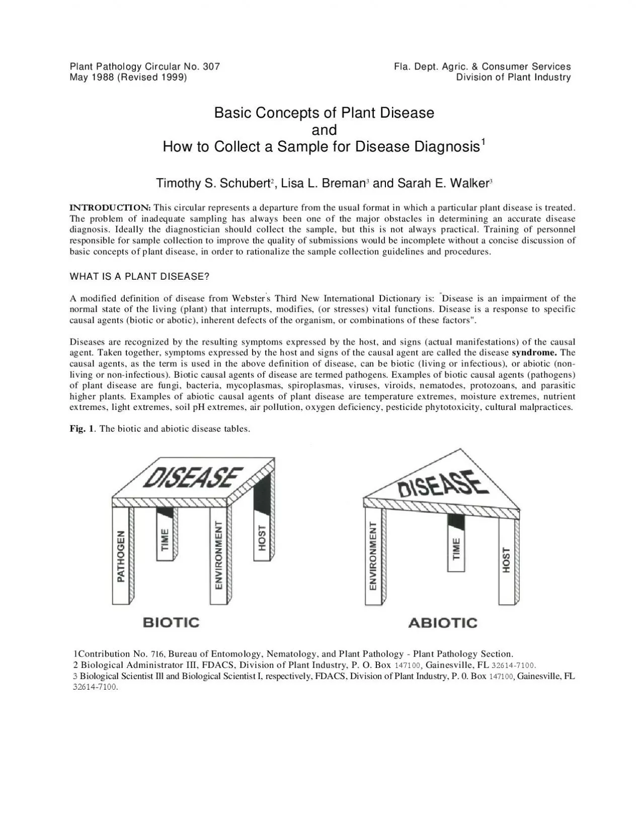
A visual aid to understanding all of the factors involved in the manifestation of both biotic and abiotic plant diseases are the plant disease tables shown in Figure 1 Each leg of the fourlegged tab ID: 955053
Download Pdf The PPT/PDF document "Plant Pathology Circular No 307 Fla Dept..." is the property of its rightful owner. Permission is granted to download and print the materials on this web site for personal, non-commercial use only, and to display it on your personal computer provided you do not modify the materials and that you retain all copyright notices contained in the materials. By downloading content from our website, you accept the terms of this agreement.


Plant Pathology Circular No. 307 Fla. Dept. Agric. & Consumer Services May 1988 (Revised 1999) Division of Plant Industry Basic Concepts of Plant Disease How to Collect a Sample for Disease DiagnosisTimothy S. Schubert, Lisa L. Breman and Sarah E. WalkerINTRODUCTION: A visual aid to understanding all of the factors involved in the manifestation of both biotic and abiotic plant diseases are the plant disease tables shown in Figure 1. Each leg of the four-legged table represents one of the essential components for biotic disease development. Where the three components of virulent pathogen, susceptible host and conducive environment coincide for a sufficient period of time, disease will occur. In the simplest interpretation, lacking any one of the components will preclude disease development. Abiotic disease development (disease caused by a non-living agent) can be similarly represented by a three-legged table. Where the two components of a susceptible plant (no longer a host in the strict sense) and harmful environment coincide for a sufficient period of time, such conditions will support the development of abiotic disease. Inherent plant defects or genetic disorders which result in disease symptoms may be expressed under stable, innocuous environmental conditions, or may require some normally harmless abiotic or biotic stimulus for symptom expression. A few cases of disease induced by exopathogens (pathogen not in intimate contact with a susceptible plant) are known, and may have been mistakenly attributed to inherent plant defects or abiotic agents in the past. Additionally, there are examples of seemingly symptomless plant diseases that are not uncovered until yield losses are compiled, or the true healthy appearance of the plant is revealed (Fox, 1993). Various symptoms of plant diseases are illustrated diagramatically in Table 1 together with possible causal agents and certain considerations to be weighed during the sample collection process. It is important to remember that even though a disease may a
ppear to be static, as a rule diseases are dynamic, and the disease syndrome will change over time. Symptoms are usually not reversible, though with some abiotic causal agents such as drought stress or nutrient deficiency, symptoms (wilting, chlorosis) can be reversible. Of course, healing of the damage done by biotic and abiotic causal agents, or even by inherent defects in the host plant can occur, though some evidence of the disease usually remains. While considerable literature has been generated on disease diagnostic techniques useful once the sample has been received in the clinic, (Burchill 1981; Dhingra and Sinclair 1985; Fox 1993; Johnston and Booth 1983; McIntyre and Sands 1977; Shurtleff and Averre III 1997; Tuite 1969; Waller, et al. 1998) , surprisingly little attention is given to the important initial step of the diagnostic process, the collection of the sample. Without an intelligent approach to the acquisition of the sample, the accuracy of the entire diagnosis is suspect. Whereas it is helpful to know the mechanisms by which causal agents and inherent plant defects cause the symptoms of a disease, this information is not essential in order to obtain a good sample for diagnosis. Good observational and detective skills are necessary. The inspector/collector should have a good concept of the normal appearance of the plants under scrutiny, so that samples of deviations from normal can be collected. Some disease is to be expected in any plant population even if (sometimes especially if!) there is human interference. If symptoms are seemingly undetectable, the disease is either not serious enough to warrant attention, or observational skills need improvement. Some suggestions to improve observational skills are given in Table 4. One common omission in inspection is examination of underground or ground level plant parts. Avoid the temptation to collect only symptomatic tissues, as these do not necessarily harbor the pathogen. Sampling techniques are unavoidably destructive to the host plant, somet
imes quite so, using even the most sophisticated diagnostic methodology. If there is ever any doubt about what normal plant appearance should be, or what to include in a sample for diagnosis, whole plants including roots and soil (about a liter of soil and roots), or representative samples of all plant parts and soil from plants that are too valuable, too large, or otherwise impractical to send to the clinic. (Other considerations for collecting samples are given in Table 1.) Some diseases will inevitably defy complete diagnosis, therefore resampling may be necessary. Even then, no thorough resolution of the problem may be forthcoming. The diagnostic team should come closer to the answer(s) with each attempt, however. Diseases of complex etiology (more than one causal agent) often fall into this refractory category, and are actually quite common (Wallace 1978). Path No. 307 May 1988 (Revised 1999) 2 TABLE 1. SYMPTOMS, POSSIBLE CAUSES, AND SAMPLING CONSIDERATIONS Possible Causes, Sampling Considerations Fungal, bacterial, or viral leaf spot pathogen, chemical phytotoxicity. Collect all stages of development. Note pesticide schedule. Root or stem dysfunction, water stress, excess soluble salts, herbicide or other chemical injury. Fungal, bacterial or other pathogenic infection. Collect whole plant with roots and soil. Note pesticide and fertilizer schedule plus other components of cultural program. Fungal, bacterial, viral or other pathogenic infection, herbicide or growth regulator injury, insect or mite damage, chemical phytotoxicity, mechanical damage. Collect representative sample of all symptoms. Note cultural program. Viral infection, growth regulator injury. Collect representative range of symptoms. If possible, keep tissue fresh or submit whole plant to permit observation and testing to take place over a period of time. Excess moisture, chemical phytotoxicity, fungal infection. Collect all representative stages. Note soil moisture, preceding weather conditions (prolonged cloudy, humid to wet weather?)
. Viral infection, growth regulator or herbicide injury. Collect all representative stages, note pesticide schedule on or near crop. Keep sample very fresh (essential for many viral diagnostic tests). Whole plant helpful. Viral infection, reaction to cold or hot water on foliage, chemical phytotoxicity. Note watering methods, pesticide schedule. Collect very fresh sample or whole plants. Fungal infection. Collect symptomatic tissues. Path No. 307 May 1988 (Revised 1999) 3 Fungal infection, insect or mite injury. Collect range of symptomatic tissues. Fungal or bacterial infection, insect or other injury, chemical damage. Collect range of symptomatic tissues. Root dysfunction, water stress, excess soluble salts, cold, heat, normal senescense, chemical or insect damage. Collect whole plants. Note recent cultural treatments. Macro- or micronutrient deficiency, low or excess light, growth regulator or herbicide injury, natural pigmentation/variegation, root dysfunction, soil pH problems. Collect representative samples of all symptoms, soil and roots. Fungal, bacterial, viral or other pathogenic wilt, root rot or canker pathogen. Water stress, excess soluble salts, high temperature, wind, cold, insect injury. Implies sudden onset. Collect whole plant with roots and soil. Slower or later expression of same factors as above, plus cold injury, insect damage. Localized or generalized branch or twig disorder caused by fungi, bacteria, insects, mechanical injury. Collect sample which includes transition zone from diseased to healthy tissue. Fungal, bacterial, viral or other pathogenic infection, nutrient deficiency, water stress, nematode or insect injury, growth regulator damage. Collect whole symptomatic and asymptomatic plant. Note cultural program. Unknown. Usually considered a genetic abnormality, sometimes associated with insect injury. Collect symptomatic tissue of all stages, whole plants if possible. Note pesticide and growth regulator schedule. Fungal, bacterial, viral, or MLO infection. Mite infestation. M
icronutrient deficiency, herbicide or growth regulator phytotoxicity. Collect whole plants if possible. Note pesticide schedule. Path No. 307 May 1988 (Revised 1999) 4 Fungal disease, mechanical constriction, ground-line heat canker, insect injury. Collect symptomatic tissues of all stages of development. Fungal or bacterial infection, mechanical injury, heat or cold injury, sunburn, chemical injury. Collect symptomatic tissues, especially transition zones between healthy and symptomatic tissue. Check pattern of symptom expression. Excess water, mechanical or chemical injury or pathogenic infection at ground line. Enough vigor to induce root primordia implies sudden onset of disorder to otherwise healthy plant. Collect whole plant or all representative plants parts. Fungal infection (rust), excess moisture (exploded lenticels). Collect symptomatic tissue. Fungal or bacterial infection, mechanical injury, hormonal disruption, insect injury. Collect samples of all stages of gall development, including surrounding soil if below ground. Fungal or bacterial infection, excess moisture, excess soluble salts, chemical injury. Collect whole plant with soil and roots. Nodulating N-fixing bacteria, actinomycetes, bluegreen algae, root-knot nematode, insect injury, fungal bacterial or viral pathogenic infection. Collect symptomatic tissue. Crown rot affects oldest tissues. Bud rot affects youngest tissue. Fungal, bacterial infection. Excess soluble salts. Collect entire plant with roots and soil. Fungal infection, mechanical injury, heat or cold damage, chemical injury. Collect representative samples of all stages. Fungal infection. Collect representative samples of all stages. Path No. 307 May 1988 (Revised 1999) 5 PROCEDURE FOR SAMPLE COLLECTION, PACKAGING AND SUBMISSION A suggested list of supplies to include in a field kit used for collecting diseased plant samples is given in Table 2. After deciding what to include in the sample, the following procedures for obtaining, packaging, and submitting the sample are
suggested: Obtain fresh material in reasonable quantity, several examples of the various symptoms being expressed. Be certain to include as many identifiable stages of the disease as are represented. Most recently developed symptoms usually afford the best material for diagnosis. Be sure to include suitable plant material for botanical identification, since occasionally field identifications may be in error or the host plant identity may not be known. Lift roots carefully so as not to leave feeder roots or rotted roots behind. Include about a liter of soil for pH, soluble salts, and possibly a nematode assay. Place samples in appropriately sized plastic bags, including a paper towel for a blotter if sample is very wet. Duplicate dry samples are recommended if the sample is succulent or fragile. Wrap a wire twist-tie around stem at ground line to keep soil off of above-ground plant parts. Accurately label samples. Place the entire sample in a paper bag or an unsealed plastic bag. Keep samples cool, protected from crushing. Gather appropriate information from the grower (see Table 3) and make pertinent observations (see Table 4). Include this information with sample submission form. Complete mailing addresses and map locations are necessary if owners want to be informed of diagnosis or site must be revisited. Also, clerical work in the clinic is greatly simplified when a properly completed submission form is included with the sample. 7) Deliver promptly to the clinic. Next day service is recommended.TABLE 2. CONTENTS OF A FIELD KIT FOR PLANT DISEASE SAMPLE COLLECTION Plastic and paper bags Pruning shears, pruning saw Wire twist ties Paper towels Knife, hatchet, increment borer Plastic/rubber gloves Trowel, shovel, soil probe Rubber bands Hand lens 5-10X Forms, pencil Labels Vials/jars with lids Padded collection boxes *Camera, film, macro lens L arge ice chest/cooler flash equipment Maps Field manuals, compendia Disinfectant for hands & tools *Light meter Portable soil pH and soluble salts meter *Optio
nal Path No. 307 May 1988 (Revised 1999) TABLE 3. EXAMPLES OF INFORMATION INSPECTOR SHOULD OBTAIN FROM GROWER Host: cultivar/variety; age; propagation method; site prep; names; dates and rates of fertilizer, pesticide history and schedule*, growth regulator applications; pruning; pinching; transplant; omissions or additions to usual/conventional culture program. Irrigation: frequency, rate, timing; water quality; determination of need. Soil/media: pH, soluble salts, texture, drainage, homogeneity, aeration, temperature, planting depth, cultivation, cropping history, earthmoving/construction, burial/disposal site, trash burning. Environmental conditions: temperature, humidity, ventilation, wind, lightning, exposure, light intensity, air quality, method of heating and cooling, etc., all preceding and during syndrome development. Date of first symptoms and rate of syndrome development, coincident with any treatment or environmental event. Recent human, animal, insect, mite activity around or on symptomatic plants. Idiosyncracies of plants in question, predisposition (plant made more susceptible) by cultural or environmental conditions. Use appropriate safety precautions when handling pesticide treated crops. TABLE 4. EXAMPLES OF OBSERVATIONS TO BE MADE BY INSPECTOR Spatial and chronological pattern of disease in crop, on individual plants. Are symptoms different or consistent, uniform or scattered? Are symptoms spreading across crop or progressing on individual plants? What is the frequency and intensity of the syndrome? Any signs of pathogen/causal agent? (fruiting structures, chemical residues, insects/mites, frass, etc.). Any evidence of host recovery? Nearby plants (same or different) show any symptoms? Are root zones shared? Inspect interior, crown, roots of plant. Cut open stems, crowns, flowers, fruits, and roots. Are there any hidden, internal symptoms/signs of disease? CONCLUSIONS: The whole process of sample collection for disease diagnosis is of pivotal importance. Unless done correctly, the entir
e diagnostic process may be side-tracked, and the subsequent recommendations for controlling or correcting the disease may be of limited effectiveness. Sample collection can be a difficult, time-consuming process, and there is no guarantee that the plant problem in question will be resolved with a single sampling. With all the variables involved in plant production, every plant problem demands different sampling techniques. By justifying and summarizing the various phases and techniques of the plant disease sample collection process, inspectors should enjoy greater efficiency, and more accurate diagnosis and resolution of the plant disease problems they encounter in the field. Path No. 307 May 1988 (Revised 1999) 7 LITERATURE CITED Burchill, R.T. (ed). 1981. Methods in Plant Pathology. Commonwealth Mycological Institute, Kew, Surrey, England. pp. Dhingra, O.D. and J.B. Sinclair. 1985. Basic Plant Pathology Methods. CRC Press, Boca Raton, FL. 355 pp. Johnston, A. and C. Booth (eds.).1983. Plant Pathologists Pocketbook. Commonwealth Mycological Institute, Kew, Survey, England. pp. Fox, R.T.V. 1993. Principles of Diagnostic Techniques in Plant Pathology. CAB International, Wallingford, UK. 213 McIntyre, J.L. and D.C. Sands. 1977. How disease is diagnosed. In "Plant Disease: An Advanced Treatise". (J. G. Horsfall and E. B. Cowling, eds.) Vol. 1, pp 35-53. Shurtleff, M.C. and C.W. Averre III. 1997. The Plant Disease Clinic and Field Diagnosis of Abiotic Diseases. American Phytopathological Society Press, St. Paul, MN. 245 pp. Tuite, John. 1969. Plant Pathological Methods. Burgess Publishing Co., Minneapolis, MN. pp 112-140. Wallace, H.R. 1978. The diagnosis of plant diseases of complex etiology. Annual Review of Phytopathology 16:379-Waller, J.M., B.J. Ritchie, and M. Holderness. 1998. Plant Clinic Handbook, IMI Technical Handbook No. 3. CAB International, Wallingford, UK. pp. Woltz, S.S. 1978. Nonparasitic plant pathogens. Annual Review of Phytopathology. 16:403-430. PI-99T-05 Path No. 307 May 1988 (Revised 199