

The Pericardium membrane surrounding heart Serous Pericardium 1 Parietal outer layer 2 Visceral inner layer part of epicardium Layers of Heart Wall Epicardium outermost layer ID: 635576
Download Presentation The PPT/PDF document "Heart Structure Heart – located in med..." is the property of its rightful owner. Permission is granted to download and print the materials on this web site for personal, non-commercial use only, and to display it on your personal computer provided you do not modify the materials and that you retain all copyright notices contained in the materials. By downloading content from our website, you accept the terms of this agreement.


Slide1
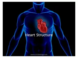
Heart StructureSlide2
Heart – located in mediastinum within the thoracic cavitySlide3
The Pericardium –
membrane surrounding heartSlide4
Serous Pericardium
= 1. Parietal - outer layer2. Visceral - inner layer (part of
epicardium
)Slide5
Layers of Heart Wall
Epicardium
– outermost layer
synonymous with visceral portion of serous membrane
Myocardium – middle layer
Cardiac muscle
Pumping chamberEndocardium – innermost layerSmooth surface to reduce frictionSlide6
Layers of Heart WallSlide7
Layers of Heart WallSlide8
Chambers of HeartSlide9
Heart Chambers and ValvesSlide10
Heart ValvesSlide11
AV Valves - structureSlide12Slide13
Heart Chambers, Valves and VesselsSlide14
Heart Chambers, Valves, and VesselsSlide15
Pulmonary vs
Systemic CirculationSlide16Slide17
Coronary CirculationSlide18
Homeostatic Imbalance:
Mycardial InfarctionsSlide19Slide20
Intrinsic Conduction System of the HeartSlide21Slide22
Intrinsic Conduction System
Also called nodal system
Sinoatrial (SA) Node = pacemaker
Atria Atria Contract
Atrioventricular
(AV) Node
Brief Delay Atrioventricular bundle (bundle of His) Bundle branches Purkinje fibers Contraction of ventricle from bottom up Blood pushed out of ventriclesSlide23
Homeostatic Imbalances
Heart block
= Damage to AV node which allows ventricles to pump at own rate, which is typically slower than normal
Fibrillation
= rapid uncoordinated shuddering of heart muscle, makes heart useless, major cause of death from heart attacks
Tachycardia
= rapid heart rate +100bpmBradycardia = slow heart rate < 60bpmSlide24
Cardiac Cycle = .8 seconds
Sequence of Events in one heartbeat
Contraction of both atria
Contraction of both ventricles
Systole = contraction – of ventricle
Diastole = relaxation – of ventricle
Lub = closing of AV valvesDub = closing of semilunar valvesSlide25
Cardiac Cycle
Systole:
Ventricles contract
AV Close = “
Lub
”
Semi-lunar openDiastole: Ventricle relaxAV valve openSemi-lunar closed= “Dub”Slide26
Cardiac CycleSlide27Slide28Slide29Slide30
Cardiac Output
Cardiac Output = amount of blood pumped out by EACH side of the heart in 1 minute
CO = HR X SV
HR = heart rate or beats per minute
SV = stroke volume = amount of blood pumped out be each ventricle with each heartbeat
If either HR or SV varies, the other tries to compensate to keep the cardiac output stableSlide31Slide32
Factors Affecting Heartrate
Autonomic NS – most important external influence on heart
Sympathetic – increase heart rate
Parasympathetic – slow and steady heartrate (
vagus
nerve)
HormonesEpinephrine and thyroxine – increase heartratePhysical factorsAge, gender, exercise, body temperatureSlide33
Factors Affecting Stroke Volume
How much cardiac muscle stretched before contraction
More stretch = more contraction
Venous return determines amount of stretch
Greater venous return = Greater SV
Increase SV
Slow HeartrateExerciseDecrease SVSevere blood lossVery Rapid heartrateSlide34Slide35
Regulation of Heart Rate =
most important external influence on heart rateSlide36
Homeostatic Imbalance: Congestive Heart Failure
When the pumping efficiency of the heart is depressed so that circulation is inadequate to meet tissue needs
Usually progressive condition that reflects weakening of the heart by atherosclerosis, persistent high blood pressure, or multiple myocardial infarctionsSlide37
Congestive Heart Failure
Pulmonary Congestions
Left side failure
Right side pumps to lungs
Left side cannot pump to body
Blood pressure inside lungs increases and fluid leaks into lung tissue
Pulmonary edemaPerson can suffocatePeripheral CongestionRight side failureBlood backs up in systemic circulation
Edema in most distal parts of bodySlide38Slide39
Review Questions – True of False
The ventricles contract in diastole
The AV valves are open during systole
The SA node is the pacemaker of the heart
Increased venous return will decrease stroke volume
Parasympathetic NS keeps heartrate slow and steadySlide40
ECG or EKG
electrical activity of the heart
depolarize
–
contract
repolarize
–relaxElectrocardiogramSlide41
Electrocardiogram = ECG
P wave
Depolarization of atria
Atria contract
QRS complex
Depolarization of ventricles
Repolarization of atriaVentricles contract (atria relax)T waveRepolarization of ventriclesVentricles relaxSlide42
Normal ECGSlide43
bradycardia
slow heart beat
tachycardia
fast heart beat
premature atrial contraction (PACs)
atria contracts before SA node
Cardiac ArrhythmiasSlide44
atrial fibrillation
atria contract faster than 350 bpm
premature ventricular contractions (PVCs)
ventricles contract too soon
ventricular tachycardia (VT)
ventricles, rather than SA node, cause beat
Cardiac ArrhythmiasSlide45
ventricular fibrillation (VF)
ventricles contract faster than 350 bpm
heart block
impulse from SA node to AV node
first
–
impulse delayedsecond–intermittently blocked third–completely blocked
Cardiac ArrhythmiasSlide46
blood vessels: the transport network
circulation: moving blood around the body
taking vital signs
know your numbers
Closed system – blood always in a vessel or heart
Blood Vessels and CirculationSlide47
tunica intima
innermost layer
Epithelial tissue
tunica media
middle layer
Smooth muscle
tunica externaoutermost layerConnective tissue The Three Layers of Blood VesselsSlide48Slide49Slide50
Blood Pathway
Heart
Arteries
Arterioles
Capillaries Venules
Veins
HeartSlide51
Arteries
Carry blood AWAY from heart
Blood usually oxygenated – exception is pulmonary artery
Thick walls, narrow lumen
Walls elastic – expand and contract with pulse
Arterioles – smaller arteries
Dilate and constrict to alter blood flowEx. Muscles dilate when running Two organs blood supply never changes = brain and kidneysSlide52
Capillaries
Smallest of all blood vessels
Only one cell thick
Narrow – blood cells flow single file
Very large surface area for gas, nutrient, and waste exchange
Blood moves slower through here to allow time for exchangesSlide53
Veins
Carries deoxygenated back to the heart
Exception: Pulmonary vein
Thinner and less elastic, more flexible
Larger veins contain valves
Prevent backflow
Skeletal muscles and breathing help return blood to heartWhen inhale, drop in pressure occurs in thorax causing large veins near heart to expand and fillSlide54Slide55
structure and function of vessels
Blood Vessels: The Transport NetworkSlide56
Factors Influencing Venous Return
Lumen of veins larger than arteries
Large veins have valves that prevent backflow
Skeletal muscles help “milk” blood through veins towards heart
When inhale, drop in pressure occurs in thorax causing large veins near heart to expand and fillSlide57
Anatomy of Capillary Bed
Two types of vessels
A.
vascular shunt
– vessel that directly connect the arteriole and
venule
at opposite ends of the bedB. true capillaries – actual exchange vessels10 – 100 per capillary bedPrecapillary sphincter surrounds each to act as cut-off valve and regulate blood flowSlide58Slide59Slide60
Capillary Exchange
Substances tend to move into and out of body cells according to their concentration gradients.
Oxygen and nutrients: blood
tissue cells
CO
2
and waster: tissue cells bloodSlide61
How Solutes Move In and Out
1. Diffusion – lipid soluble
2. Endocytosis and Exocytosis – lipid insoluble
3. Intercellular clefts – special diffusion through gaps between cell membranes of adjoining cells
4. Fenestrated capillaries – special capillaries filled with pores. Found in areas where absorption and filtration importantSlide62
Fluid Movement -
due to differences in pressure
Blood pressure highest - arterial end
Fluid moves out of capillaries
Osmotic pressure highest -
venule
endFluid moves into capillariesSlide63
Homeostatic Imbalance – Varicose VeinsSlide64
Thrombophlebitis
Complication of varicose veins
Inflammation of a vein that results when a clot forms in vein with poor circulation
Common Consequence: clot detachment and pulmonary embolismSlide65
Special Circulations
Arterial Supply of Brain and Circle of Willis –
Supplied by 2 sets of arteries
These blood supplies are united by special arteries
Result is complete circle of connecting blood vessels called the circle of Willis
Protects the brain because it provides more than one route for blood to reach the brainSlide66
Special Circulations
Hepatic Portal Circulation
Veins drain the digestive organs and deliver blood to liver
Liver processes nutrients before make it to systemic circulation
Fetal Circulation
All nutrient, excretory, and gas exchange occur through placenta
Umbilical veins and arteries