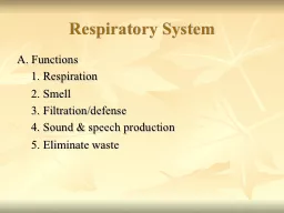
/
Respiratory system Development Respiratory system Development

Respiratory system Development - PowerPoint Presentation

patricia
patricia . @patricia
Follow
27 views
Uploaded On 2024-02-09

Respiratory system Development - PPT Presentation

By Ass Lec Reham saad Kadhum Formation of the Lung Bud The embryo is 4th week old the respiratory diverticulum lung bud appears as an outgrowth from the ID: 1045186

respiratory lung development laryngeal lung respiratory laryngeal development bronchi trachea foregut bud lungs cells mesoderm pleura alveolar diverticulum nerve

Share:

Link:

Embed:

Download Presentation from below link

Download Presentation The PPT/PDF document "Respiratory system Development" is the property of its rightful owner. Permission is granted to download and print the materials on this web site for personal, non-commercial use only, and to display it on your personal computer provided you do not modify the materials and that you retain all copyright notices contained in the materials. By downloading content from our website, you accept the terms of this agreement.


Presentation Transcript

1. Respiratory system Development By Ass. Lec. Reham saad Kadhum

2. Formation of the Lung BudThe embryo is 4th week old,the respiratory diverticulum (lung bud)appears as an outgrowth from the ventral wall foregut,The appearance and location of the lung bud are dependent upon: retinoic acid (RA) produced by adjacent mesoderm,The epithelium of the respiratory tract is derived from the endoderm of the respiratory diverticulum, while the cartilages, connective tissues, and muscles of the respiratory tree are derived from the visceral layer of the lateral plate of the mesoderm.

3. Formation of the Lung Bud Initially, the lung bud is in open communication with the foregut.When the diverticulum expands caudally, however, two longitudinal ridges, the tracheoesophageal ridges, separate it from the foregut. These ridges fuse to form the tracheoesophageal septum . the foregut is divided into two parts by the tracheoesophageal septum; these are a ventral part called the respiratory diverticulum (trachea & lung bud) and a dorsal part called the esophagus.The respiratory primordium communicates with the pharynx through the laryngeal orifice

4. DEVELOPMENT OF THE LARYNXInternal lining of the larynx originates from endoderm. Cartilages; muscles originate from mesenchyme of the 4th& 6thpharyngeal arches. As a result of rapid proliferation of the mesenchyme,the laryngeal orifice changes from a sagittal slit to a T-shaped opening. Characteristic adult shape of the laryngeal orifice can be recognized when mesenchyme of the two arches transforms into the thyroid;cricoid; and arytenoid cartilages. The laryngeal epithelium also proliferations rapidly ,resulting in a temporary occlusion of the laryngeal lumen occurs due to the proliferation of laryngeal epithelium.

5. DEVELOPMENT OF THE LARYNX Recanalization produce a pair of lateral recesses, the laryngeal ventricles.These recesses are bounded by folds of tissue that differentiate into the falseand true vocal cords.Since musculature of the larynx is derived from mesenchyme of the 4th & 6th pharyngeal arches, all laryngeal muscles are innervated by branches of the 10thcranial nerve, the vagus nerve. The superior laryngeal nerve innervates derivatives of the 4thpharyngeal arch, and the recurrent laryngeal nerve innervates derivatives of the 6thpharyngeal arch.

6. TRACHEA, BRONCHI, AND LUNG : During its separation from the foregut,the lung bud forms the tracheaand two lateral outpocketing, the bronchial buds.At the beginning of the 5th week, each of these buds enlarges to form right and left main bronchi. The right forms three secondary bronchi(three lobes).The left forms twosecondary bronchi(two lobes).

7. TRACHEA, BRONCHI, AND LUNGThe lobar (secondary) bronchidivided forming tentertiary (segmental) bronchi in the right & lift lung, which become surrounded by masses of vesical mesoderm creating the bronchopulmonary segments of the adult lung. By the end of the 6thmonth, approximately 17 generations of subdivisions have forming. The developing lungs deviate caudally so that the tracheal bifurcation reaches the level of the 4 th thoracic vertebra.

8. TRACHEA, BRONCHI, AND LUNG : The division of the bronchioles continuesup to the 7th month of development, some of the cells cuboidal respiratory bronchioles become flat cells. These cells intimately associated with numerous blood and lymphatic capillaries, and the surrounded space are now known as terminal sacs or primitive alveoli that will serve the function of gaseous exchange after birth. Therefore, the lungs of the premature infant born at the 7th month of development are able for gas exchange and thus the infant can survive.

9. TRACHEA, BRONCHI, AND LUNG : The formation of the primitive alveolar sacwill continue several years after birth. Epithelial alveolar sacs show two types of cells at the 6th month of development; namely type Iand type IIof epithelial alveolar cells. Type II produce surfactant, phospholipid-rich fluid capable of lowering surface tension at the alveolar-air interface.The amount of surfactant secretion increases inside the fluid filled fetal lung inside the uterus, especially during the last two weeks before birth. After birth, the fluid filling the lung is expelled out of the lung and is also absorbed by the lung vesselsto allow replacement of this fluid by the inspired air.

10. The lung of the premature baby•The premature baby may be born beforethe development of the surfactant substance in a sufficient amount inside the lungs, the air-blood surface tension of the alveoli will not be reduced due to this low surfactant in the lungs. •The premature born baby may therefore develop collapseof his alveoli and the condition is called respiratory distress syndrome or called hyaline membrane disease.

11. Development of Pleura : The space for the lungs, the pericardioperitoneal canal, are narrow.They lie on each side of the foregut and are gradually filled by the expanding lung buds.Ultimately, pleuroperitoneal and pleuropericardialfolds separate the pericardioperitoneal canals from the peritoneal and pericardial cavities, respectively, and the remaining spaces form the primitive pleural cavities.

12. Development of pleura : The splanchnic mesoderm, which covers the outside of the lung, develops into the visceral pleura.The somatic mesoderm layer, covering the body wall from the inside, becomes the parietal pleura.The space between the parietaland visceral pleura is the pleural cavity.

13. Clinical Correlate : 1.Blind ended trachea with absence of the lungs.2.Increased number of the lung lobules.3.Ectopic lung lobes arising from the trachea or esophagus from additional respiratory buds of the foregut that develop independently of the main respiratory system.4.Congenital cysts of the lung, which are formed by dilation of terminal or larger bronchi. These cysts may be small and multipleformation leading to a honeycombappearance of the lung X-ray.

14.