

Lecture Overview Week 1 Embryonic Development Gastrulation is a phase early in the embryonic development of most animals during which the singlelayered blastula is reorganized into a multilayered structure known as the gastrula ID: 778983
Download The PPT/PDF document "Nervous System Development" is the property of its rightful owner. Permission is granted to download and print the materials on this web site for personal, non-commercial use only, and to display it on your personal computer provided you do not modify the materials and that you retain all copyright notices contained in the materials. By downloading content from our website, you accept the terms of this agreement.


Slide1
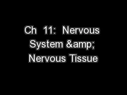
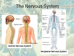
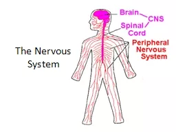
Nervous System Development
Slide2Lecture Overview
Slide3Week 1
Slide4Embryonic Development
Gastrulation is a phase early in the embryonic development of most animals, during which the single-layered blastula is reorganized into a multilayered structure known as the gastrula.
Slide5Week 3
Inner Cell Mass
Inner Cell Mass
Slide6Epiblast Cells: From inner cell mass, will ultimately give rise to the three germ layers and the entire embryo.
Hypoblast Cells: These cells are the first to migrate and eventually disintegrate.
The Inner Cell Mass of the Blastocyst
Differentiates into the Epiblast and the Hypoblast
Slide7The 3 Primary Germ Layers
The Blastocyst has three primary layers that undergo many interactions in order to evolve into organ, bone, muscle, skin or neural tissue.
The cross-section on the right illustrates three germ layers:
The ectoderm
The mesoderm
The endoderm
Slide8Week 4
Slide9The Blastocyst
ECTODERM
- The outside layer is the ectoderm. This becomes the epidermal (skin) layers, the brain and the spinal cord.
MESODERM
- The middle layer is the mesoderm. This becomes the connective tissues including the bones and the blood, as well as the gonads and the kidneys.
ENDODERM
- Inner layer is the endoderm. This becomes the digestive systems and the respiratory system.
Slide10Gastrulation
Gastrulation is a phase early in the embryonic development during which the single-layered blastula (blastocyst) is reorganized into a multilayered structure known as the gastrula.
Slide115 Weeks
Slide12Neurulation
Neurulation is the process of neural tube formation and its development into the spinal cord and the brain. Any complication during this complex process can result in neural tube defects.
The development of the nervous system begins during the third week of gestation.
Primary neurulation is the process that forms the functional central nervous system.
Slide13Neurulation
Neurulation
Begins with the Development of the Neural Plate
Three to four weeks after conception, a
flat neural plate
grows.
Slide14HEAD
TAIL BONE
The Neural Plate Elongates
Slide15The central nodal cord has induced the overlying ectoderm to thicken and form the neural plate.
The cross-section helps to visualize the folding of the neural plate leading to the formation of the neural tube.
Slide16By the end of the third week the edges of the neural plate extend upward to become the neural folds
Slide17As the edges of the neural plate extend upward to form the neural folds, the depressed region becomes the neural groove.
Slide18The Neural Folds curve inwards to make the NEURAL TUBE
Within a few days, the ridges
fold in toward
each other and fuse to form the
hollow neural tube
.
Slide19The neural crest cells migrate throughout the body to form a wide variety of cell types including
Supporting cells of the nervous system – Glial Cells
The coverings of the nervous system – The Meninges
The NEURAL TUBE then Closes and the Neural Crest Forms
Slide20Neural Tube and Crest Formation
The neural folds then grow toward each other to fuse at the midline. The neural groove then is transformed into a completely closed, but hollow neural tube.
Around this same, the neural folds become a separate population of cells called the neural crest.
Slide21Slide22Sonic hedgehog
Sonic hedgehog
is a protein that in humans is encoded by the
SHH
("
s
onic
h
edge
hog") gene.Sonic hedgehog is one of three proteins in the mammalian signaling pathway family called hedgehog, the others being desert hedgehog (DHH) and Indian hedgehog (IHH).
Slide23Sonic Hedgehog Disorders
More than 100 mutations in the
SHH
gene have been found to cause
nonsyndromic
holoprosencephaly.
This condition occurs when the brain fails to divide into two hemispheres during early development.
SHH
gene mutations are the most common cause of
nonsyndromic holoprosencephaly. These mutations reduce or eliminate the activity of Sonic Hedgehog.
Slide24Sonic Hedgehog Disorders
Without the correct activity of this protein, the eyes will not form normally and the brain does not separate into two hemispheres.
The development of other parts of the face is affected if the eyes do not move to their proper position.
The signs and symptoms of
nonsyndromic
holoprosencephaly are caused by abnormal development of the brain and face.
Slide25Before the fusion of the neural tube is complete, the cephalic and caudal ends have openings called the cranial and caudal neuropores.
The Cranial
Neurpore
is at the head end of the developing embryo
The Caudal Neuropore is at the “tail bone” end of the developing embryo.
https://www.youtube.com/watch?v=FhhWG3XzARY
Slide26Neural Tube Formation Summary
Slide27Week 5
Slide283 primary vesicles of the Neural Tube
The top of the tube thickens into three bulges that form the hindbrain, midbrain and forebrain.
The first signs of the eyes and then the hemispheres of the brain appear later.
Slide293 primary vesicles of the Neural Tube
Then the neural tube develops into three primary vesicles
The forebrain or prosencephalon
The midbrain or mesencephalon
The hindbrain or rhombencephalon
Slide30The Five Secondary Vesicles
These three develop later into five secondary vesicles the forebrain develops into
The prosencephalon (forebrain) develops into
The Telencephalon
The Diencephalon
The mesencephalon (midbrain) does not change names, it stays the mesencephalon (midbrain).
the Rhombencephalon (hind brain) develops into
The
Metencephalin
The Myelencephalin
Slide31Week 7
Telencephalon
Di
encephalon
Mes
encephalon
Met
encephalon
My
encephalon
Slide32Slide33The five secondary vesicles -
The Telencephalon
Telencephalon
is also known as the cerebrum.
The
telencephalon
refers to the region of the brain that includes the cerebral cortex and the hippocampus and basal ganglia.
Slide34The five secondary vesicles-
The Diencephalon
These three develop later into five secondary vesicles the forebrain develops into
The prosencephalon (forebrain) develops into
The Telencephalon
The Diencephalon
Becomes structures for regulating growth, hormones, sleep/wake cycles, and more.
Slide35The five secondary vesicles –
The mesencephalon
Midbrain, also called
mesencephalon
, region of the developing vertebrate brain that is composed of the tectum and tegmentum.
The midbrain serves important
functions
in motor movement, particularly movements of the eye, and in auditory and visual processing.
The mesencephalon
Slide36Metencephalon
Cerebellum (coordination)
Pons
The cerebellum coordinates voluntary movements such as posture, balance, coordination, and speech, resulting in smooth and balanced muscular activity.
Slide37Metencephalon
Cerebellum (coordination)
Pons
The
Pons
serves as a message station between several areas of the brain. It helps relay messages from the cortex and the cerebellum.
Slide38Myelencephalon
-
The medulla oblongata helps regulate
breathing, heart and blood vessel function,
digestion, sneezing,
respiration
and circulation.
Slide39Cell Differentiation
DNA has all of the instruction needed for the cell INCLUDING...
What type of cell it will become
All of the functions of the cell
All of the cells of your body
(almost all)
have the same DNA.
So HOW do cells develop from stem cells without an identity, to different types of cells (skin cells, nerve cells, blood cells, muscle cells, etc.)
Slide40Cellular differentiation
Cellular differentiation
is the process where a
cell
changes from one
cell
type to another. The
cell
changes to a more specialized type.
Differentiation
occurs numerous times during the development of a multicellular organism as it changes from a simple zygote to a complex system of tissues and
cell
types.
Slide41Embryonic stem cells
Embryonic stem cells
(
ES cells or ESCs
) are pluripotent stem cells derived from the inner cell mass of a blastocyst, an early-stage pre-implantation embryo.
Human embryos reach the blastocyst stage 4–5 days post fertilization, at which time they consist of 50–150 cells.
Slide42Stem Cell Types
Among dividing cells, there are multiple levels of cell potency, the cell's ability to differentiate into other cell types.
A greater potency indicates a larger number of cell types that can be derived.
A cell that can differentiate into all cell types, including the placental tissue, is known as
totipotent.
This Photo
by Unknown Author is licensed under
CC BY-SA
Slide43Stem Cell Types
Human development begins when a sperm fertilizes an egg and the resulting fertilized egg creates a single totipotent cell, a zygote.
In the first hours after fertilization, this zygote divides into identical totipotent cells, which can later develop into any of the three germ layers of a human (endoderm, mesoderm, or ectoderm), or into cells of the placenta
This Photo
by Unknown Author is licensed under
CC BY-NC-ND
Slide44Stem Cell Types
After reaching a 16-cell stage, the totipotent cells of the morula differentiate into cells that will eventually become either the blastocyst's Inner cell mass or the outer trophoblasts. Approximately four days after fertilization and after several cycles of cell division, these totipotent cells begin to specialize.
The inner cell mass, the source of embryonic stem cells, becomes pluripotent.
This Photo
by Unknown Author is licensed under
CC BY-NC-ND
Slide45Sonic Hedgehog
SHH is the best studied ligand of the hedgehog signaling pathway.
It plays a key role in regulating vertebrate organogenesis, such as in the growth of digits on limbs and organization of the brain.
Sonic hedgehog is a molecule that diffuses to form a concentration gradient and has different effects on the cells of the developing embryo depending on its concentration.
Slide46TAXIS: Cells can be attracted to certain chemicals or stimuli
Neurons in early development, behave somewhat like Amoeba…
In approaching and avoiding various chemicals.
But rather than the whole cell moving, neural growth involves the outreach of the cell’s connecting pathway (axon) towards its downstream partner neurons.
Slide47Axon guidance mechanisms
Axonal growth is led by growth cones
Filopodia (growing from axons) are able to sense the environment ahead for chemical markers and cues.
Mechanisms are fairly old in evolutionary terms.
Intermediate chemical markers
Guideposts studied in invertebrates
Short and long range cues
Short range chemo-attraction and chemo-repulsion
Long range chemo-attraction and chemo-repulsion
Gradient effects
Slide48Slide49Slide50Slide51Slide52NEURAL TUBE DEFECTS
Slide53Neural tube defects
Neural tube defects can occur due to…
Chromosomal disorders
Environmental exposure
Folic acid deficiency
Certain drugs that block folic acid
Carbamazepine
Phenytoin
Trimethoprim
Slide54NEURAL TUBE DEFECTS – Anencephaly
Anencephaly is the failure of the neural tube to spontaneously close at the cranial end hence the brain does not develop. Anencephaly means “no brain”. This condition is incompatible with life.
Slide55NEURAL TUBE DEFECTS – Microencephaly
Some infants with congenital
Zika virus
infection who do not have microcephaly at
birth
may later experience slowed head growth and develop postnatal microcephaly. Recognizing that
Zika
is a cause of certain
birth defects
does not mean that every pregnant woman infected with
Zika
will have a baby with a
birth defect
.
Microencephaly means “small brain”.
Slide56Spina bifida
Spina bifida is the failure of the neural tube to spontaneously close at the caudal end.
Vertebra overlying the area of the defect do not fully develop causing the vertebral arch to remain open.
Slide57Slide58Spina Bifida OccultaSpina bifida occulta is an asymptomatic defect caused by failure of the two halves of the vertebral arch to fuse at the midline. The only evidence of its presence may be a small tuft of hair over the defect
Slide59Spina Bifida Occulta
In this picture, hair tuft is shown on the skin just above the vertebra with a bony defect. A red circle is made around the hair tuft.
Spina bifida occulta only affects the vertebral arch leaving the spinal cord intact.
Slide60Spina Bifida OccultaA hair tuft is visible on the skin just above the vertebra with a bony defect. A red circle is made around the hair tuft. This is the most common sign of spina bifida occulta.
Slide61BRAIN DEVELOPMENT. The human brain and nervous system begin to develop at three weeks’ gestation as the closing neural tube (left). By four weeks, major regions of the human brain can be recognized in primitive form, including the forebrain, midbrain, hindbrain, and optic vesicle (from which the eye develops). Irregular ridges, or convolutions, are clearly seen by six months.
Slide62Slide63Folic acid
Why is folic acid needed before and during pregnancy?
Neural tube defects occur in the earliest weeks of pregnancy – before many women even know they're pregnant.
It's important for women to begin taking folic acid before you start trying to conceive.
Slide64Folic acid
It's critically important to get enough folic acid because it helps prevent neural tube defects (NTDs), such as…
spina bifida (which affects the spinal cord)
Spina Bifida Occulta
Anencephaly (which affects the brain)
Microcephaly
The neural tube is the part of the embryo from which your baby's spine and brain develop.
Slide65Neural tube defectsAccording to the Centers for Disease Control (CDC), Neural tube defects affect about 3,000 pregnancies a year in the United States. Women who take the recommended daily dose of folic acid starting at least one month before conception and during the first trimester of pregnancy can reduce their baby's risk of neural tube defects by up to 70 percent.
This Photo
by Unknown Author is licensed under
CC BY-SA
Slide66Folic acid
Folic acid is a form of vitamin B9, also known as folate.
This vitamin helps prevent certain birth defects.
When the nutrient comes directly from food sources, it's called folate.
When it's manufactured for use as a supplement or to fortify foods, it's called folic acid.
Slide67Folic acid
Folic acid is needed to make normal red blood cells and prevent a type of anemia called folate-deficiency anemia.
It's essential for the production, repair, and functioning of DNA – our genetic map and a basic building block of cells.