PPT-INSTRUMENT CONTROLLED MICROSCOPY
Author : payton | Published Date : 2024-02-03
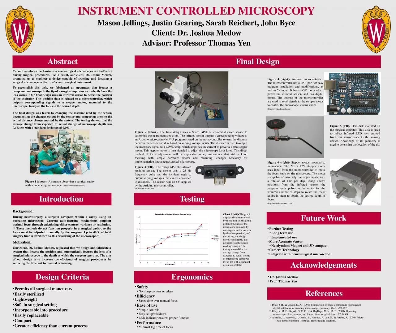
Mason Jellings Justin Gearing Sarah Reichert John Byce Client Dr Joshua Medow Advisor Professor Thomas Yen Introduction Design Criteria Final Design Testing
Presentation Embed Code
Download Presentation
Download Presentation The PPT/PDF document "INSTRUMENT CONTROLLED MICROSCOPY" is the property of its rightful owner. Permission is granted to download and print the materials on this website for personal, non-commercial use only, and to display it on your personal computer provided you do not modify the materials and that you retain all copyright notices contained in the materials. By downloading content from our website, you accept the terms of this agreement.
INSTRUMENT CONTROLLED MICROSCOPY: Transcript
Download Rules Of Document
"INSTRUMENT CONTROLLED MICROSCOPY"The content belongs to its owner. You may download and print it for personal use, without modification, and keep all copyright notices. By downloading, you agree to these terms.
Related Documents