
Spinal Cord Location Begins at the foramen magnum Ends as conus medullaris at L 1 vertebra Functions Provides twoway communication to and from the brain Contains spinal reflex centers Figure 1230 ID: 928583
Download Presentation The PPT/PDF document "The Spinal Cord and Spinal Nerves" is the property of its rightful owner. Permission is granted to download and print the materials on this web site for personal, non-commercial use only, and to display it on your personal computer provided you do not modify the materials and that you retain all copyright notices contained in the materials. By downloading content from our website, you accept the terms of this agreement.


Slide1
The Spinal Cord and Spinal Nerves
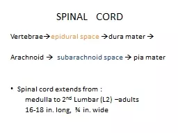
Slide2Spinal Cord
Location
Begins at the foramen magnum
Ends as conus medullaris at L
1
vertebra
Functions
Provides two-way communication to and from the brain
Contains spinal reflex centers
Slide3Figure 12.30
Ligamentum
flavum
Supra-
spinous
ligament
Lumbar puncture
needle entering
subarachnoid
space
Filum
terminale
Inter-
vertebral
disc
T
12
L
5
Cauda equina
in subarachnoid
space
Dura
mater
L
5
L
4
S
1
Arachnoid
matter
Slide4Figure 12.29a
Cervical
enlargement
Dura and
arachnoid
mater
Lumbar
enlargement
Conus
medullaris
Cauda
equina
Filum
terminale
Cervical
spinal nerves
Lumbar
spinal nerves
Sacral
spinal nerves
Thoracic
spinal nerves
(a) The spinal cord and its nerve
roots, with the bony vertebral
arches removed. The dura mater
and arachnoid mater are cut
open and reflected laterally.
Slide5Spinal Cord
Spinal nerves
31 pairs
Cervical and lumbar enlargements
The nerves serving the upper and lower limbs emerge hereCauda equinaThe collection of nerve roots at the inferior end of the vertebral canal
Slide6Spinal Cord
Gray matter – more centrally located; looks like a butterfly. Consists of nerve cell bodies and dendrites
White matter – surrounds the gray matter and is composed of white, myelinated fibers (axons)
Central canal – center of the gray matter; contains cerebrospinal fluid
Slide7Spinal Nerves
Formed from the
posterior (dorsal) root
and
anterior (ventral) rootThe posterior (dorsal) root carries sensory fibersThe anterior (ventral) root carries motor fibersThe posterior (dorsal) root ganglion consists of somatic sensory neuron cell bodies that are unipolarA plexus is a braided network of the anterior branches from some spinal nerves (cervical, brachial, lumbar and sacral)
Slide8Figure 12.31b
(b) The spinal cord and its meningeal coverings
Dorsal funiculus
Dorsal median sulcus
Central canal
Ventral median
fissure
Pia mater
Arachnoid mater
Spinal dura mater
Gray
commissure
Dorsal horn
Gray
matter
Lateral horn
Ventral horn
Ventral funiculus
Lateral funiculus
White
columns
Dorsal root
ganglion
Dorsal root
(fans out into
dorsal rootlets)
Ventral root
(derived from several
ventral rootlets)
Spinal nerve
Slide9Figure 12.32
Somatic
sensory
neuron
Dorsal root (sensory)
Dorsal root ganglion
Visceral
sensory
neuron
Somatic
motor neuron
Spinal nerve
Ventral root
(motor)
Ventral horn
(motor neurons)
Dorsal horn (interneurons)
Visceral
motor
neuron
Interneurons receiving input from somatic sensory neurons
Interneurons receiving input from visceral sensory neurons
Visceral motor (autonomic) neurons
Somatic motor neurons