
httpyoutubeaDvbAvBLQuM Ligaments and Vertebral Discs Muscular Anatomy Neurological Anatomy Prevention of Injuries to the Spine Cervical Spine Muscle Strengthening Muscles of the neck resist hyperflexion hyperextension and rotational forces ID: 774871
Download Presentation The PPT/PDF document " Chapter 20: The Spine Anatomy of the Sp..." is the property of its rightful owner. Permission is granted to download and print the materials on this web site for personal, non-commercial use only, and to display it on your personal computer provided you do not modify the materials and that you retain all copyright notices contained in the materials. By downloading content from our website, you accept the terms of this agreement.


Slide1
Chapter 20: The Spine
Slide2Anatomy of the Spine
http://youtu.be/aDvbAvBLQuM
Slide3Slide4Slide5Ligaments and Vertebral Discs
Slide6Muscular Anatomy
Slide7Neurological Anatomy
Slide8Prevention of Injuries to the Spine
Cervical Spine
Muscle Strengthening
Muscles of the neck resist hyperflexion, hyperextension and rotational forces
Prior to impact the athlete should brace by “bulling” the neck (isometric contraction of neck and shoulder muscles)
Exercises can be used to strengthen the neck
Range of Motion
Must have full ROM to prevent injury
improved through stretching exercises
Slide9Prevention of Injuries to the Spine
Using Correct Technique
Athletes should be taught and use correct technique to reduce the likelihood of cervical spine injuries
Avoid using head as a
weapon
Spearing in football, diving
into shallow water
Lumbar Spine
Avoid Stress
Avoid unnecessary stresses and strains of daily living
Avoid postures and positions that can cause injury
Strength and Flexibility
Basic
conditioning should emphasize trunk flexibility
Spinal
and Core strength
should be stressed in order to maintain proper alignment
Slide10Prevention of Injuries to the Spine
Using Correct Lifting Techniques
Weight lifters can minimize injury of the lumbar spine by using proper technique
Incorporation of appropriate breathing techniques can also help to stabilize the spine
Weight belts can
be
useful in providing
stabilization
Use
spotters
when lifting
Core Stabilization
Core stabilization, dynamic abdominal bracing and maintaining neutral position can be used to increase
lumbo
-pelvic-hip
stability
Increased stability helps the athlete maintain the spine and pelvis in a comfortable and acceptable mechanical position (prevents
microtrauma
)
Slide11Assessment of the Spine
History
Mechanism of injury (rule out spinal cord injury)
What happened? Did you hit someone or did someone hit you? Did you lose consciousness
Pain in your neck? Numbness, tingling, burning?
Can you move your ankles and toes?
Do you have equal strength in both hands
Positive responses to any of these questions will necessitate extreme caution when the athlete is moved
Slide12Assessment of the Spine
Other general questions
Where is the pain and what kind of pain are you experiencing?
What were you doing when the pain started?
Did the pain begin immediately and how long have you had it?
Positions or movements that increase/decrease pain?
Past history of back pain
Sleep position and patterns, seated positions and postures
Slide13Assessment of the Spine
ObservationsBody typePostural alignments and asymmetries should be observed from all viewsAssess height differences between anatomical landmarks
Slide14Assessment of the Spine
Palpation
Should be performed with athlete prone
Head and neck should be slightly flexed, pillow under hips if suffering from low back pain
Spinous and transverse processes of each vertebrae should be palpated along with sacrum and coccyx
Muscles should be palpated bilaterally
Be aware of the possibility of referred pain
Slide15Assessment of the Spine
Special Tests
Test for lumbar spine should be performed standing, sitting, supine, side-lying and prone
Assess levels of pain and motion restriction during the following tests
Forward and backward bending
Side-bending and rotation
Slide16SLR Test
Straight Leg RaisesApplies pressure to SI joint and may indicate problems with sciatic nerve, SI joint or lumbar spine
Slide17SI Compression and Distraction Tests
Used for pathologies involving the SI jointDistraction Compression
Slide18Brachial Plexus Traction/Compression Test
Slide19Hoover Test
Athlete Position: SupineAthletic Trainer Position: Supporting the ankle of each leg while standing at athlete’s feet.Procedure: Athlete is asked to perform a unilateral straight leg raise actively on the involved extremity.Test is positive if: The athlete does not push down on the uninvolved leg or athlete does not attempt this maneuver. Implications: The athlete is malingering.
Slide20Faber’s Test
FABER’S
: Flexion Abduction External Rotation
Assess SI joint pathology
Slide21Recognition and Management of Specific Injuries and Conditions
Slide22Cervical Spine Injuries
Cervical Fractures
Cause of Injury
Generally an axial load w/ some degree of cervical flexion
Addition of rotation may result in dislocation
Signs of Injury
Neck point tenderness, restricted motion, cervical muscle spasm, cervical pain, pain in the chest and extremities, numbness in the trunk and or limbs, weakness in the trunk and/or limbs, loss of bladder and bowel control
Care
Treat like an unconscious athlete until otherwise
ruled
out - use extreme
care
Spine Board and transport
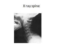
Slide23Cervical Fracture and Dislocation
Slide24Cervical Spine Injuries
Cervical Dislocation
Cause of Injury
Usually the result of violent flexion and rotation of the head
Signs of Injury
Considerable pain, numbness, weakness, or paralysis
Unilateral dislocation causes the head to be tilted toward the dislocated side with extreme muscle tightness on the elongated side
Care
Extreme care must be used - more likely to cause spinal cord injury than a fracture
Slide25Cervical Spine Injuries
Acute Strains of the Neck and Upper Back
Cause of Injury
Sudden turn of the head, forced flexion, extension or rotation
Generally involves upper
trapezius
Signs of Injury
Localized pain and point tenderness, restricted motion, reluctance to move the neck in any direction
Care
RICE and application of a cervical
collar in severe cases
Follow-up care will involve ROM exercises, isometrics which progress to a full isotonic strengthening program,
cryotherapy
and superficial thermotherapy, analgesic medications
Slide26Cervical Spine Injuries
Cervical Sprain (Whiplash)
Cause of Injury
Generally the same mechanism as a strain, just move violent
Involves a snapping of the head and neck
Signs of Injury
Similar signs and symptoms to a strain - however, they last longer
Tenderness over the transverse and spinous processes
Pain will usually arise the day after the trauma (result of muscle spasm)
Management
Rule out fracture, dislocation, disk injury or cord injury
RICE for first 48-72 hours, possibly bed rest if severe enough
Analgesics and NSAID’s, mechanical traction
Slide27Cervical Spine Injuries
Acute
Torticollis
Cause of Injury
Pain on one side of the neck upon wakening
Result of synovial capsule impingement w/in a facet
Signs of Injury
Palpable point tenderness and muscle spasm, restricted ROM, muscle guarding,
Care
Variety of techniques including traction, superficial heat and cold treatments, NSAID’s
Use of a soft collar can be helpful as well
May last 2-3 days
Gradual strengthening and stretching exercises should be utilized for neck and shoulders for prevention
Slide28Cervical Spine Injuries
Pinched Nerve (Brachial Plexus Injury)
Cause of Injury
Result of stretching or compression of the brachial plexus
Referred to as stinger or burner
Signs of Injury
Burning sensation, numbness and tingling as well as pain extending from the shoulder into the hand
Some loss of function of the arm and hand for several minutes
Symptoms rarely persist for several days
Repeated injury can result in neuritis, muscular atrophy, and permanent damage
Care
Return to activity once S&S have returned to normal
Strengthening and stretching program
Padding to limit neck ROM during impact
Slide29Lumbar Spine Conditions
Slide30Lumbar Spine Conditions
Low Back Pain
Cause of Injury
Congenital/biomechanical
anomalies
Mechanical defects of the spine (posture, obesity and body mechanics)
Back trauma
Recurrent and chronic low back pain
Signs of Injury
Pain, possible weakness,
antalgic
gait,
ligament
sprain, muscle strains and bony defects
Neurological signs and symptoms if it becomes disk related
Care
Correct alignments and body
mechanics (better posture, lifting techniques, lose weight, etc)
Strengthening and stretching – avoid unnecessary stresses and strains associated with daily living
Slide31Lumbar Spine Conditions
Lumbar Vertebrae Fracture and Dislocation
Cause
Compression fractures or fracture of the spinous or transverse processes
Compression fractures are usually the result of trunk hyperflexion or falling from a height
Fractures of the processes are generally the result of a direct blow
Stress from improper lifting or too much weight
Dislocations tend to be rare
Slide32Lumbar Fractures
Slide33Lumbar Spine Conditions
Signs of Injury
Compression fractures will require X-rays for detection
Point tenderness over the affected area
Palpable defects over the
spinous
and transverse processes
Localized swelling and guarding
Care
X-ray and physician referral
Transport with extreme caution and care to minimize movement of the segments
Utilize a spine board
Slide34Lumbar Spine Conditions
Low Back Muscle Strain
Cause of Injury
Sudden extension contraction overload generally in conjunction w/ some type of rotation
Chronic strain associated with posture and mechanics
Signs of Injury
Pain may be diffuse or localized; pain w/ active extension and passive flexion
Care
RICE to decrease spasm; followed by a graduated stretching and strengthening program
Elastic wrap/back brace may be useful for support and compression
Complete bed rest may be necessary if it is severe enough
NSAID’s
Slide35Lumbar Spine Conditions
Lumbar Strains
Cause of Injury
Forward bending and twisting can cause injury
Chronic or repetitive in
nature
Improper lifting techniques
Signs of Injury
Localized pain lateral to the
spinous
process
Pain becomes sharper w/ certain movements or postures
Care
RICE, joint
mobes
, strengthening for abdominals, stretching in all directions
Trunk stabilization exercises
Braces should be worn early to provide support
Will require time for healing
Slide36Lumbar Spine Conditions
Back Contusions
Cause of Injury
Significant impact or direct blow to the back
Signs of Injury
Pain, swelling, discoloration, muscle spasm and point tenderness
Management
RICE for the first 72 hours
Ice massage combined with gradual stretching
Recovery generally last 2 days to 2 weeks
Slide37Lumbar Spine Conditions
Sciatica
Cause of Injury
Inflammatory condition of the sciatic nerve
Nerve is vulnerable to torsion or direct blows that place abnormal amounts of stretching or pressure on nerve
Signs of Injury
Arises abruptly or gradually; produces sharp shooting pain, tingling and numbness
Sensitive to palpation with straight leg raises intensifying the pain
Care
Rest is essential acutely – recovery = 2-3 weeks
Treat the cause of inflammation; traction if disk protrusion is suspected; NSAID’s
Slide38Sciatica / Sciatica Nerves
Slide39Lumbar Spine Conditions
Herniated DiskCause of InjuryCaused by abnormal stresses and degeneration due to use (forward bending and twisting)Improper lifting techiquesOver weight
Slide40Lumbar Spine Conditions
Signs of Injury
Centrally located pain that radiate unilaterally in
dermatomal
pattern
Symptoms are worse in the morning
Onset is sudden or gradual, pain may increase after the athlete sits and then tries to resume activity
Forward bending and sitting increase pain, while back extension reduces pain
Straight leg raise to 30 degrees is painful
Care
Rest and ice for pain management
Extension exercises may be comfortable
Core stabilization exercises should be integrated as athlete improves
Slide41Lumbar Spine Conditions
Spondylolysis
and
Spondylolisthesis
Cause of Injury
Spondylolysis
refers to degeneration of the vertebrae due to congenital weakness (stress fracture results)
Slipping of one vertebrae above or below another is referred to as
spondylolisthesis
and is often associated with a
spondylolysis
Signs of Injury
Pain and persistent aching, low back stiffness with increased pain after activity
Frequent need to change position or “pop” back to reduce pain
Localized tenderness to one segment
Slide42Spondylolysis
Slide43Spondylolisthesis
Slide44Lumbar Spine Conditions
Care
Bracing and occasionally bed rest for 1-3 days will help to reduce pain
Major focus should be on exercises directed as controlling or stabilizing hypermobile segments
Progressive trunk strengthening, dynamic core strengthening, concentration on abdominal work
Braces can also be helpful during high level activities
Increased susceptibility to lumbar strains and sprains and thus vigorous activity may need to be limited
Slide45Sacroiliac and Coccyx Injuries
Sacroiliac Sprain
Cause of Injury
Result of twisting with both feet on the ground, stumbles forward, falls backward, steps too far down, heavy landings on one leg, bends forward with knees locked during lifting
Signs of Injury
Palpable pain and tenderness over the joint, medial to the PSIS w/ some muscle guarding
Pelvic asymmetries are possible
Slide46Sacroiliac and Coccyx Injuries
Care
Ice can be used to reduce pain
Bracing can be helpful in acute sprains
Strengthening exercises should be used to stabilize the joints
Slide47Sacroiliac and Coccyx Injuries
Coccyx Injuries
Cause of Injury
Generally the result of a direct impact which may be caused by forcibly sitting down, falling, or being kicked by an opponent
Signs of Injury
Pain is often prolonged and at times chronic
Tenderness over the bone and pain with sitting
Care
Analgesics and a ring seat to relieve pressure while sitting
Pain from a fractured coccyx could last months
May require protective padding to prevent further injury