
Chapter 12 Skin anatomy withMeshergraft thicknessSkin graftHumby knifeTying the dressing Using the dermatomein placeUsing the Humby knifeDefatting the FTSG 98Practical Plastic Surgery for Nonsurgeons ID: 355076
Download Pdf The PPT/PDF document "SKKIINN GGRRAAFFTTSSAskin graft involve..." is the property of its rightful owner. Permission is granted to download and print the materials on this web site for personal, non-commercial use only, and to display it on your personal computer provided you do not modify the materials and that you retain all copyright notices contained in the materials. By downloading content from our website, you accept the terms of this agreement.

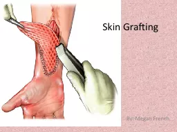


SKKIINN GGRRAAFFTTSSAskin graft involves taking a piece of skin from an uninjured area ofthe body (called the donor site) and using it to provide coverage for anopen wound. When primary closure is impossible because of softtissue loss and closure by secondary intention is contraindicated, a skingraft is the next rung on the reconstructive ladder. It is not a technicallydifficult procedure but does require some surgical skills. For a success-ful result, you need a thorough understanding of how skin grafts healand how to perform the procedure.The epidermis is the top portion of the skin. The outer layers of the epi-dermis are formed by essentially dead, nonreplicating cells. The inner-most layer contains the cells capable of replication, which are responsibleImmediately below the epidermis is the dermis. It is made primarily of col-lagen and is much thicker than the epidermal layer. The dermal-epi Chapter 12 Skin anatomy withMeshergraft thicknessSkin graftHumby knifeTying the dressing Using the dermatomein placeUsing the Humby knifeDefatting the FTSG 98Practical Plastic Surgery for Nonsurgeons junction is irregular and has the appearance of ridges. This anatomicarrangement accounts for the skins strength and prevents injury fromnormal shear forces. Nerve endings, hair follicles, and sweat glands arelocated in the dermis. The subcutaneous fatty tissue below the dermis provides padding formany important nerves for pressure sensation, reside in the subcuta-the subcutaneous tissue as a layer of the skin. However, it is not in-cluded in a skin graft. Fat attached to the graft interferes with transportof nutrients to the important upper skin layers. Therefore, no fatWhen the skin graft is harvested from the donor site, it is completelyseparated from its blood supply. In its new position covering the openwound, the graft initially survives by diffusion of nutrients from thewound bed into the graft. Diffusion of nutrients keeps the skin graft two different germ layers). The relative thickness of skin grafts is shown. TheCohen M (ed): Mastery of Plastic and Reconstructive Surgery. Boston, Little, Skin Grafts99 alive for, at most, 3 5 days. During this period, blood vessels begin togrow from the wound bed into the graft. By the time the graft is nolonger able to survive by diffusion of nutrients alone, this vascular net-work has formed and becomes the primary mechanism for providingIn the first several weeks after the procedure, the skin graft looks quitered and irregular compared with normal surrounding skin. Reassurethe patient that the appearance will improve dramatically over the nextseveral months, but the skin-grafted area will never look completelygraft. See chapter 15, Scar Formation, for more detailsAwound will accept a skin graft when there is no overlying deadtissue and the wound is clean, beefy red (from granulation tissue), andwithout surrounding infection. Skin grafts heal well over muscle.Therefore, if muscle is exposed in the wound, skin can be grafted atAwound that has exposed tendon or bone can be successfully coveredwith a skin graft only if the thin layer of tissue connecting the tendon or TTable 1.Compensating for Factors that Interfere with Graft SurvivalGranulation tissue is the beefy red tissue that develops as a wound heals. It has an excellent bloodsupply but also contains bacteria in its crevices. Dirty wound (e.g.,Debride the wound and treat it with wet-to-dry dressingssurrounding in-until the wound looks clean. Use antibiotics to clearfection, necroticsigns of surrounding infection. The skin graft can betissue over wound)done once the wound has improved in appearance andFat in base of woundFat has a poor blood supply and may not be able to sup-port the graft. Treat the wound with wet-to-dry dressingsuntil granulation tissue* begins to appear. Then do theShear forces betweenMovement of the graft over the wound interferes with vas-graft and base ofcular ingrowth. The graft must be kept well secured towoundthe wound by the dressing. If the graft is on an extremity,Blood or serumFluid collection under the graft prevents the ingrowth ofgraftcan be prevented by cutting holes in the graft and keep- Skin Grafts101 Athin layer of skin (epidermis with some underlying dermis) isWatson knife). Adermatome is powered by air or electricity, but it isnot available in all hospitals, especially in rural settings. Remember:you are justed to set the thickness of the graft. Place the settings at 0.011 0.015inch (0.25 0.4 mm). Unfortunately, these settings are often unreliable.Another technique to ensure proper thickness of the graft is to adjustAlways check the knife settings just before you take the graft.This safety check prevents the accidental taking of too thick or too thinAn assistant should help to spread and flatten out the donor site byplacing tension on the skin with gauze or tongue depressors.1.Turn on the power while the dermatome is in the air before it comes2.Hold the dermatome at a 45°angle with the skin and hold it firmly3.Slowly move down the donor site until you have taken the properly4.At this point do turn off the power. Remove the dermatomefrom the skin with the power on so that the graft is completely freedfrom the donor site.5.The entire movement is evocative of landing an airplane and takingoff again right away. Skin-graft (Humby) knife. (From Padgett Instruments, Inc., with permission.) 102Practical Plastic Surgery for Nonsurgeons 1.Hold it with the sharp edge at about a 45°angle with the skin.2.With a back-and-forth motion run the knife over the tight skin.3.When you have taken a large enough graft, continue the back-and-forth motion, and twist your wrist into supination to remove theknife from the skin. Another option is to stop the knife movementand then use a scalpel to cut the skin graft from the donor site at the Cohen M (ed): Mastery of Plastic and Reconstructive Surgery. Boston, Little, (ed): Plastic Surgery. Philadelphia, W.B.Saunders, 1990, with permission.) Skin Grafts107 FTSGs are rarely done, because the wound must begraft to survive. Most often they are used for a small wound, usuallyone created surgically (such as a wound on the face created by excisionthe hands and fingers. These areas may scar too tightly if the thinnerThe best donor site is usually just above the inguinal crease on thelower abdomen. If the graft is needed to cover a facial wound, extraskin of a reasonable color match often can be taken from the supraclav-icular area in the neck or from behind the ear.An ellipse is drawn at the donor site. Make sure that it is large enoughto cover the defect but not too large to close the donor site.You can tell how large a graft you can take by seeing how much skinyou can pinch or pull up at the donor site. At the inguinal area, you canflex the patients hip to decrease tension on the closure. After a fewdays the patient will be able to extend the hip fully. This approachcauses no long-term problems.Because you are taking a relatively small graft, the FTSG can be harvestedwith a local anesthetic. Lidocaine and marcaine work equally well.1.The ellipse of skin is excised with the full layer of dermis. To facili-tate the procedure, take the graft with some underlying fat attached.2.You must remove the attached fat, which will interfere with graft1.The skin graft should be placed under tension. Place clamps on thehang freely. 2.Use scissors to remove the fat on the dermis. Place the scissors flushwith the skin, and cut away the fat. Do not worry if you take a littledermis or cut into small areas of the epidermis.