
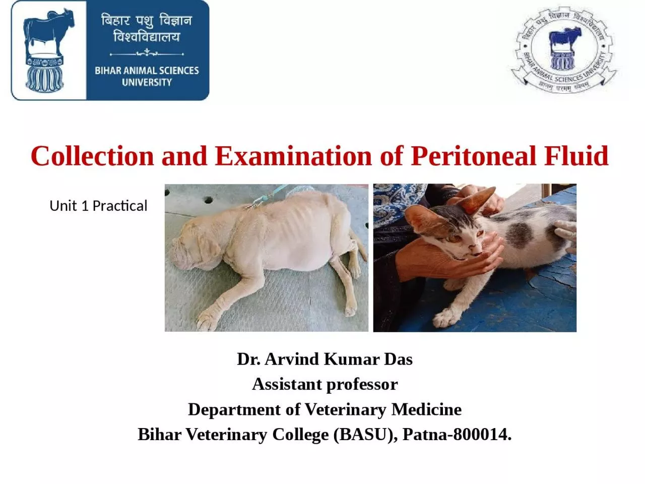
Dr Arvind Kumar Das Assistant professor Department of Veterinary Medicine Bihar Veterinary College BASU Patna800014 Unit 1 Practical Introduction Collection of peritoneal fluid is a useful to diagnose the diseases of the peritoneum and the abdominal segment of the alimentary tract ID: 1011511
Download Presentation The PPT/PDF document "Collection and Examination of Peritoneal..." is the property of its rightful owner. Permission is granted to download and print the materials on this web site for personal, non-commercial use only, and to display it on your personal computer provided you do not modify the materials and that you retain all copyright notices contained in the materials. By downloading content from our website, you accept the terms of this agreement.


1. Collection and Examination of Peritoneal Fluid Dr. Arvind Kumar DasAssistant professorDepartment of Veterinary MedicineBihar Veterinary College (BASU), Patna-800014.Unit 1 Practical
2. IntroductionCollection of peritoneal fluid is a useful to diagnose the diseases of the peritoneum and the abdominal segment of the alimentary tract. Peritoneal fluid analysis is used to investigate the cause of fluid buildup in the abdomen (ascites) and inflammation of the peritoneum (peritonitis). These are two main reasons for fluid accumulation. An initial set of tests is used to differentiate between the two types of fluid that may be transudate or exudate. Peritoneal fluid reflects the pathophysiological state of the parietal and visceral mesothelial surfaces of the peritoneum.Determining the presence of: Peritonitis (chemical or infectious), infarction of a segment of gut wall, perforation of the alimentary tract wall, rupture of the urinary bladder, leakage from the biliary system, intraperitoneal hemorrhage and peritoneal neoplasia.
3. Peritoneal fluidNormal peritoneal fluid is a transudate with properties. It has functions similar to those of other tissue fluids. It contains mesothelial cells, lymphocytes, neutrophils, a few erythrocytes and occasional monocytes and eosinophils. It is of vital importance in horses in the differential diagnosis and prognosis of colic and in cattle in the diagnosis of peritonitis.
4. Collection SitesCattle: Ventral abdominal wall, between the forestomach, abomasum, diaphragm and liver. These are usually caudal to the xiphoid sternum and 4-10 cm lateral to the midline.Horse: Ventral midline, 25 cm caudal to the xiphoid (or midway between the xiphoid and the umbilicus)Canines: Area surrounding the umbilicus
5. Collection of Peritoneal FluidFollowing surgical preparation and subcutaneous infiltration of an anaesthetic, a stab incision is made through the skin and subcutaneous tissues and into the linea alba. A 9 cm long blunt-pointed bovine teat cannula, or similar metal catheter, with the tip wrapped in a sterile swab to avoid blood and skin contamination, is inserted into the wound and manipulated until the incision into the linea alba can be felt. With a quick thrust the cannula is pushed through the linea alba into the peritoneal cavity. A 'pop' is often heard on entry into the peritoneal cavity.Failure to incise into the linea alba first will cause many cannulas to bend and break.The technique most likely to cause bowel penetration is the use of a sharp needle instead of the blunt cannula recommended, and forcibly thrusting the cannula through the linea alba without a prior incision.
6. Collection… continuedWhen the suggested incision is made in the linea alba, the cannula can be pushed gently through rotating it.In most horses (about 75%) a sample of fluid is readily obtained. But, sometimes it takes a moment before the fluid runs out, usually spurting synchronously with the respiratory movements. Applying suction with a syringe may yield some fluid if there is no spontaneous flow. Normal fluid is clear, yellow and flows easily through an 18-gauge needle. Two samples are collected, one in a plain tube and one in a tube with an anticoagulant. In case the fluid clots readily a few drops should be placed and smeared out on a glass slide and allowed to dry for staining purposes.
7. Collection… continuedIf first attempt of paracentesis causes penetration of the gut, it is recommended to repeat the attempt, if necessary two or three times, at more posterior sites. Repeated abdominocentesis does not cause alterations in peritoneal fluid constituents and any significant changes are likely due to alterations in the disease state present.
8. Risks in CollectionAbdominocentesis is not without some danger, especially the risk of introducing needle.Puncture of a devitalized loop of intestine may cause a leakage of intestinal contents and cause acute diffuse peritonitis, which is fatal. This is a common occurrence in the later stages of intestinal obstruction. Presence of faecal contents into the peritoneal cavity causing peritonitis. This appears to be of major importance only if there are loops of distended atonic intestine situated on the ventral abdominal wall. Penetration of a normal loop of intestine occurs often enough to lead to the conclusion that it appears to have no ill effects.
9. Examination of Peritoneal FluidPhysical characteristics:It can be examined in terms of physical characteristics, especially colour, translucence, specific gravity, clotting time, biochemical composition, cell volume, cell morphology and cell type.The reaction of the peritoneum varies with time and a single examination can be dangerously misleading. A series of examinations may be necessary, in acute cases at intervals of as short as an hour.
10. Changes in peritoneal fluidA significant reaction in a peritoneal cavity may be quite localized. So, fluid collected at one point is not representative of the entire cavity.Changes in peritoneal fluid (chemical composition, e.g. lactate) -systemic change. The examination of peripheral blood sample determine the changes.
11. Peritoneal Fluid Analysis
12. Clinicopathological examinationResults must be interpreted on the basis of history and clinical findings.Specific properties of peritoneal fluid (normal and abnormal) Turbidity: Presence of increased leukocytes and protein-fine strands of fibrin in fluid. Colour: Normal fluid-Crystal clear, Straw coloured to yellow. Green colour- food materialIntense orange-green- Rupture of the biliary system. Pink-red colour - Presence of haemoglobin, Degenerated erythrocytes or entire erythrocytes Damaged vascular system by infarction, Perforation or hydrostatic pressure.
13. Clinicopatho….. Exam… ContinuedRed brown colour- Late stages of necrosis of the gut wall, Presence of degenerated blood and haemoglobin Damage of gut wall with hemorrhage.Whole blood- Clear fluid streaked with blood or heavily blood stained fluid- sample of spleen or blood vessel or there is hemoperitoneum. Rupture of the uterus or bladder or Dicoumarol poisoning are also possibilities.Dark green- sample with motile protozoa with very few leukocytes and no mesothelial cells- sample from the gut lumen.
14. Effect of EnterocentesisEnterocentesis has little clinical effect in normal horses. Transient fever puncture of a devitalized loop of intestine- extensive leakage of gut contents -fatal peritonitis. Enterocentesis of normal gut- increase the neutrophilic count of peritoneal fluid -persists for several days.Surgical manipulation of the intestinal tract and viscera-injury of mesothelial surfaces-postoperative peritoneal inflammatory reaction. In this condition ↑TLC, DLC, RBC, TP and fibrinogen from first day after the surgery up to 7 days.In cattle, exploratory celiotomy and omentopexy- ↑ total nucleated cell count or specific gravity and TP from two days after surgery up to 6 days.
15. Cellular and other propertiesParticulate matter in peritoneal fluid - Either fibrin clots/strands or gut contents- caused by leakage from a perforated or ruptured gut wall.High specific gravity and high protein content- Vascular damage-leakage of plasma protein- due to peritonitis or mural infarction.The volume and viscosity of fluid varies. Normal flow: 1-5 ml per sample. Continuous flow with 10-20 ml per sample- Excess fluid due to ruptured bladder/ or ascites (clear yellow), acute diffuse peritonitis (yellow, turbid), infarction or necrosis of gut wall (thin, red- tinged).As the peritoneal fluid shifts- transudate to inflammatory exudate- ↑protein content and ↑ viscosity.Highly viscous fluid may clot.
16. Cellular.....continuedA rapid staining method, using a modified Wright's stain, gives a stained slide ready for examination within 5 minutes. Indicating the number of leukocytes and other cells present, and differentiating the types of cell.↑ TLC of the fluid including disproportionate number of polymorphonuclear cells- acute inflammation-infectious or non-infectious.An increase in mononuclear phagocytes from the peritoneum in peritoneal fluid- chronic peritonitis. Degenerate and toxic neutrophils- possibility of presence of infection.
17. Cellular..... continuedIncrease in mesothelial cells with the distinctive presence of actively dividing mitotic figures-neoplasia.Bacteria as phagocytosed inclusions body in leukocytes, or by culture of fluid- Infective peritonitis- by haematogenous spread. If there is leakage from a peritoneal abscess the same condition as above,If there is leakage through a segment of devitalized or perforated bowel wall- Mixed infection- by particulate matter of bowel contents.
18. Cellular..... continuedThe blood is likely to be concentrated- if sufficient time for fluid resorption across the peritoneum. Splenic blood has a higher packed cell volume (PCV) also, but there is no erythrophagocytosis evident in the sample. A PCV of less than 5% in peritoneal fluid suggests extravasation of blood from an infarcted or inflamed gut wall and more than 20% suggests a significant haemorrhage.Total leukocyte counts ranging from 10 000-150 000 has recorded in peritonitis and in infarction of the intestine in horses.
19. Collection IndicationPeritonitis Hemoperitoneum Biliary Peritonitis Uroperitoneum
20. Thank You