
spinal cord spinal segment Dermatomes Dr Gábor GERBER cervical thorac ic lumba r sacral coccygeal Intumescentia cervicalis C5T1 Intumescentia lumbalis L2L3 ID: 916741
Download Presentation The PPT/PDF document "Gross anatomy of the" is the property of its rightful owner. Permission is granted to download and print the materials on this web site for personal, non-commercial use only, and to display it on your personal computer provided you do not modify the materials and that you retain all copyright notices contained in the materials. By downloading content from our website, you accept the terms of this agreement.


Slide1
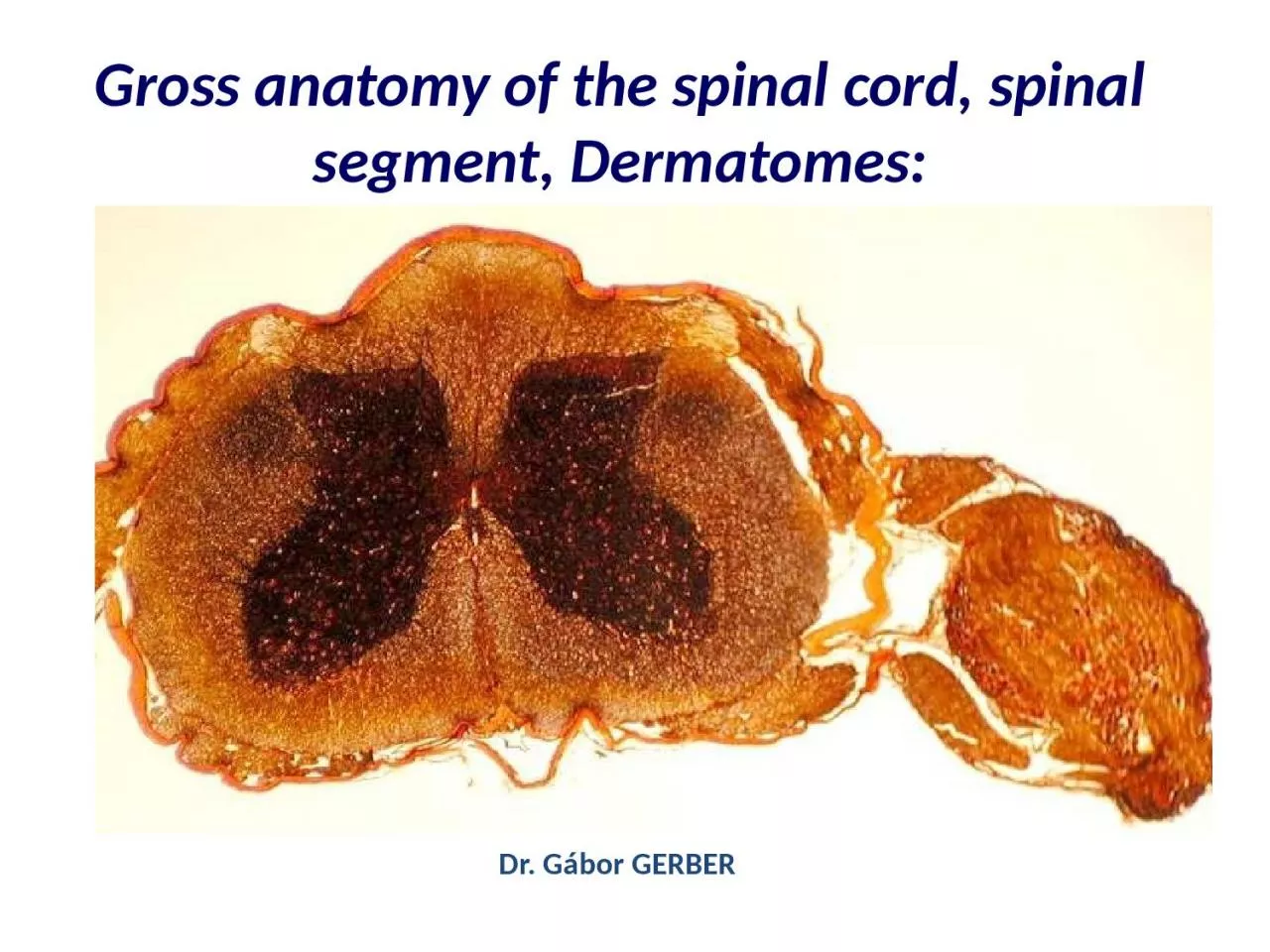
Gross
anatomy of the spinal cord, spinal segment, Dermatomes:
Dr. Gábor GERBER
Slide2cervical
thoraciclumbarsacral
coccygeal
Slide3Slide4Intumescentia
cervicalis
(C5-T1):
Intumescentia
lumbalis
(L2-L3):
Slide5cervicalis
thoracalislumbosacralis
Slide6Slide7Dura
mater spinalisArachnoidea materPia mater Ligg. denticulata
Endorachis
Denticulate
ligaments
(
purple
)
are
bilateral
thickenings
of
the
collagen
component
of
pia mater.
Slide8Slide9Spinal nerve
Root vs. Ramus !Ventral ramus plexus
Slide10DermatomArea
of skin - innervated by the same spinal nerve
Slide11Head’s
Zones (referred pain)
Slide12Herpes Zoster (
shingles)viral infection
Slide13Blood
supplyAnterior spinal artery anterior 2/3posterior spinal arteries posterior 1/3
Slide14Intercostal
, segmental arteriesAdamkiewicz artery T10
Slide15S2: end of
dural sacfilum terminale pars duralis
Slide16Slide17Slide18Lumbar puncture
Slide19Spinal, Epidural, Caudal
anesthesia
LAYERS TO PENETRATE
Skin
Supraspinal ligament
Interspinal ligament
Lig. flavum
Endorachis
Epidural space
Dura mater spinalis
Arachnoid mater
Subarachnoid space
Slide20Thank
you for your attention
Slide21Slide22SPINAL MENINGES
1) DURA MATER
Envelops the cord, descends
to S2
vertebra, between
L2 and S2 the sac contains only the cauda equina.
Each nerve passes through the
intervertebral foramen
retaining its dural cover, it continues as
perineurium
.
Endorachis
-
connective tissue „outer layer” of the
spinal dura mater
-
epidural space
(content: fat,
internal vertebral venous plexus)
2) ARACHNOID MATER
Lines the dural cavity, rather large subarachnoideal,
but insignificant subdural space.
LUMBAR PUNCTION - below L2
3) PIA MATER
Adheres to the spinal cord and roots, its lateral aspect gives rise to the
denticulate ligament
(21 on each side) reaching the arachnoid mater. It lies between the dorsal and ventral nerve roots.