
Dr Sushma Tomar Associate Professor Department of Anatomy Lesson Plan Introduction Parts of Nasal Cavity Vestibule Nasal Cavity Proper Roof of Nasal Cavity Floor of Nasal Cavity Nasal Septum ID: 910631
Download Presentation The PPT/PDF document "Nasal Cavity-I Presented by:-" is the property of its rightful owner. Permission is granted to download and print the materials on this web site for personal, non-commercial use only, and to display it on your personal computer provided you do not modify the materials and that you retain all copyright notices contained in the materials. By downloading content from our website, you accept the terms of this agreement.


Slide1
Nasal Cavity-I
Presented by:-
Dr.
Sushma
Tomar
Associate Professor
Department of Anatomy
Slide2Lesson Plan
Introduction
Parts of Nasal Cavity:
Vestibule
Nasal Cavity Proper
Roof of Nasal Cavity
Floor of Nasal Cavity
Nasal Septum:
Introduction
Parts
Arterial Supply
Venous Drainage
Nerve Supply
Lymphatic Drainage
Applied Aspects
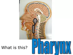
Slide3Introduction
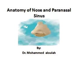
Internal nose is divided into right and left nasal cavities by a nasal septum.
Each nasal cavity communicates with the exterior through
nostril
(
naris).Each nasal cavity communicates with the nasopharynx through posterior nasal aperture (choana).
Nostri
l
Slide4Slide5Parts of Nasal Cavity
2
parts:
Vestibule
Nasal cavity proper
Vestibule-A small anteroinferior part.Lined by skin.Nasal Cavity Proper-
Large posterosuperior part.Lined by mucosa.
Slide6Vestibule
Its upper limit on lateral wall of nasal cavity is marked by
Limen
nasi
.Its medial wall is formed by columella.
Slide7Nasal Cavity Proper
Boundaries-
Roof.
Floor.
Medial wall (Nasal Septum).
Lateral wall.Lateral Wall
RoofFloor
Nasal Septum
Slide8Roof of Nasal Cavity
Divided into 3 parts:
Anterior 1/3
rd
.
Middle 1/3rd .Posterior 1/3rd .Anterior 1/3rd –Slopes downwards and forwards.
Formed by:Nasal spine of Frontal bone.Nasal bone.
Junction of Septal and Lateral cartilages
Nasal spine of Frontal bone
Nasal bone
Junction of
Septal and Lateral cartilages
Slide9Roof of Nasal Cavity contd…
Middle 1/3
rd
–
Horizontal.
Formed by:Cribriform plate of Ethmoid bone.Through the cribriform plate, olfactory nerves enter the cranial cavity.
Slide10Roof of Nasal Cavity contd…
Posterior 1/3
rd
–
Slopes downwards and backwards.
Formed by:Anterior surface of body of Sphenoid bone.
Slide11Floor of Nasal Cavity
Almost horizontal.
Formed by:
Upper surface of hard palate.
Anterior 3/4
th –By palatine process of Maxilla.Posterior 1/4th –Horizontal plate of Palatine bone.
Slide12Nasal Septum
Slide13Introduction
A median
osseocartilaginous
partition between two nasal cavities.
Forms the medial wall of nasal cavity.
Nasal Septum
Nasal Septum
Slide14Parts of Nasal Septum
3
parts:
Bony part.
Cartilaginous part.
Membranous part.Framework of Bony Part-Ethmoid- perpendicular plateVomer
Frontal - nasal spine
Nasal- crestSphenoid- crest
Maxilla- palatine process
Palatine- horizontal plate
Slide15Cartilaginous Part
Framework of Cartilaginous Part-
Septal
cartilage
Medial
crura (septal processes) of 2 major alar cartilages.
Slide16Cartilaginous Part contd
…
Columellar
Septum-
Medial
crura of 2 major alar cartilages are united together in the midline by a fibrous tissue to form columellar septum (columella).
Slide17of
Major
Alar
Cartilage
Slide18Membranous Part
Between the columella and caudal border of septal cartilage, a small portion of septum is made up of double layer of skin and is referred as
membranous septum.
Both columellar and membranous parts of the septum are freely movable from side to side.
Slide19Arterial Supply of Nasal Septum
Septal
b/o
Anterior
Ethmoidal
arterySeptal b/o Posterior Ethmoidal arterySeptal
b/o Sphenopalatine artery
Septal b/o Greater Palatine artery
Septal b/o Superior labial artery
Slide20Little’s Area
An area in anteroinferior part of nasal septum just above the vestibule.
It is highly vascular.
In this area, an arterial plexus is formed by the following arteries:
Septal
b/o Anterior Ethmoidal
arterySeptal b/o
Sphenopalatine arterySeptal b/o
Greater Palatine arterySeptal b/o Superior labial artery.
This plexus is known as
Kiesselbach’s plexus.
In Little’s area,
submucous
venous plexus is also more marked.
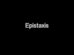
Applied aspect-
Little’s area is the
commonest site of
epistaxis
(bleeding through the nasal cavity) in children and young adults usually due to
finger nail trauma
during
pricking of nose.
Slide21Slide22Slide23Venous Drainage of Nasal Septum
In nasal septum, veins accompany the arteries.
A venous plexus is formed beneath the mucosa.
Veins drain into:
Facial vein.
Pterygoid venous plexus.Pharyngeal venous plexus.
Slide24Nerve Supply of Nasal Septum
Anterosuperior
Part-
Internal nasal
nerve (a branch of Anterior Ethmoidal Nerve).Anteroinferior Part- Anterior Superior Alveolar Nerve (a branch of Infraorbital
nerve).Posterosuperior Part-
Medial posterior superior nasal nerves ( branches of
Pterygopalatine ganglion).Posteroinferior Part-
Nasopalatine nerve ( a branch of
Pterygopalatine ganglion).Olfactory Part (upper 1/3
rd of nasal septum just below cribriform plate)-
Olfactory Nerves.
Slide25Lymphatic Drainage of Nasal Septum
Anterior ½-
Drains into:
Submandibular
lymph nodes.
Posterior ½-Drains into:Retropharyngeal lymph nodes.
Slide26Applied Aspects
Nasal Septum is seldom exactly in median plane.
Usually it bulges to right or left side,
more frequently to
the
right.DNS [Deviated Nasal Septum]-An important cause of nasal obstruction.Sex Predilection-Male>Female.Etiology-Trauma.Developmental error.Clinical Features-
Mechanical nasal obstruction.Sinusitis.Headache etc.Treatment-
Surgical correction-SMR (Submucosal resection).
Septoplasty.
Slide27Applied Aspects contd…
Supratip
depression of External Nose-
Septal
cartilage provides support to dorsum of anterior 2/3
rd of nose.Excessive removal of septal cartilage during submucosal resection can lead to Supratip
depression of External Nose.Destruction of septal
cartilage due to disease may result into this deformity.
Slide28Slide29