
ypotonia Clinical Approach to Floppy Baby H ypotonia in the newborn is a common presenting feature of systemic illness or neurologic dysfunction at any level of the central or peripheral nervous ID: 961058
Download Pdf The PPT/PDF document "Neonatal H" is the property of its rightful owner. Permission is granted to download and print the materials on this web site for personal, non-commercial use only, and to display it on your personal computer provided you do not modify the materials and that you retain all copyright notices contained in the materials. By downloading content from our website, you accept the terms of this agreement.


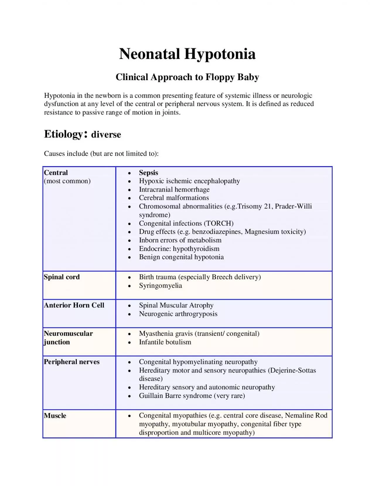
Neonatal H ypotonia Clinical Approach to Floppy Baby H ypotonia in the newborn is a common presenting feature of systemic illness or neurologic dysfunction at any level of the central or peripheral nervous system. It is defined as reduced resist ance to passive range of motion in joints. Etiology : diverse Causes include (but are not limited to): Central (most common) Sepsis Hypoxic ischemic encephalopathy Intracranial hemorrhage Cerebral malformations Chromosomal abnormalities (e.g.Trisomy 21, Prader - Willi syndrome) Congenital infections (TORCH) Drug effects (e.g. benzodiazepines , Magnesium toxicity ) Inborn errors of metabolism Endocrine: hypothyroidism Benign congenital hypotonia Spinal cord Birth trauma (especially Breech delivery) Syringomyelia Anterior Horn Cell Spinal Muscular Atrophy Neurogenic arthrogryposis Neuromuscular junction Myasthenia gravis (transient/ congenital) Infantile botulism Peripheral nerves Congenital hypomyelinating neuropathy Hereditary motor and sensory neuropathies (Dejerine - Sottas disease) Hereditary sensory and autonomic neuropathy Guillain Barre syndrome (very rare) Muscle Congenital myopathies (e.g. central core disease , Nemaline Rod myopathy, myotubular myopathy, congenital fiber type disproportion and multicore myopathy) Congenital muscular dystrophies ( merosin deficient, Walk
er - Warburg disease, muscle - eye - brain disease, Fukuyama disease Muscular dystrophies (inc. congenital myotonic dystrophy) Metabolic myopathies and multisystem disease Disease of Glycogen Metabolism Acid maltase deficiency Severe neonatal Phosphofructokinase deficiency Severe neonatal phosphrylase deficiency Debrancher deficiency Peroxisomal disorder Neonatal adrenoleukodystrophy Cerebrohepatorenal syndrome(Zellweger’s) Disease of Creatine metabolism Mitochondrial myopathies Cytochrome - c - oxidase deficiency Primary Carnitine deficiency The first goal in diagnosing the source of neonatal hypotonia is to ascertain if it is central (upper motor neuron) or peripheral (lower motor neuron). Central causes are the most common. Thi s delineation will determine the investigations most likely to yield a diagnosis. History Any significant family history – affected parents or siblings, consanguinity, stillbirths, childhood deaths Maternal disease – diabetes, epilepsy, myotonic dystrophy (may not be recognized) Pregnancy and delivery history – drug or teratogen exposure Decreased fetal movements Abnormal presentation Polyhydramnios/ oligohydramnios Apgar scores Resuscitation requirements Cord gases History since delivery o Respiratory effort o Ability to feed o Level of alertness o Level of spontan
eous activity o Character of cry Physical Examination A detailed physical examination should be performed, assessing muscle tone, any asymmetry, the infant’s strength, deep tendon reflexes (DTR), and any dysmorphic or unusual features. Central Anterior Horn Cell Nerve Neuromuscular Junction Muscle normal strength proxima�ldistal weakness dista�lproximal weakness, bulbar weakness proxima�ldistal weakness, normal/ increased DTRs + decreased/ absent DTRs decreased/ absent DTRs normal DTRs decreased DTRs +/ - seizures + fasciculation +/ - fasciculation no fasciculation +/ - dysmorphic features often described as alert At times babies with profound central hypotonia may have abse nt DTR, therefore absent DTR at least in the first few days of life would not rule out a central cause for the hypotonia Note that the presence of profound weakness as well as hypotonia suggests a disorder of the lower motor neuron. A sign of this may be a weak cry. Weakness is uncommon in central hypotonia except in the acute stages. Arthrogryposis (the fixation of joints at birth) may be associated with neonatal hypotonia, more commonly with lower motor neuron unit or multisystem abnormalities. Additional clues which may direct to a specific diagnosis: Hepatosplenomegaly – storage disorders, congenital infections Renal cysts, high forehead, wide fontanelles – Zellweger’s syndrome Hepatomegaly,
retinitis pigmentosa – neonatal adrenoleukodystrophy Congenital cataracts, glaucoma – oculocerebrorenal (Lowe) syndrome Abnormal odor – metabolic disorders Hypo pigmentation, undesceded testes – Prader Willi Examination of the mother is also important in suspected cases of congenital myotonic dystrophy or myasthenia g ravis. Most studies have found that central causes account for 60 - 80% of cases and that the diagnosis can usually be made by a careful history and examination. However, there may be a mixed picture. Infants with a peripheral cause for their hypotonia may b e at increased risk for problems during labor, delivery and resuscitation and develop hypoxic ischemic encephalopathy. Investigations Further investigation needs to be guided by history and examination. If the infant is hypotonic but has a degree of stren gth, a central cause is most likely and investigations should be directed toward this. If the infant is hypotonic and weak a peripheral cause is possible and an early review by the neurology service is warranted. Central causes Neuroimaging o Ultrasounds scan in the first instance. o MRI may be indicated if a structural abnormality of brain development is suspected and to exclude other abnormalities (for example, evidence of HIE) EEG: prognostic information as to brain function, useful clinically if seizures suspected Karyotype and Microarray (if dysmorphic features) TORCH screen
DNA methylation studies or FISH for Prader - Willi syndrome (if clinically indicated after a genetics review) Metabolic workup Peripheral causes Cervical myelopathies are an infrequent cause of hypotonia. The diagnosis is made by history and examination. Diagnostic studies are of limited value. Molecular genetics – CTG repeats, deletions in SMN gene Creatine kinase (levels need to be interpreted with caution in the newborn, as levels tend to be high at birth and increase in the first 24 hours, they also increase with acidosis). If elevated in an early sample, repeat after a few days. Nerve conductio n studies and muscle biopsy (Depending on clinical situation, may be delayed until around 6 months of age as neonatal results are difficult to interpret) Suggested readings: Fenichel GM. Neonatal Neurology 3rd edition. Churchill Livingston Inc. 1990 Paro - Panjan D, Neubauer D. Congenital hypotonia: is there an algorithm? Journal of Child Neurology; Jun2004, Vol.19 (6): 439 - 43 Prasad AN, Prasad C. The floppy infant: contribution of genetic and metabolic disorders. Brain and Development; Oct 2003, Vol.25(7): 457 - 7 Royden - Jones H, Devivo D, Darras BT. Neuromuscular disease Of infancy, childhood And adolescence: a clinician’s approach. Philsdelphia:Butterworth - Heinemann:2003 Dubowitz V. Muscle disorders in Childhood. 2 nd ed. Philadelphia: W.W. Saun ders; 1995 Volpe JJ. Neurology of the newborn. 4 th ed. Philadelphia; W.B. saunders;20