

Brain amp eyeball Basal nuclei The term basal nuclei is applied to a collection of masses of gray matter situated within each cerebral hemisphere They are the corpus striatum the amygdaloid nucleus and the claustrum ID: 909740
Download Presentation The PPT/PDF document "Basal Ganglia, Internal capsule, extra p..." is the property of its rightful owner. Permission is granted to download and print the materials on this web site for personal, non-commercial use only, and to display it on your personal computer provided you do not modify the materials and that you retain all copyright notices contained in the materials. By downloading content from our website, you accept the terms of this agreement.


Slide1
Basal Ganglia, Internal capsule, extra pyramidal system & limbic system
Brain & eyeball
Slide2Basal nuclei
The term basal nuclei is applied to a collection of masses of gray matter situated within each cerebral hemisphere. They are the corpus striatum, the amygdaloid nucleus, and the claustrum.
Corpus Striatum
The corpus striatum is situated lateral to the thalamus and is almost completely divided by a band
of nerve fibers, the internal capsule, into the caudate nucleus and the lentiform nucleus. The term striatum is used here
because of the striated appearance produced by the strands of gray matter passing through the internal capsule and connecting the
caudate nucleus to the putamen of the lentiform nucleus
Slide3Slide4Slide5Basal nuclei
Caudate Nucleus
The caudate nucleus is a large C-shaped mass of gray matter that is closely related to the lateral ventricle and lies lateral to the thalamus. The lateral surface of the nucleus is related to the internal capsule, which separates it from the lentiform nucleus. For purposes of description, it can be divided into a head, a body, and a tail.
The head of the caudate nucleus is large and rounded and forms the lateral wall of the anterior horn of the lateral ventricle. The head is continuous inferiorly with the putamen of the lentiform nucleus (the caudate nucleus and the putamen are sometimes referred to as the neostriatum or striatum). Just superior to this point of union, strands of gray matter pass through the internal capsule, giving the region a striated appearance, hence the term corpus striatum.
The body of the caudate nucleus is long and narrow and is continuous with the head in the region of the interventricular foramen. The body of the caudate nucleus forms part of the floor of the body of the lateral ventricle.
The tail of the caudate nucleus is long and slender and is continuous with the body in the region of the posterior end of the thalamus. It follows the contour of the lateral ventricle and continues forward in the roof of the inferior horn of the lateral ventricle. It terminates anteriorly in the amygdaloid nucleus.
Slide6Basal nuclei
Slide7Basal nuclei
Lentiform Nucleus
The lentiform nucleus is a wedge-shaped mass of gray matter whose broad convex base is directed laterally and whose blade is directed medially. It is buried deep in the white matter of the cerebral hemisphere and is related medially to the internal capsule, which separates it from the caudate nucleus and the thalamus. The lentiform nucleus is related laterally to a thin sheet of white matter, the external capsule, which separates it from a thin sheet of gray matter, called the claustrum. The claustrum, in turn, separates the external capsule from the subcortical white matter of the insula. A vertical plate of white matter divides the nucleus into a larger, darker lateral portion, the putamen, and an inner lighter portion, the globus pallidus. The paleness of the globus pallidus is due to the presence of a high concentration of myelinated nerve fibers. Inferiorly at its anterior end, the putamen is continuous with the head of the caudate nucleus.
Slide8Basal nuclei
Slide9Basal nuclei
Amygdaloid Nucleus
The amygdaloid nucleus is situated in the temporal lobe close to the uncus. The amygdaloid nucleus is considered to be part of the limbic system. Through its connections, it can influence the body's response to environmental changes. In the sense of fear, for example, it can change the heart rate, blood pressure, skin color, and rate of respiration.
Substantia Nigra and Subthalamic Nuclei
The substantia nigra of the midbrain and the subthalamic nuclei of the diencephalon are functionally closely related to the activities of the basal nuclei and are described elsewhere. The neurons of the substantia nigra are dopaminergic and inhibitory and have many connections to the corpus striatum. The neurons of the subthalamic nuclei are glutaminergic and excitatory and have many connections to the globus pallidus and substantia nigra.
Claustrum
The claustrum is a thin sheet of gray matter that is separated from the lateral surface of the lentiform nucleus by the external capsule. Lateral to the claustrum is the subcortical white matter of the insula. The function of the claustrum is unknown.
Slide10Functions of the Basal Nuclei
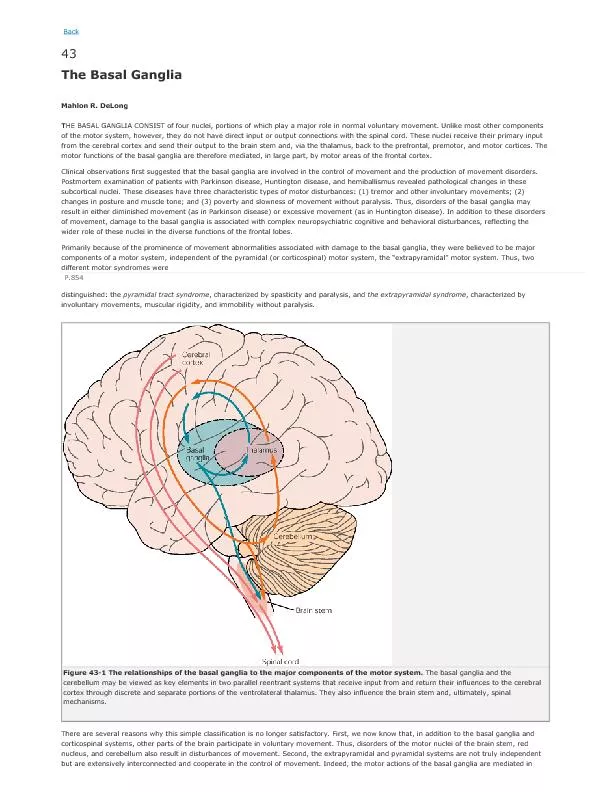
The basal nuclei are joined together and connected with many different regions of the nervous system by a very complex number of neurons.
Basically, the corpus striatum receives afferent information from most of the cerebral cortex, the thalamus, the subthalamus, and the brainstem, including the substantia nigra. The information is integrated within the corpus striatum, and the outflow passes back to the areas listed above.
The activity of the basal nuclei is initiated by information received from the premotor and supplemental areas of the motor cortex, the primary sensory cortex, the thalamus, and the brainstem. The outflow from the basal nuclei is channeled through the globus pallidus, which then influences the activities of the motor areas of the cerebral cortex or other motor centers in the brainstem. Thus, the basal nuclei control muscular movements by influencing the cerebral cortex and have no direct control through descending pathways to the brainstem and spinal cord. In this way, the basal nuclei assist in the regulation of voluntary movement and the learning of motor skills.
Slide11Functions of Corpus Striatum1.The corpus striatum regulates muscle tone and thus helps in smoothening voluntary movements.
2.It controls automatic associated movements, like the swinging of arms during walking. Similarly, it controls the coordinated movements of different parts of the body for emotional expression.
3.It influences the precentral motor cortex which is supposed to control the extrapyramidal activities of the body.
4.Lesions of the corpus striatum result in Parkinsonism. The rigidity and tremors associated with this condition can be controlled both medically and surgically
Slide12INTERNAL CAPSULE
The internal capsule is a large band of
fibres
, situated in the inferomedial part of each cerebral hemisphere.
In horizontal sections of the brain, it appears V shaped with its concavity directed laterally. The concavity is occupied by the lentiform nucleus.
The internal capsule contains
fibres
going to and coming from the cerebral cortex. It can be compared to a narrow gate where the
fibres
are densely crowded.
Small lesions of the capsule can give rise to widespread derangements of the body.
When traced upwards, the
fibres
of the capsule diverge and are continuous with the corona radiata.
When traced downwards its
fibres
converge and many of them are continuous with the cerebri of the midbrain.
Slide13Internal capsule
Slide14The internal capsule is divided into the following.
1. The anterior limb lies between the head of the nucleus and the lentiform nucleus.
2. The posterior limb lies between the thalamus and the lentiform nucleus.
3. The genu is the bend between the thalamus & lentiform nucleus.
Slide15Slide16Slide17Arterial Supply of Other Parts of Cerebral Hemisphere
Internal capsule.
It is supplied by the central branches of (
i
) the middle cerebral artery; (ii) the anterior cerebral artery; (iii) the posterior communicating artery; and (iv) the anterior choroidal artery.
Corpus striatum:
(
i
) Chiefly by central branches of the middle cerebral artery; (ii) partly by the anteromedial central branches from the anterior cerebral and anterior communicating arteries.
Slide18Slide19Extrapyramidal system
Slide20Slide21Slide22Slide23Extrapyramidal system
Slide24Slide25Slide26Slide27Slide28Slide29Slide30Slide31Slide32Slide33Thank you