
Silvina G Horovitz PhD Human Motor Control Section Medical Neurology Branch National Institute of Neurological Disorders and Stroke National Institutes of Health Outline EEG overview Why simultaneous EEGfMRI ID: 1000516
Download Presentation The PPT/PDF document "Multi-modal imaging: simult..." is the property of its rightful owner. Permission is granted to download and print the materials on this web site for personal, non-commercial use only, and to display it on your personal computer provided you do not modify the materials and that you retain all copyright notices contained in the materials. By downloading content from our website, you accept the terms of this agreement.


1. Multi-modal imaging: simultaneous EEG-fMRI Silvina G Horovitz, PhDHuman Motor Control SectionMedical Neurology BranchNational Institute of Neurological Disorders and StrokeNational Institutes of Health
2. OutlineEEG overview Why simultaneous EEG-fMRI? How? Technical considerations When? Examples
3. EEG (electroencephalography) measure of synchronous activity of population of neurons, primarily reflects postsynaptic potentials.
4. EEG measuresrecording device Acquisition PCElectrodes and conductive mediaIsolated amplifiersfilters A/D converter1 sEyes closed
5. montageInternational 10-20 System of Electrode Placement F - Frontal lobe T - Temporal lobeC - Central lobe P - Parietal lobeO - Occipital lobe"Z" refers to an electrode placed on the mid-line. Odd: leftEven: rightElectrode configurationReferential (Si vs. Ref; Sk vs. Ref)Bipolar(Si vs.. Sk )
6. Data processingTime domainEvent Related Potentials (ERPs)pre-processing:detrend - filtering epochbaseline correctionocular artifact reduction(common grounded, artifact rejection)time-locked averaging
7. Data processingFrequency domainPower at different bandsPower spectra density (FFT)Cross-spectra (correlation among different electrodes)Coherence (measure of stability of the phase shift between electrodes) Event related desynchronization
8. Human EEG Spontaneous Evoked Clinical Applicationsepilepsy, head trauma, drug overdose, brain infection, sleep disorder, coma, stroke, Alzheimer’s disease, brain tumor, multiple sclerosis, surgical monitoring Scalp Depth ECoG Transient Steady StateCognitive Sciencesensory pathways, stimulus encoding, motor process, spatial task, verbal task, mathematics, short term memory, memory encoding, selective attention, task context, general intelligence, dynamic brain theory PL Nunez, EEG, Encyclopedia of the Brain, 2003
9. Why do we want to measure EEG and fMRI simultaneously?
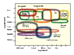
10. NeuroimagingBRAIN ACTIVITY f(x,t)Metabolic/vascularresponsesg[f(x,t)]fMRI k{g[f(x,t)]}electricalEEG, MEG m[f(x,t)] Good time resolution (ms)Poor spatial resolutionPoor time resolution (s)Good spatial resolution (mm)
11. EEG fMRIEEG is the gold standard for sleep studies, epilepsy, some cognitive tasks, etcP1N170P2
12. Data PubMed search July 14, 2015YEARNr. of publicationsHOW often measured together?
13. EEG setupEEGstudyStimulus PCEEG amplifierAcquisition PC
14. EEG-fMRI setupScanner clockSynchronization Adapter Optic fiberAcquisition PCAmplifierScanner triggerStimulus PCto projectorEEG-fMRIstudy
15. Technical IssuesElectromagnetism 101Maxwell's Laws. A changing magnetic field produces an electric field A changing electric field or current produces a magnetic field. Luckily, the magnetic field change Form the EEG does not affect the image quality! BIG PROBLEM
16. THE not so good NEWSMRI is noisy Electrical noise MRI and EEG were not meant for each other …Remember Maxwell's Law?
17. Simultaneous EEG-fMRI - Technical issuesSources of artifacts: gradient artifact physiological noise: ballistocardiogramExample fromBOLD & Perfusion MRI sequenceoptimized for EEG-fMRI acquisition 5 slices
18. Simultaneous EEG-fMRI - Technical issuesApproximate values of different signalsGradient artifact : ± 10mVEEG: ± 150mVBC artifact: ± 200mVBlink: ± 150mVMovement: < 1mVECG: ± 20mVEMG: ± 50mVHelium pump: 40-60Hz and
19. THE good NEWS MRI compatible EEGequipment, leads and electrodesSafe for the scannerSafe for the subjectMore on safety later Careful setup: Equipment CablesSubject head
20. DATA acquisitionSample EEG at 5 kHz (or more)Slice TR at a frequency that is not of interest (and a round number)Low Pass Filter at 250Hz~0.01 Hz high pass to avoid saturation (use DC only if enough range )Volume (or slice) markerResolution: 0.5mV (make sure dynamic range covers the signal, depends on scanner and configuration)Clock synchronization
21. EEG DATA acquisitionMake sure amplifiers do not saturateAdjust amplifier resolutionKeep electrodes’ impedance low (unless using high impedance equipment)Keep cabling safe and fixedHave a good cardiac signalAdjust MR sequenceAdjust experiment (ISI <> TR)
22. Gradient artifact removalArtifact removal (Matching filter)Create a template of the artifactSubtract average artifactIf proper timingand no movement works great!Raw EEG during EPI acquisition
23. RawDr Jen EvansGradient artifact corrected
24. Ballistocardiogram artifact removalMatching filter (BV Analyzer) (Allen et al, 2000):Detect R Create a templateSubtract (allows for amplitude adjustment)Single Value Decomposition (Neuroscan)Run classificationRemove componentsReconstruct time seriesOptimal base set (EEGLAB Niazy, 2005)PCA to create basesFitting (adaptive algorithm)SubtractionCombinationsi.e Liu, 2012 use ICA, SVD & mutual information (based on Peng, IEEE 2005) software download: http://amri.ninds.nih.gov/cgi-bin/software
25. Gradient artifact correctedCardioballistogram correctedDr Jen Evans
26. Gradient artifact correctedCardioballistogram corrected
27. SAFETY considerationsSimultaneous Electroencephalography-Functional MRI at 3 T: An Analysis of Safety Risks Imposed by Performing Anatomical Reference Scans With the EEG Equipment in PlaceUlrike Nöth, Laufs, Stoermer, and Deichmann JMRI 2012SAR: Specific Absorption Rate (or the energy deposited in the body by the radio frequency transmission)
28. SAFETY considerationsSequences EPIs (in most cases ok to run an MPRAGE for localization)be aware of high res short TR EPIs (pay attention to SAR)Special sequences require special safety testing Set upCables straight and in the center. Avoid loopsEquipment as far back from iso-center as possible(far front for EMG)All scanners are not equal; gradients and coils affect electrodes’ temperatureBe aware different body shapes and weight load coil differently
29. Interim SummaryEEG measurements have: Good temporal resolution Poor spatial resolution (when measure non invasively)Electrical and hemodynamic responses are relatedSimultaneous EEG-fMRI requires special equipment SAFETY PROCEDURES ARE KEYDimensionality reduction is needed for data integration
30. When do we want to measure EEG and fMRI simultaneously?
31. When is it important to measure simultaneously? State dependent analysisAlertness State vs Traitunderstanding of BOLD signal/EEG origingsPhysiological markers defined by EEGSeizuresSleep stages
32. Type of studiesCorrelations of EEG and fMRIIn time domainIn frequency domain Multivariate methodsICAInforming one with the otherSorting data and perform analysis in one modalityMix analysis
33. EEG parameter as regressor
34. BOLD-EEG band-power correlations Goldman et al. 2002Simultaneous EEG and fMRI of the alpha rhythm.
35. How to link time and space information? Parametric studies and correlational analysis Correlation maps between fMRI signal change and P300 amplitude. Composite of 7 subjects.Horovitz et al MRI, 2002OLD DAYS: SAME SUBJECTS, EEG AND FMRI ON SEPARATE SESSONS
36. Assessing the spatiotemporal evolution of neuronal activation with single-trial event-related potentials and functional MRITom Eichele PNAS 2005 vol. 102 no. 49
37. Single-Trial Analysis of Oddball Event-RelatedPotentials in Simultaneous EEG-fMRIBenar et al. Human Brain Mapping 28:602–613 (2007)
38. DAYTIME 1 hour PARADIGMHorovitz et al HBM, 2008Correlation between Amplitude of BOLD fluctuations and alertness Index derived from EEG
39. EEG to define states
40. Changes in the level of consciousness Use EEG to sort fMRI data
41. Do changes in connectivity over time have a physiological origin?
42. EEG-vigilance and BOLD effect during simultaneous EEG/fMRI measurementS. Olbrich et al. / NeuroImage 45 (2009) 319–
43. Spatiotemporal dynamics of the brain at rest-exploring EEG microstates as electrophysiological signatures of BOLD resting state networks. Yuan H, Zotev V, Phillips R, Drevets WC, Bodurka J.Neuroimage. 2012 
44. EEG to understand BOLD signal
45. Correlations of simultaneously acquired SSVEPs with BOLD fMRI response J.W Evans et al OHBM 2015
46. EEG & fMRI to study disease
47. Widespread epileptic networks in focalepilepsies—EEG-fMRI studyFahoum et al Epilepsia, 53(9), 2012Between April 2006 and December 2010, 168 consecutive 3T EEG-fMRI scans were performed in focal epilepsy patients FLE group (n = 14) TLE group (n = 32)Group analysis results for hemodynamic response functions peaking at 3, 5, and 7 s after the interictal epileptic dischargesDifferent epileptic syndromes result in unique and widespread networks related to focal IEDs.
48. intracranial recordings - fMRISimultaneous intracranial EEG–fMRI in humans: Protocol considerations and data quality D.W. Carmichael (2012)
49. Simultaneous EEG-fMRI summarySafety first!Quality control at experiment setup & data collection Equipment setupPulse sequenceTask designEEG pre-processing Gradient & ballistocardiogram artifacts Data integrationDimensionality reductionSpatial correlationsRegressionsSorting data based on stateSome applicationsUnderstanding BOLD signalUnderstanding DiseaseOrigins of EEG signalsState dependent studies