
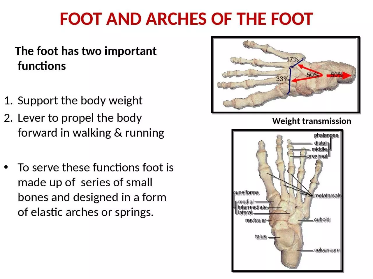
The foot has two important functions 1 Support the body weight 2 Lever to propel the body forward in walking amp running To serve these functions foot is made up of series of small bones and designed in a form of elastic arches or springs ID: 1032001
Download Presentation The PPT/PDF document "FOOT AND ARCHES OF THE FOOT" is the property of its rightful owner. Permission is granted to download and print the materials on this web site for personal, non-commercial use only, and to display it on your personal computer provided you do not modify the materials and that you retain all copyright notices contained in the materials. By downloading content from our website, you accept the terms of this agreement.


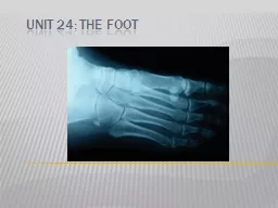
1. FOOT AND ARCHES OF THE FOOT The foot has two important functions 1. Support the body weight2. Lever to propel the body forward in walking & runningTo serve these functions foot is made up of series of small bones and designed in a form of elastic arches or springs.Weight transmission
2. FUNCTIONS OF THE ARCHESHelp in proportionate distribution of weight -weight distributed equally through the anterior and the posterior part of the foot -heads of five metatarsals posses six weight bearing pointsArched foot acts as a segmental levelPlantar concavity prevents compression of neurovascular structures of the footArched foot is dynamic and pliableInvertors and evertors help in shifting weight distributionNormal Foot Foot print
3. ARCHES OF FOOTLongitudnal arches- -medial longitudnal arch -lateral longitudnal arch 2. Transverse archSupports during standing- - plantar ligaments, plantar aponeurosis bear maximum stressSupports during locomotion- - muscles are active - windlass action of plantar aponeurosis
4.
5.
6. Medial part
7. Lateral part
8. FDB
9. Flexor dig Brevis
10. Flex hallucis longusFlex dig longus
11. FDLAB HAL
12. Flex accessorius
13. F AcceSLumbiricals
14.
15.
16. Long planter ligPeronues longus
17.
18. P Longus
19. FACTORS MAINTAINING THE ARCHESShape of bonesStaplesSlingsTie beam
20. MEDIAL LONGITUDNAL ARCHSummit -trochlear surface of talusAnterior pillar -heads of medial three metatarsalsPosterior pillar - medial tubercle of calcaneusVulnerable part of arch - head of talus (keystone)Characteristic feature of arch - resiliency
21. FACTORS MAINTINING MEDIAL ARCHShape of bones - wedge shaped bones - keystone (head of talus)Staples - plantar ligaments - most important plantar calcaneonavicular (spring ligament)Tie beam - plantar aponeurosis, abductor hallucis, flexor hallucis longus and brevis tendon, medial part of flexor digitorum longus and brevisSlings - tibialis anterior tendon, deltoid ligament and tibialis posterior tendon
22. LATERAL LONGITUDNAL ARCHSummit - subtalar jointAnterior pillar - head of fourth and fifth metatarsalsPosterior pillar - medial tubercle of calcaneusVulnerable part of arch - calcaneocuboid jointCharacteristic feature of arch - rigidity
23. FACTORS MAINTINING LATERAL ARCHShape of bones - calcanean angle of cuboid maintains upward tilt of calcaneusStaples - long and short plantar ligamentsTie beam - plantar aponeurosis, abductor digiti minimi, flexor digiti minimi brevis, lateral part of flexor digitorum longus and brevis tendonsSlings - Peroneus brevis , peroneus tertius and peroneus longus
24. TRANSVERSE ARCHAnterior transverse archis formed by heads of the five metatarsal bones is completePosterior transverse archis formed by greater parts of tarsus & metatarsusis incomplete – only the lateral end comes in contact with the ground
25. FACTORS MAINTINING TRANSVERSE ARCHShape of bones - wedge shaped cuneiforms and bases of middle three metatarsalsStaples - deep transverse ligaments, intrinsic plantar ligaments, dorsal interossei, oblique and transverse heads of adductor hallucisTie beam - tendons of peroneus longus and tibialis posteriorSlings - peroneus brevis and tertius laterally - tibialis anterior tendon medially
26. APPLIED ANATOMY OF FOOTPlantar fasciitis: Inflammation in the plantar fascia ligament Pain in the heel and arch, worst in the morning, are symptoms.Osteoarthritis of the feet: wear and tear cause the cartilage to wear out. Pain, swelling, and deformity in the feet.Gout: An inflammatory condition causing severe pain and swelling big toe is often affectedAthlete's foot: A fungal infection of the feet, causing dry, flaking, red, and irritated skin. Rheumatoid arthritis: An autoimmune form of arthritis that causes inflammation and joint damageBunions (hallux valgus): A bony prominence next to the base of the bigDiabetic foot infection: diabetics are vulnerable , such as redness, warmth, swelling, and pain.
27. Swollen feet (edema): can be normal after prolonged standing common in people with varicose veins, can also be a sign of heart, kidney, or liver problems.Calluses: A buildup of tough skin over an area of frequent friction or pressure on the feet, usually develop on the balls of the feet or the heels and may be uncomfortable or painful.Corns: corns consist of excessive tough skin buildup at areas of excessive pressure on the feet,typically have a cone shape with a point, and can be painful.Heel spurs: An abnormal growth of bone in the heel, which may cause severe pain during walking or standing. Ingrown toenails: One or both sides of a toenail may grow into the skin. Ingrown toenails may be painful or lead to infections. Fallen arches (flat feet): The arches of the feet flatten during standing or walkingNail fungal infection (onychomycosis): Fungus creates discoloration or a crumbling texture in the fingernails or toenails. Nail infections can be difficult to treat.
28. Mallet toes: The joint in the middle of a toe may become unable to straighten, causing the toe to point down. Irritation and other feet problems may develop without special footwear to accommodate the mallet toe.Metatarsalgia: Pain and inflammation in the ball of the foot. Strenuous activity or ill-fitting shoes are the usual causes.Claw toes: Abnormal contraction of the toe joints, causing a claw-like appearance. Claw toe can be painful and usually requires a change in footwear.Fracture: The metatarsal bones are the most frequently broken bones in the feet, either from injury or repetitive use. Pain, swelling, redness, and bruising may be signs of a fracture.Plantar wart: A viral infection in the sole of the foot that can form a callus with a central dark spot. Plantar warts can be painful and difficult to treat.Morton's neuroma: A growth consisting of nerve tissue often between the third and fourth toes. A neuroma may cause pain, numbness, and burning and often improves with a change in footwear.
29. Hammer toeA hammer toe occurs when a toe bends down at the middle toe joint, metatarsophalangeal joint,causing the middle toe joint to be raised slightly. There may also be a deformity at the distal interphalangeal jointClaw toeClaw toe often affects all toes at the same time (except the big toe), causing them to bend downward at both the middle joints and the joints nearest the tip so that the toes curl down. The toes bend up at the joints where the toes and the foot meet (MTP joint).Mallet toeA mallet toe often affects the longest toe (generally the second toe), but it may affect the other toes as well. The toe bends down at the joint closest to the tip (DIP joint).
30. Talipes – congenital deformity of the foot, usually marked by a curled shape or twisted position of the ankle, heel and toes. the most common is known as Congenital Talipes Equinovarus (CTEV) or clubfoot. The four different forms are Talipes equinusTalipes calcaneus Talipes valgus Talipes varus
31.
32. PES CAVUS deformity characterised by an abnormally high medial longitudinal archPES PLANUS
33. 3 ways in which the Achilles can rupture are: -Pushing off on the weight bearing foot while extending the flexed knee e.g. out of the blocks in a sprint, or jumping. -Suddenly and unexpectedly dorsiflexing the foot, which would happen when stepping into a hole -Violent dorsiflexion of the foot when it is plantarflexed, such as in falling from a height. usually person feels sudden pain in the heel, and may mistakenly think they have been shot or kicked in the heel. will have weakness when plantar flexing Thompson test. Squeezing the calf as shown will plantarflex the foot (black dotted line) if the tendon is intact. There will be no movement with Achilles tendon rupture.
34. Walking Cycle
35. Propulsive Mechanism of the footIn walking: Transmission of weight borne by the foot is successively transferred along the heel, lateral border & the ball of foot and anterior pillar of the medial longitudinal arch and medial 3 digits.Increase in the height of medial arch by “windlass action” of plantar aponeurosis.Extension of great toe causes elongation of FH & FDL which increases the force of subsequent contraction.Both long and short flexor muscles of toes increase the force of take-off.Prevention of buckling of toes is done by lumbricals.In running: Heel remains above the ground.Take-off point is maintained by the anterior pillar of the medial longitudinal arch.
36. Walking CycleA person makes about 1700-1800 foot strikes in every mile.The average man walks 70,000 miles in his life-time.It consists of series of swing and stance phases in succession.Swing phase: when the lower limb is off the ground.Stance phase: the foot strikes the ground and bears weight.Walking Cycle: the period from the heel-strike of one foot to the heel-strike of the same foot.Bobbing: Head is displaced upward twice in stance phase, by alternate uses of lower limbs and undergoes corresponding downward movement in swing phase. The amount of vertical displacement is about 5 cm.During locomotion lateral displacement of 5 cm is evident when viewed from front or behind.In swing phase, a person bends the trunk to the side on which he stands for balancing.The trunk displacement is reversed when the other lower limb bears the weight alternately.Simultaneously the arm undergoes alternate forward swing with the opposite leg.
37. Sequence of movements in walking cycleIn first part of swing phase: Hip, knee and ankle are flexed.The limb then begins to extend and is fully extended until the heel of the advancing foot strikes the ground.Hip flexors are most active during the early part of the swing, and the hip extensors are maximally active at heel-strike.Dorsiflexors of the foot are active at the beginning of swing phase to clear the foot from the ground, and after heel-strike to prevent it from slapping the ground.
38. Sequence of movements in walking cycle (contd.)At the beginning of stance phase: the knee flexes slightly when the weight is fully borne.It again undergoes full extension at the end of the stance phase.Foot is then bent at the metatarso-phalangeal joints.Plantar flexors of the foot are most active during the later half of stance.The toes tend to flex and grip the ground during the same period.Long extensors and intrinsic muscles of the foot stabilize the toes and provide fixed origins for the long flexors and extensors to act on the leg.Invertors and evertors of foot are important stabilizers in stance phase.
39. Sequence of movements in walking cycleIn first part of swing phase: Hip, knee and ankle are flexed.The limb then begins to extend and is fully extended until the heel of the advancing foot strikes the ground.Hip flexors are most active during the early part of the swing, and the hip extensors are maximally active at heel-strike.Dorsiflexors of the foot are active at the beginning of swing phase to clear the foot from the ground, and after heel-strike to prevent it from slapping the ground.
40. Control and RegulationWalking is a laboriously learned and automatic activity.It depends upon the reflex patterns mediated by the spinal cord and controlled by the brain.Walking reflexes are regulated by a variety of sensory informations of tactile, ligamentous, articular and musculo-tendinous nerve endings.The pattern of walking may be altered by individual style, poor posture, overweight, foot wear etc.Disturbances of gait are important signs in many disorders of the central nervous system.
41. Disturbances of GaitAtaxic gaitHemiplegic gaitScissors gaitStaggering gaitWaddling gaitCerebellar gaitPropulsion gaitLimping gait
42. Multiple choice Questions
43. The arrow marked structure is the tendon ofQ. 1A. Peroneus BrevisB. Peroneus LongusC. Tibialis AnteriorD. Tibialis Posterior
44. Which of the following ligament supports the transverse arch -A. Long plantar ligamentB. Short plantar ligamentC. Deep metatarsal ligament D. Plantar Aponeurosis Q. 2
45. The keystone of the lateral longitudinal arch isA. NavicularB. Lateral CuneiformC. CalcaneumD. CuboidQ. 3
46. Medial longitudinal arch of the foot is not formed by –A. CuboidB. CalcaneusC. TalusD. NavicularQ. 4
47. Q. 5A 32 years old patient goes to the clinic with a noticeable pain in the first and second metatarsal heads of the left foot, and in the first metatarsal head of the right foot. The patient works as a hairdresser, she stands up about 8 hours a day in static position and walking. The following symptoms could be associated withA. Pes cavusB. Pes planusC. TalipesD. Hallux valgus